Review
. 2015 Jun;30(3):163-82.
doi: 10.1177/0748730414563537. Epub 2015 Jan 13.

The circadian clock in skin: implications for adult stem cells, tissue regeneration, cancer, aging, and immunity

Affiliations
Review

The circadian clock in skin: implications for adult stem cells, tissue regeneration, cancer, aging, and immunity

Maksim V Plikus et al. J Biol Rhythms. 2015 Jun.

Abstract

Historically, work on peripheral circadian clocks has been focused on organs and tissues that have prominent metabolic functions, such as the liver, fat, and muscle. In recent years, skin has emerged as a model for studying circadian clock regulation of cell proliferation, stem cell functions, tissue regeneration, aging, and carcinogenesis. Morphologically, skin is complex, containing multiple cell types and structures, and there is evidence for a functional circadian clock in most, if not all, of its cell types. Despite the complexity, skin stem cell populations are well defined, experimentally tractable, and exhibit prominent daily cell proliferation cycles. Hair follicle stem cells also participate in recurrent, long-lasting cycles of regeneration: the hair growth cycles. Among other advantages of skin is a broad repertoire of available genetic tools enabling the creation of cell type-specific circadian mutants. Also, due to the accessibility of skin, in vivo imaging techniques can be readily applied to study the circadian clock and its outputs in real time, even at the single-cell level. Skin provides the first line of defense against many environmental and stress factors that exhibit dramatic diurnal variations such as solar ultraviolet (UV) radiation and temperature. Studies have already linked the circadian clock to the control of UVB-induced DNA damage and skin cancers. Due to the important role that skin plays in the defense against microorganisms, it also represents a promising model system to further explore the role of the clock in the regulation of the body's immune functions. To that end, recent studies have already linked the circadian clock to psoriasis, one of the most common immune-mediated skin disorders. Skin also provides opportunities to interrogate the clock regulation of tissue metabolism in the context of stem cells and regeneration. Furthermore, many animal species feature prominent seasonal hair molt cycles, offering an attractive model for investigating the role of the clock in seasonal organismal behaviors.

Keywords: UV exposure; aging; autoimmune diseases; cancer; cell cycle; hair follicle; immunity; regeneration; skin; stem cells.


Figures

Figure 1
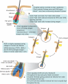
Figure 1. The complex anatomical organization of skin and its circadian clocks
The schematic drawing depicts the three principle layers of the skin: the keratinocyte-containing epidermis (brown), the fibroblast-rich dermis (purple) and the fat cell-containing adipose tissue (yellow). The interfollicular epidermis is a stratified squamous epithelium containing stem/progenitor cells in the basal layer (BL) and post-mitotic differentiated cells in the suprabasal layers (SBL). The top layer of the epidermis, the stratum corneum (SC), is composed of dead cells with a tough cell envelope that is sealed by an impermeable intercellular lipid layer. Also, highlighted are hair follicles with the associated lipid-rich sebaceous glands, as well as sweat glands; both are keratinocyte-containing mini organs which develop as outgrowths from the epidermis. The skin is also inhabited by melanocytes, melanin-producing pigment cells that confer color to hair and skin. In mice, melanocytes are primarily found in hair follicles while in humans melanocytes are also prominent at the epidermal-dermal junction. In addition, several types of resident and transient immune cells are found within the skin. These include Langerhans cells and dendritic cells (DCs), antigen-presenting cells residing in the epidermis and dermis, respectively, as well as lymphocytes, macrophages and mast cells. The skin is also richly vascularized and innervated; cells within these structures likely have their own circadian clock that could modify their functions including sensory responses, heat regulation and oxygenation. The surface of the skin is populated by a large number of commensal bacteria (microbiota) that may play a role in skin function and disease. There is evidence for active circadian clock in all cell types of the skin, and it is highly likely that distinct functions are modulated in different cell types. It is also known that circadian clock activity in skin is coordinated by the suprachiasmatic nucleus, presumably through neuronal and hormonal mediators although this remains to be defined in skin.
Figure 2
Figure 2. The circadian clock modulates skin responses to environmental stresses
Protection against a variety of environmental insults -- these include UVB radiation, temperature, chemical and physical injury and microbial infections -- is a major function of the skin. (A, C) There are prominent diurnal rhythms in DNA replication, DNA repair mechanisms and cell division in progenitor/stem cells of the epidermis -- features that depend on an intact circadian clock within epidermal keratinocytes. Exposure of mouse epidermis to UVB at night, during the replicative burst when DNA excision repair is at its nadir, causes more DNA damage and higher incidence of skin cancer than during the day. Based on these mouse studies, and the fact that in diurnal humans the epidermis is more proliferative during the day, it has been proposed that the epidermis could be most susceptible to UVB-induced DNA damage during the day, the time of maximum solar exposure (Gaddameedhi et al., 2011; Geyfman et al., 2012). If correct, this hypothesis implies that the circadian clock confers regulation that promotes rather than protects against UVB-induced skin cancer formation and skin aging in humans. (B) Epidermal barrier functions, such as transepidermal water loss and stratum corneum hydration are also prominently regulated by the circadian clock and become largely arrhythmic in Clock mutant mice. Diurnal cycle in epidermal barrier functions at least in part depend on the daily changes in Aquaporin 3 expresion levels -- the putative clock output gene in basal epidermal keratinocytes. (D) Disruption of the circadian clock in the germline leads to aberrant wound healing in mice. Wound healing is a very complex process involving most, if not all, major cell types in the skin and the underlying mechanisms for clock modulation of wound healing remain to be defined. However, it is known that during injury-induced wound healing, the circadian clock contributes to the replicative slowdown of myofibroblasts (aka senescence), a process important for conversion from scar production to scar remodeling and its proper maturation. (E) The immune system is under control of the circadian clock. The influx of immune cells into tissues has been shown to be regulated by the circadian clock. In the skin, studies in hamsters have shown circadian variation in the trafficking of antigen presenting cells. In mice, both acute and delayed-type inflammatory responses have been linked to clock regulation. Many immune-related genes in skin have circadian rhythmicity, suggesting that skin responses to infection and autoimmune insults can be time-of-day dependent.
Figure 3
Figure 3. The circadian clock modulates hair follicle regeneration
(A) Circadian clock activity in the infundibulum and isthmus of hair follicles is similar to that of the interfollicular epidermis. In these progenitor/stem cell populations, the clock intrinsic to keratinocytes is required for diurnal variation in DNA replication. (B) There is heterogeneity in circadian output in bulge stem cells. High clock activity correlates with the high expression of WNT and TGFβ pathway genes, components of signaling pathways involved in stem cell activation. The functional consequence of clock-WNT/TGFβ connection is unclear, because deletion of Bmal1 within keratinocytes does not affect stem cell activation and timing of the first two hair growth cycles (Geyfman et al., 2012). Clock regulation of these signaling pathways, however, could affect the aging and cancer forming susceptibility of these stem cells. (C) Epithelial hair germ progenitors display prominent circadian activity prior to and during anagen onset. Whereas germline deletion of Bmal1 delays anagen initiation, such effects are not found in mice deleted for Bmal1 in keratinocytes. These findings suggest that the influence of the clock on anagen initiation is through other cell types, for example dermal papilla cells, or through more global regulation, such as modulation of systemic hormones that affect hair growth. (D) Silencing on clock genes in human hair follicles in vitro prolongs active growth phase. Due to technical limitations, the influence of the clock on other aspects of human hair follicle growth could not be studied. (E) During active growth phase in mouse hair follicles, circadian clock gates cell cycle progression in the epithelial matrix cells at the G2/M checkpoint. Administration of ionizing radiation to mice leads to more severe hair loss in the morning, during the mitotic peak.
Figure 4
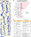
Figure 4. Many immune genes in skin display circadian rhythmicity
(A) A heat map showing the expression of immune-related genes in telogen over two days based on previously published whole skin microarray datasets generated in mice (Geyfman et al., 2012). Multiple genes with established immune function exhibit circadian expression. The day and night periods are indicated at the top and the gene expression strength indicated at the bottom. (B) Shown is the gene ontology for the circadian immune genes. The number at the end of bars refers to the number of genes with the specific function. Genes that peak at day or night are indicated with the pink and purple color. (C) The function of selective immune genes is indicated. These genes are marked with an asterix in A.

Similar articles

Cited by

References

    1. Abe T, Sakaue-Sawano A, Kiyonari H, Shioi G, Inoue K, Horiuchi T, Nakao K, Miyawaki A, Aizawa S, Fujimori T. Visualization of cell cycle in mouse embryos with Fucci2 reporter directed by Rosa26 promoter. Development. 2013;140:237–246. - PubMed
    1. Akashi M, Soma H, Yamamoto T, Tsugitomi A, Yamashita S, Yamamoto T, Nishida E, Yasuda A, Liao JK, Node K. Noninvasive method for assessing the human circadian clock using hair follicle cells. Proceedings of the National Academy of Sciences of the United States of America. 2010;107:15643–15648. - PMC - PubMed
    1. Al-Nuaimi Y, Hardman JA, Biro T, Haslam IS, Philpott MP, Toth BI, Farjo N, Farjo B, Baier G, Watson RE, Grimaldi B, Kloepper JE, Paus R. A meeting of two chronobiological systems: circadian proteins Period1 and BMAL1 modulate the human hair cycle clock. The Journal of investigative dermatology. 2014;134:610–619. - PubMed
    1. Ansari N, Agathagelidis M, Lee C, Korf HW, von Gall C. Differential maturation of circadian rhythms in clock gene proteins in the suprachiasmatic nucleus and the pars tuberalis during mouse ontogeny. The European journal of neuroscience. 2009;29:477–489. - PMC - PubMed
    1. Arjona A, Silver AC, Walker WE, Fikrig E. Immunity's fourth dimension: approaching the circadian-immune connection. Trends in immunology. 2012;33:607–612. - PMC - PubMed

Publication types