. 2015 Jan 13;56(2):1097-107.
doi: 10.1167/iovs.14-15596.

Degeneration and regeneration of corneal nerves in response to HSV-1 infection

Affiliations

Degeneration and regeneration of corneal nerves in response to HSV-1 infection

Ana J Chucair-Elliott et al. Invest Ophthalmol Vis Sci. .

Abstract

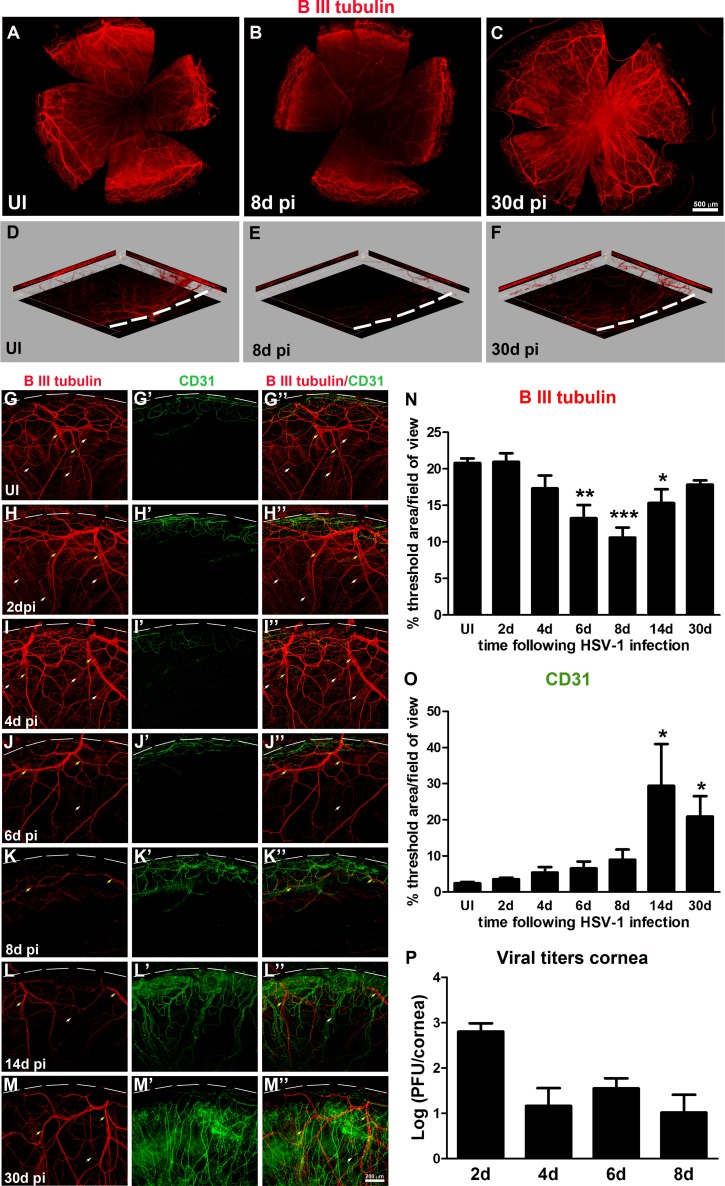
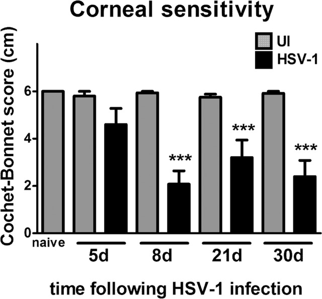
Purpose: Herpes simplex virus type 1 (HSV-1) infection is one cause of neurotrophic keratitis, characterized by decreases in corneal sensation, blink reflex, and tear secretion as consequence of damage to the sensory fibers innervating the cornea. Our aim was to characterize changes in the corneal nerve network and its function in response to HSV-1 infection.

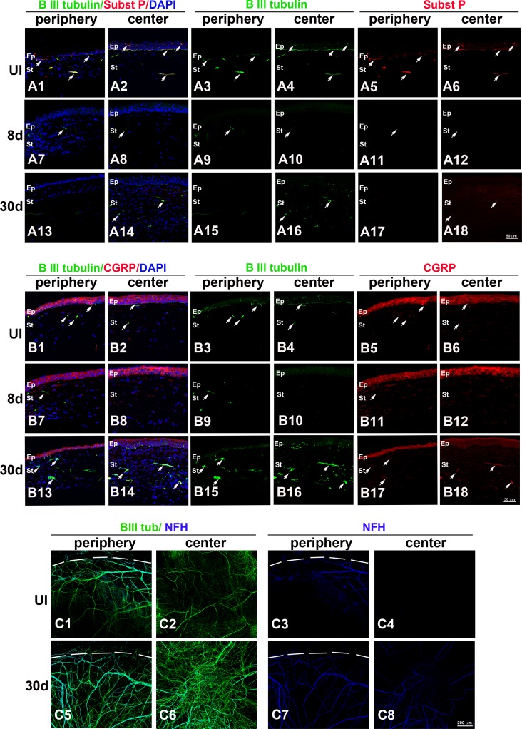
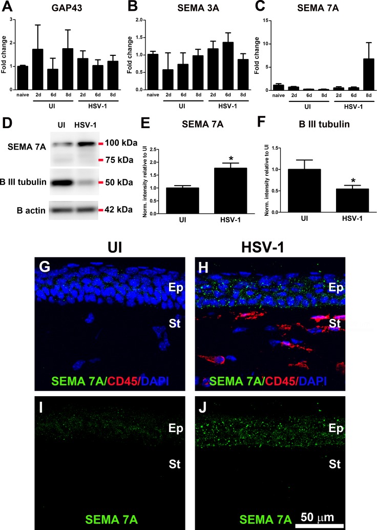
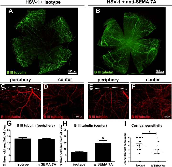
Methods: C57BL/6J mice were infected with HSV-1 or left uninfected. Corneas were harvested at predetermined times post infection (pi) and assessed for β III tubulin, substance P, calcitonin gene-related peptide, and neurofilament H staining by immunohistochemistry (IHC). Corneal sensitivity was evaluated using a Cochet-Bonnet esthesiometer. Expression of genes associated with nerve repair was determined in corneas by real time RT-PCR, Western blotting, and IHC. Semaphorin 7A (SEMA 7A) neutralizing antibody or isotype control was subconjunctivally administered to infected mice.

Results: The area of cornea occupied by β III tubulin immunoreactivity and sensitivity significantly decreased by day 8 pi. Modified reinnervation was observed by day 30 pi without recovery of corneal sensation. Sensory fibers were lost by day 8 pi and were still absent or abnormal at day 30 pi. Expression of SEMA 7A increased at day 8 pi, localizing to corneal epithelial cells. Neutralization of SEMA 7A resulted in defective reinnervation and lower corneal sensitivity.

Conclusions: Corneal sensory nerves were lost, consistent with loss of corneal sensation at day 8 pi. At day 30 pi, the cornea reinnervated but without recovering the normal arrangement of its fibers or function. SEMA 7A expression was increased at day 8pi, likely as part of a nerve regeneration mechanism.

Keywords: HSV-1; corneal nerves; neurotrophic keratitis; reinnervation; semaphorin 7A.


Figures

Figure 1
Figure 1
HSV-1 infection results in deinnervation and subsequent reinnervation, consistent with progressive neovascularization of the cornea. Mouse eyes were infected with 103 PFU of HSV-1 or left UI as controls. At 2, 4, 6, 8, 14, and 30 days pi, corneas were processed for IHC staining with β III tubulin (red) and CD31 (green) antibodies. (A–C) Representative corneal whole-mount images. In comparison to UI (A), displaying intact corneal innervation, HSV-1 infection resulted in loss of corneal nerves more pronounced in the center of a 6- and 8-day pi (B) and in the reinnervation process evident at 30 days pi (C). A unique feature observed in some corneas at 30 days pi was abnormal and disorganized hyperinnervation in the center of the tissue. (D–F) Three-dimensional view of representative images showing loss of overall innervation at day 8 pi (E) and regaining innervation at day 30 pi (F). Notice the red signal from β III tubulin-stained nerves, absent throughout the thickness of the optical section (E), and nerve recovery (P). (G–M'') Representative confocal images of UI and infected cornea quadrants oriented with the limbal margins at the top (white dashed lines). Compared to UI (G–G”) displaying intact innervation with thick stromal nerves (yellow arrows) and fine sub-basal nerve bundles (white arrows), HSV-1 infection caused progressive loss of nerves and complete loss of sub-basal nerves at day 8 pi, consistent with the onset of neovascularization of the cornea (K–K''). This event was followed by reinnervation that coexisted with persistent neovascularization (L–L'', M–M''). Corneal innervation and neovascularization were quantified as percentage of threshold area positive for β III tubulin signal (N) and for CD31 signal (O) in representative confocal images (G–M''). Bars represent the percentage of threshold area/field of view ± SEM (n = at least 4 total replicates from 3 independent experiments for each group in (N); and n = at least 3 replicates from 2 independent experiments for each group in (O). *P < 0.01; **P < 0.001; ***P < 0.0001, compared to UI group as determined by ANOVA followed by Newman-Keuls multiple comparison test. (P) Viral content in corneas at 2, 4, 6, and 8 days pi as assessed by viral plaque assay and expressed as log (PFU/cornea) ± SEM (n = 6 total replicates from 2 independent experiments per time point).
Figure 2
Figure 2
HSV-1 infection results in loss of corneal sensory fibers and a deficient or abnormal reinnervation process. Mouse eyes were infected with 103 PFU HSV-1 or left UI as controls. At 8 and 30 days pi, corneas were processed for IHC costaining with β III tubulin (green) with SP (red), CGRP (red), or NFH (blue) antibodies. (A1–A18) Sagittal frozen sections of corneas costained with β III tubulin and SP. In the UI cornea, both periphery (A1, A3, A5) and center (A2, A4, A6) showed β III tubulin+ nerves in the stroma and rich subbasal innervation penetrating the layers of epithelial cells that colocalized with expression of SP (white arrows). Loss of sub-basal innervation was observed at 8 days pi (A7–A12) with remaining peripheral nerves that did not colocalize with SP (A7, A9, A11). The reinnervation of the cornea at 30 days pi (A13–A18) involved poor sub-basal reinnervation and hyperinnervation of the central stroma without recovery of SP+ signal (white arrows). (B1–B18) Sagittal frozen sections of corneas costained with β III tubulin and CGRP. In the UI cornea, CGRP expression was found mostly within the epithelial cells (B1–B6) and was occasionally associated with nerves at the sub-basal and stromal locations. At 8 days pi, CGRP continued to be expressed in the epithelium and was not associated with remaining nerves at the peripheral cornea (B7–B12). At day 30 pi, CGRP reactivity at the central epithelium region was largely lost, whereas some of the nerves that reinnervated the stroma colocalized with CGRP expression (B13–B18). (C1–C8) Corneal whole mounts show hyper-reinnervation at the center of the cornea at 30 days pi that included the abnormal presence of NFH+ nerves. DAPI, nuclei counterstain in blue; Ep, epithelium; St, stroma. Figure represents n = 3 mice/time point or UI controls from two independent experiments.
Figure 3
Figure 3
Corneal reinnervation following HSV-1 infection does not correlate with recovery of corneal sensitivity. Mouse eyes were infected with 103 PFU HSV-1, were left UI as controls, or were not subjected to the scarification procedure (naive). At 5, 8, 21, and 30 days pi, corneal sensitivity levels were measured by Cochet-Bonnet esthesiometer and compared among groups. At day 8 pi, there was a significant loss of corneal sensitivity recorded in corneas of infected mice that did not recover by day 30 pi. Bars represent average Cochet-Bonnet score ± SEM (n = at least 10 for the HSV-1 groups and n = at least 6 for the UI groups from 2 independent experiments). ***P < 0.0001 compared to UI group at each time point as determined by ANOVA followed by Newman-Keuls multiple comparison test.
Figure 4
Figure 4
HSV-1 infection of the cornea induces up-regulation of SEMA 7A. Mouse eyes were infected with 103 PFU HSV-1, left UI as controls, or were not subjected to the scarification procedure (naive). (A–C) At 2, 6, and 8 days pi, corneas were harvested and processed for RNA isolation, followed by cDNA synthesis and real time RT-PCR analysis of genes associated with nerve damage or regeneration. (A) GAP43, (B) SEMA 3A, and (C) SEMA 7A. Although no significant changes were observed in the expression of GAP43 and SEMA3A transcripts, a strong trend toward increase in SEMA 7A was observed at day 8 pi (n = at least 3 replicates for each group from 2 independent experiments for GAP43 and SEMA 3A and from 1 experiment for SEMA 7A analyzed by ANOVA). (D) Representative blots from protein extracts in which infected corneas showed significant increase in SEMA 7A content, consistent with decreased content of β III tubulin at 8 days pi. (E, F) Densitometric analysis of SEMA 7A and β III tubulin proteins. Internal normalizations were done for β actin (loading control), and results are normalized intensity values relative to mean UI ± SEM (n = 4 for UI and n = 7 for HSV-1 groups from 2 independent experiments; *P < 0.05 as determined by unpaired t-test). (G–J) Representative confocal images of frozen cross-sections of corneas immunostained with SEMA 7A (green) and CD45 (red) antibodies. At day 8 pi, the constitutive expression of SEMA 7A was increased mostly in epithelial cells. Faint or absent staining was found within stromal and/or inflammatory cells. DAPI, nuclei counterstain in blue; Ep, epithelium; St, stroma. Images are representative for two corneas/mice per group.
Figure 5
Figure 5
Neutralization of SEMA 7A affects reinnervation of the central cornea and decreases corneal sensitivity. Mice were infected with 103 PFU of HSV-1 in their corneas and at day 7 pi were given subconjunctival doses of 5 μg of anti-SEMA 7A or isotype control (IgG2B) antibody at repeated doses every 3 days. At 24 days pi, mice were subjected to cornea sensitivity measurements, and their corneas were harvested and processed for IHC analysis. (A, B) Representative β III tubulin staining of corneal whole mount from infected mouse treated with isotype (HSV-1+ isotype) (A) or with anti-SEMA 7A (B) antibody shows aberrant innervation at the center of the anti-SEMA 7A-treated corneas. In the peripheral area, confocal images show nerves (red) recovered to a similar extent in isotype- (C) and anti-SEMA 7A antibody-treated (E) corneas. At the center, the anti-SEMA 7A antibody-treated corneas show aberrant hyperinnervation (F) compared to isotype-treated corneas (D). (G, H) Corneal innervation was quantified as percentage of threshold area positive for β III tubulin signal in representative confocal images (C–F). Bars represent the percentage of threshold area/field of view ± SEM (*P < 0.05; n = 11 for (G) and n = 12 total replicates per group for (H) from two independent experiments). (I) At 24 days pi, corneal sensitivity levels were measured by a Cochet-Bonnet esthesiometer and compared among groups. Corneal sensitivity was significantly impaired in anti-SEM7A antibody-treated corneas compared to that in isotype-treated mice by an unpaired t-test (*P < 0.05). Individual scores recorded for each individual cornea with their respective average ± SEM per experimental group are indicated in the graph (n = 11 for HSV-1+ isotype; and n = 12 for HSV-1+ anti-SEMA 7A from two independent experiments).

Comment in

Similar articles

Cited by

References

    1. Muller LJ, Marfurt CF, Kruse F, Tervo TM. Corneal nerves: structure, contents and function. Exp Eye Res. 2003; 76: 521–542. - PubMed
    1. Radner W, Mallinger R. Interlacing of collagen lamellae in the midstroma of the human cornea. Cornea. 2002; 21: 598–601. - PubMed
    1. Shaheen BS, Bakir M, Jain S. Corneal nerves in health and disease. Surv Ophthalmol. 2014; 59: 263–285. - PMC - PubMed
    1. Beuerman RW, Schimmelpfennig B. Sensory denervation of the rabbit cornea affects epithelial properties. Exp Neurol. 1980; 69: 196–201. - PubMed
    1. Marfurt CF, Cox J, Deek S, Dvorscak L. Anatomy of the human corneal innervation. Exp Eye Res. 2010; 90: 478–492. - PubMed

Publication types