Prevention of glucocorticoid induced bone changes with beta-ecdysone
- PMID: 25585248
- PMCID: PMC4355031
- DOI: 10.1016/j.bone.2015.01.001
Prevention of glucocorticoid induced bone changes with beta-ecdysone
Abstract
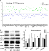
Beta-ecdysone (βEcd) is a phytoecdysteroid found in the dry roots and seeds of the asteraceae and achyranthes plants, and is reported to increase osteogenesis in vitro. Since glucocorticoid (GC) excess is associated with a decrease in bone formation, the purpose of this study was to determine if treatment with βEcd could prevent GC-induced osteoporosis. Two-month-old male Swiss-Webster mice (n=8-10/group) were randomized to either placebo or slow release prednisolone pellets (3.3mg/kg/day) and treated with vehicle control or βEcd (0.5mg/kg/day) for 21days. GC treatment inhibited age-dependent trabecular gain and cortical bone expansion and this was accompanied by a 30-50% lower bone formation rate (BFR) at both the endosteal and periosteal surfaces. Mice treated with only βEcd significantly increased bone formation on the endosteal and periosteal bone surfaces, and increased cortical bone mass were their controls to compare to GC alone. Concurrent treatment of βEcd and GC completely prevented the GC-induced reduction in BFR, trabecular bone volume and partially prevented cortical bone loss. In vitro studies determined that βEcd prevented the GC increase in autophagy of the bone marrow stromal cells as well as in whole bone. In summary, βEcd prevented GC induced changes in bone formation, bone cell viability and bone mass. Additional studies are warranted of βEcd for the treatment of GC induced bone loss.
Keywords: Autophagy; Beta-ecdysone (βEcd); Bone formation; Glucocorticoid.
Copyright © 2015 Elsevier Inc. All rights reserved.
Figures


Similar articles
-
β-Ecdysone Augments Peak Bone Mass in Mice of Both Sexes.Clin Orthop Relat Res. 2015 Aug;473(8):2495-504. doi: 10.1007/s11999-015-4246-5. Clin Orthop Relat Res. 2015. PMID: 25822452 Free PMC article.
-
A novel flavonoid C-glucoside from Ulmus wallichiana preserves bone mineral density, microarchitecture and biomechanical properties in the presence of glucocorticoid by promoting osteoblast survival: a comparative study with human parathyroid hormone.Phytomedicine. 2013 Nov 15;20(14):1256-66. doi: 10.1016/j.phymed.2013.07.007. Epub 2013 Aug 6. Phytomedicine. 2013. PMID: 23928508
-
Sclerostin-antibody treatment of glucocorticoid-induced osteoporosis maintained bone mass and strength.Osteoporos Int. 2016 Jan;27(1):283-294. doi: 10.1007/s00198-015-3308-6. Epub 2015 Sep 18. Osteoporos Int. 2016. PMID: 26384674 Free PMC article.
-
Histomorphometric analysis of glucocorticoid-induced osteoporosis.Micron. 2005;36(7-8):645-52. doi: 10.1016/j.micron.2005.07.009. Epub 2005 Sep 2. Micron. 2005. PMID: 16243531 Review.
-
Pre-receptorial regulation of steroid hormones in bone cells: insights on glucocorticoid-induced osteoporosis.J Steroid Biochem Mol Biol. 2008 Feb;108(3-5):292-9. doi: 10.1016/j.jsbmb.2007.09.018. Epub 2007 Sep 14. J Steroid Biochem Mol Biol. 2008. PMID: 17950597 Review.
Cited by
-
Bad to the Bone: The Effects of Therapeutic Glucocorticoids on Osteoblasts and Osteocytes.Front Endocrinol (Lausanne). 2022 Mar 31;13:835720. doi: 10.3389/fendo.2022.835720. eCollection 2022. Front Endocrinol (Lausanne). 2022. PMID: 35432217 Free PMC article. Review.
-
β-Ecdysone Augments Peak Bone Mass in Mice of Both Sexes.Clin Orthop Relat Res. 2015 Aug;473(8):2495-504. doi: 10.1007/s11999-015-4246-5. Clin Orthop Relat Res. 2015. PMID: 25822452 Free PMC article.
-
β‑Ecdysterone promotes autophagy and inhibits apoptosis in osteoporotic rats.Mol Med Rep. 2018 Jan;17(1):1591-1598. doi: 10.3892/mmr.2017.8053. Epub 2017 Nov 14. Mol Med Rep. 2018. PMID: 29138818 Free PMC article.
-
The Potential of Natural Compounds Regulating Autophagy in the Treatment of Osteoporosis.J Inflamm Res. 2023 Dec 8;16:6003-6021. doi: 10.2147/JIR.S437067. eCollection 2023. J Inflamm Res. 2023. PMID: 38088943 Free PMC article. Review.
-
A soluble bone morphogenetic protein type 1A receptor fusion protein treatment prevents glucocorticoid-Induced bone loss in mice.Am J Transl Res. 2019 Jul 15;11(7):4232-4247. eCollection 2019. Am J Transl Res. 2019. PMID: 31396331 Free PMC article.
References
-
- Goemaere S, Liberman UA, Adachi JD, Hawkins F, Lane N, Saag KG, Schnitzer T, Kaufman JM, Malice MP, Carofano W, Daifotis A. Incidence of nonvertebral fractures in relation to time on treatment and bone density in glucocorticoid-treated patients: a retrospective approach. J Clin Rheumatol. 2003;9:170–5. - PubMed
-
- Silverman SL, Lane NE. Glucocorticoid-induced osteoporosis. Curr Osteoporos Rep. 2009;7:23–6. - PubMed
-
- Chiodini I, Carnevale V, Torlontano M, Fusilli S, Guglielmi G, Pileri M, Modoni S, Di Giorgio A, Liuzzi A, Minisola S, Cammisa M, Trischitta V, Scillitani A. Alterations of bone turnover and bone mass at different skeletal sites due to pure glucocorticoid excess: study in eumenorrheic patients with Cushing's syndrome. J Clin Endocrinol Metab. 1998;83:1863–7. - PubMed
-
- Michaud K, Forget H, Cohen H. Chronic glucocorticoid hypersecretion in Cushing's syndrome exacerbates cognitive aging. Brain Cogn. 2009;71:1–8. - PubMed
-
- Sorva R, Turpeinen M, Juntunen-Backman K, Karonen SL, Sorva A. Effects of inhaled budesonide on serum markers of bone metabolism in children with asthma. J Allergy Clin Immunol. 1992;90:808–15. - PubMed
Publication types
MeSH terms
Substances
Grants and funding
LinkOut - more resources
Full Text Sources
Other Literature Sources
Medical
Miscellaneous

