. 2015 Jul;166(1):89-102.
doi: 10.1016/j.trsl.2014.11.010. Epub 2014 Dec 20.

Intermediate-conductance calcium-activated potassium channel KCa3.1 and chloride channel modulate chemokine ligand (CCL19/CCL21)-induced migration of dendritic cells

Affiliations

Intermediate-conductance calcium-activated potassium channel KCa3.1 and chloride channel modulate chemokine ligand (CCL19/CCL21)-induced migration of dendritic cells

Zhifei Shao et al. Transl Res. 2015 Jul.

Abstract

The role of ion channels is largely unknown in chemokine-induced migration in nonexcitable cells such as dendritic cells (DCs). Here, we examined the role of intermediate-conductance calcium-activated potassium channel (KCa3.1) and chloride channel (CLC3) in lymphatic chemokine-induced migration of DCs. The amplitude and kinetics of chemokine ligand (CCL19/CCL21)-induced Ca(2+) influx were associated with chemokine receptor 7 expression levels, extracellular-free Ca(2+) and Cl(-), and independent of extracellular K(+). Chemokines (CCL19 and CCL21) and KCa3.1 activator (1-ethyl-1,3-dihydro-2H-benzimidazol-2-one) induced plasma membrane hyperpolarization and K(+) efflux, which was blocked by 1-[(2-chlorophenyl)diphenylmethyl]-1H-pyrazole, suggesting that KCa3.1 carried larger conductance than the inward calcium release-activated calcium channel. Blockade of KCa3.1, low Cl(-) in the medium, and low dose of 4,4'-diisothiocyano-2,2'-stilbenedisulfonic acid (DIDS) impaired CCL19/CCL21-induced Ca(2+) influx, cell volume change, and DC migration. High doses of DIDS completely blocked DC migration possibly by significantly disrupting mitochondrial membrane potential. In conclusion, KCa3.1 and CLC3 are critical in human DC migration by synergistically regulating membrane potential, chemokine-induced Ca(2+) influx, and cell volume.


Figures

Figure 1
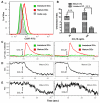
Figure 1. CCR7 expression and CCL19/CCL21-induced cellular changes
A, CCR7 expression in DCs matured with LPS, OVA and TNF-α cocktail as compared to immature DCs. B, Chemotaxis in response to CCL19 in immature and mature DCs. C, intracellular Ca2+ Kinetics: Ca2+ mobilization and influx in response to CCL19 and CCL21 in immature and mature DCs. Stimuli were added at 90 sec. D, CCL19/CCL21 decreased the side-scatter in mature DCs suggesting some intracellular reorganization. E, forward scatter also decreased in the matured DCs with CCL19/CCL21 stimulation suggesting a decrease in the cell size. Data are representatives of 3 independent experiments.
Figure 2
Figure 2. Extracellular cations and CCL19/CCL21-induced intracellular Ca2+ increase
CCL21 (1µg/ml) was added at 90 sec. A, intracellular Ca2+ Kinetics: Ca2+ mobilization and influx in response to CCL19 (left panel) and CCL21 (right panel) in the presence of L15 medium or Ca2+-free PBS in mature DCs. B: intracellular Ca2+ Kinetics: Ca2+ mobilization and influx in response to CCL19 (left panel) and CCL21 (right panel) in the presence of L15 medium, or K+-free (CsCl2) solution. Data are the representatives of 3 independent experiments.
Figure 3
Figure 3. CCL21-induced K+ efflux is carried by KCa3.1
A, kinetics of plasma membrane potential: in response to CCL21. CCL21 (1µg/ml) was added at 90 sec. (The figure is a representative of 6 independent experiments using CCL21 or CCL19 as stimulus) B, Kinetics of plasma membrane potential in response to KCa3.1 opener/activator, 1-EBIO. The 1-EBIO was added at 90 seconds with final concentration of 500mM and led to membrane hyperpolarization indicated by a decreased fluorescence intensity of DiBAC4(3). (The figure is a representative of 3 independent experiments) C, CCL21 (1µg/ml) was added at 90 sec to controls or 2µM TRAM-34-treated mature DCs and intracellular K+ kinetics was recorded as indicated by PBFI fluorescence intensity over the time. The figure is a representative of 3 independent experiments. D, mRNA expression KCa3.1 by quantitative real-time PCR (n=3) E, Western blot of KCa3.1 protein expression in mature DCs (n=3).
Figure 4
Figure 4. KCa3.1 modulates CCL19/21-induced Ca2+ influx and migration in mature DCs
A, CCL21 or CCL19 (1 µg/ml) was added at 90 sec to controls or 2µM TRAM-34-treated mature DCs. Intracellular Ca2+ kinetics were measured by indo-1 fluorescence over the time. B, The overall calcium loading indicated by the area under the curve (AUC) in control and 2µM TRAM-34-treated mature DCs in the context of CCL19/CCL21-induced Ca2+ mobilization and influx (n=5, *p<0.05, paired student t-test). C, DC migration to CCL19/CCL21 with the presence/absence of KCa3.1 blocker TRAM-34 as measured by Transwell (n=6, *p<0.05). Final concentration of chemokines and blocker: CCL19, 100ng/mL; CCL21, 100ng/mL; TRAM34, 2µM. D. Mitochondrial membrane potential as indicated by red fluorescence (575 nm) of JC-1 staining. Mitochondrial membrane potential disrupter, CCCP (carbonyl cyanide 3-chlorophenylhydrazone) serves as controls. The figure is a representative of three independent experiments.
Figure 5
Figure 5. TRAM-34 does not change KCa3.1 expression in mature DCs
Mature DCs were treated with 2µM TRAM-34 for 2 and 6 hours. A, Amplification curve (in log phase) showed the cycles at which KCa3.1 and 18S were amplified. B, Melt peak indicated the formation of specific products corresponding to KCa3.1 and 18S. C, No statistical change was observed in the mRNA levels of KCa3.1 in mature DCs without TRAM-34 and mature DCs with TRAM-34 treatment (2µM) for 2 hours or 6 hours (n=3, *p<0.05).
Figure 6
Figure 6. Extracellular Cl-modulates CCL21-induced cell volume change, Ca2+ influx, and chemotaxis
CCL21 was added at 90 sec. A, Intracellular Ca2+ kinetics in response to CCL21 (1 µg/ml) in the presence/absence of extracellular Cl in mature DCs. B, Kinetics of average cell size of mature DCs in response to CCL21 (1µg/ml) in the presence/absence of extracellular Cl and in the presence of TRAM-34 treatment for 6 hours, indicated by forward scatter values over the time. C, Kinetics of membrane potential in response to CCL21 (1µg/ml) in the absence of extracellular Cl (Cl/gluconate replacement) as indicated by DiBAC4(3) fluorescence over the time. D, Chemotaxis in response to 100 ng/ml CCL21 in medium containing 2mM EGTA, and medium in the presence and absence of Cl.
Figure 7
Figure 7. DIDS abolishes CCL19/CCL21-induced Ca2+ influx and migration in mature DCs
A, mature DCs were treated with various concentrations of DIDS (0, 1, 10, 100µM) were used to measure CCL19/CCL21-induced intracellular Ca2+ mobilization and influx indicated by indo-1 fluorescence (upper panel) and cell size change indicated by forward scatter values (lower panel) over the time. CCL19/CCL21 was added at 90 seconds. B, mature DCs were treated with various concentrations of DIDS (0, 10, 100µM) for 2 hours and the chemotaxis were examined with Transwell (n=3, ***<0.001, error bars indicate SEM). Final chemokine concentration: CCL19, 100ng/mL; CCL21, 100ng/mL. C. Mitochondrial membrane potential as indicated by red fluorescence (575 nm) of JC-1 staining. Valinomycin, a potassium channel ionophore, was used as a positive control for depolarization. D, Expression of annexin-V in DIDS-treated mature DCs. The figures are a representative of three independent experiments.

Similar articles

Cited by

References

    1. Gaurav R, Agrawal DK. Clinical view on the importance of dendritic cells in asthma. Expert review of clinical immunology. 2013;9:899–919. - PMC - PubMed
    1. Bharadwaj AS, Agrawal DK. Transcription factors in the control of dendritic cell life cycle. Immunologic research. 2007;37:79–96. - PubMed
    1. Agrawal DK, Shao Z. Pathogenesis of allergic airway inflammation. Current allergy and asthma reports. 2010;10:39–48. - PMC - PubMed
    1. Braun A, Worbs T, Moschovakis GL, Halle S, Hoffmann K, Bolter J, et al. Afferent lymph-derived T cells and DCs use different chemokine receptor CCR7-dependent routes for entry into the lymph node and intranodal migration. Nature immunology. 2011;12:879–87. - PubMed
    1. Ricart BG, John B, Lee D, Hunter CA, Hammer DA. Dendritic cells distinguish individual chemokine signals through CCR7 and CXCR4. Journal of immunology. 2011;186:53–61. - PubMed

Publication types

MeSH terms