. 2015;6(2):133-43.
doi: 10.1080/19491034.2014.1003509. Epub 2015 Jan 8.

The distinct functions of CENP-C and CENP-T/W in centromere propagation and function in Xenopus egg extracts

Affiliations

The distinct functions of CENP-C and CENP-T/W in centromere propagation and function in Xenopus egg extracts

Iva Krizaic et al. Nucleus. 2015.

Abstract

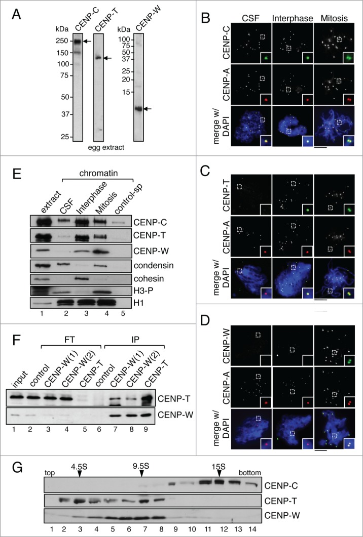
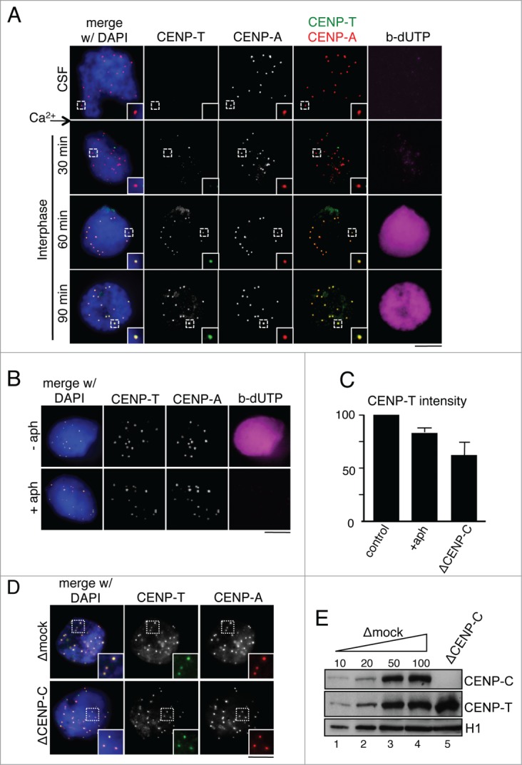
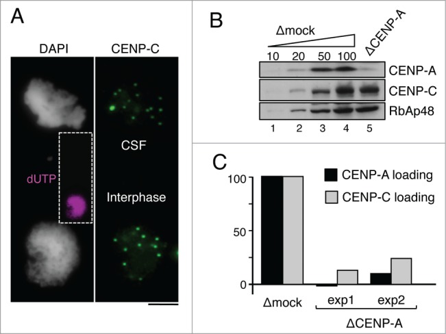
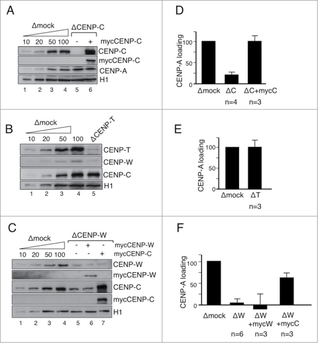
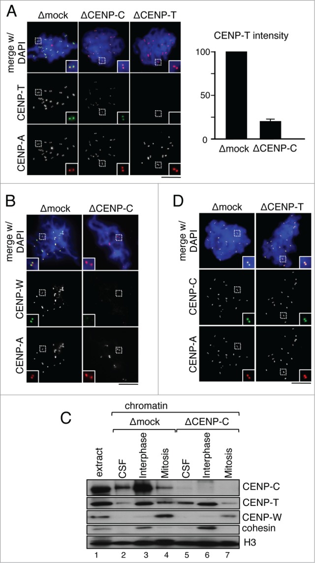
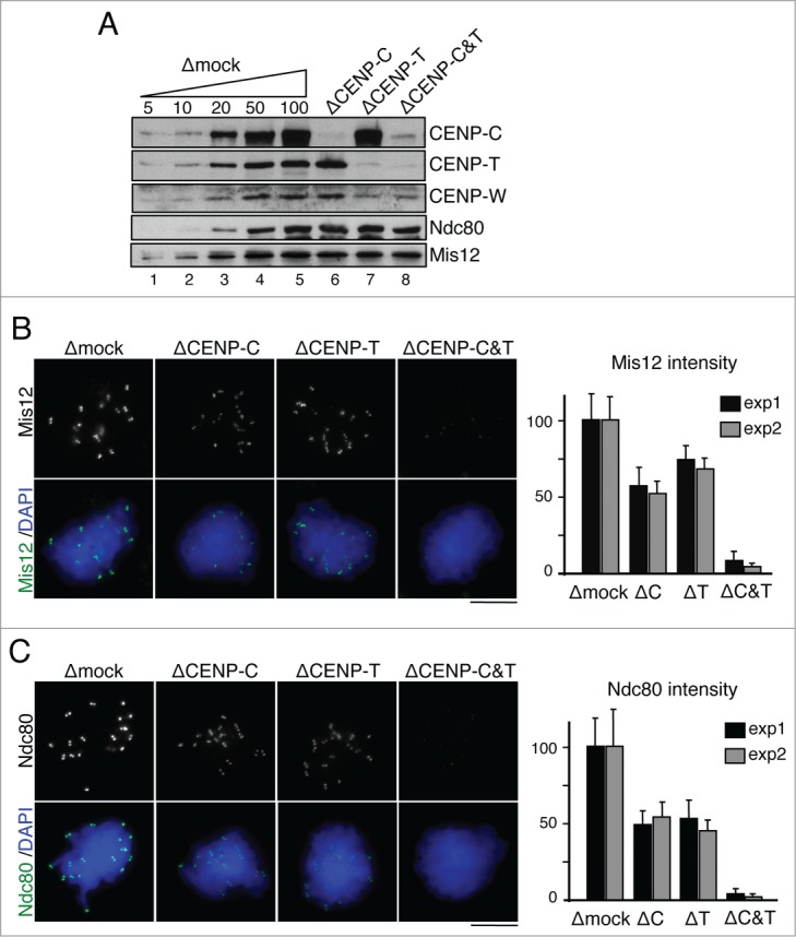
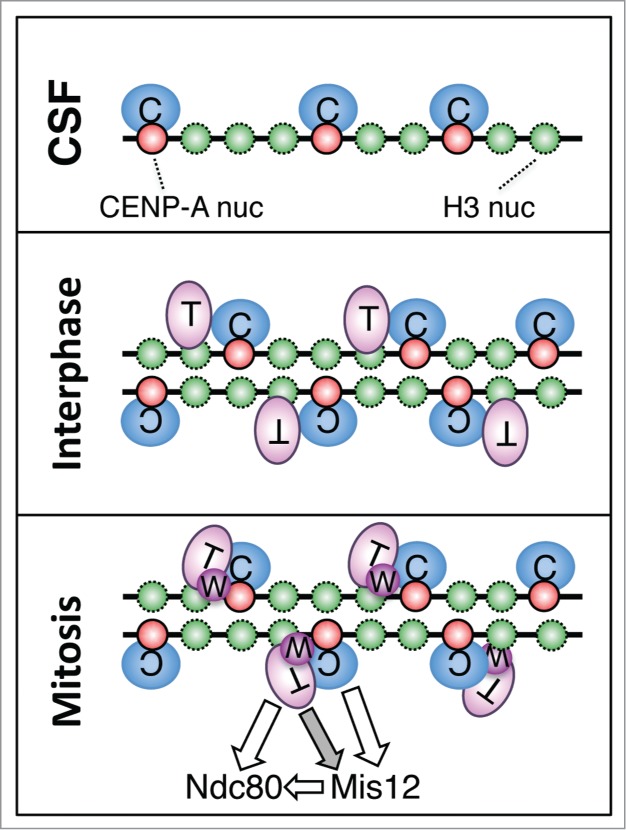
The centromere is the chromosomal region in which the kinetochore is assembled to orchestrate chromosome segregation. It is defined by the presence of a histone H3 variant called Centromere Protein A (CENP-A) or CenH3. Propagation of centromere identity entails deposition of new CENP-A upon exit from mitosis in vertebrate cells. A group of 16 proteins that co-immunoprecipitate with CENP-A, the Constitutive Centromere Associated Network or CCAN, contribute to kinetochore assembly and function. For most of them it is still unclear how and when they are recruited to centromeres and whether they have a role in CENP-A deposition. Taking advantage of the Xenopus egg cell-free system, we have addressed these issues for CCAN proteins CENP-C, CENP-T and CENP-W. CENP-C recruitment occurs as soon as sperm DNA, containing CENP-A, is added to the egg extract, and continues after de novo incorporation of CENP-A in early interphase. In contrast, centromeric recruitment of CENP-T occurs in late interphase and precedes that of CENP-W, which occurs in mitosis. Unlike CENP-C, CENP-T and CENP-W do not participate in CENP-A deposition. However, like CENP-C, they play a major role in kinetochore assembly. Depletion of CENP-C results in reduced amount of CENP-T at centromeres, an effect more prominent in mitosis than in interphase. In spite of this, kinetochores can still be assembled under this condition although the recruitment of Ndc80 and Mis12 is decreased. Our results support the existence of 2 pathways for kinetochore assembly directed by CENP-C and CENP-T/W, which can be reconstituted in Xenopus egg extracts.

Keywords: CCAN; CCAN, Constitutive Centromere Associated Network; CENP, centromere protein; CENP-A; CenH3; kinetochore.


Figures

Figure 1.
Figure 1.
CENPC, CENP-T and CENP-W are recruited to chromatin at different times. (A) Immunoblot analysis of an egg extract with the indicated antibodies. For CENP-W, 2 different antibodies were raised, CENP-W(1) and CENP-W(2) (see Methods for description). While both worked for immunoprecipitation (see Fig. 1F), CENP-W(1) worked best for immunoblot and immunofluorescence. (B-D) Immunofluorescence analysis of CSF (unreplicated) chromosomes, interphase nuclei and mitotic (replicated) chromosomes with antibodies against CENP-C (B), CENP-T (C) and CENP-W (D), in green, and Cy3-labeled anti-CENP-A in red to label centromeres. DNA was counterstained with DAPI (blue). Colored images are shown for the insets and the merge with DAPI while single antibody stainings appear as gray scale images. Scale bar is 10μm. (E) Immunoblot analysis of chromatin assembled in CSF, interphase and mitotic extracts and purified by centrifugation through a sucrose cushion. Chromatin purified in the same way from a mock assembly reaction without sperm serves as control (control-sp, lane 5). Cohesin (Rad21) is a marker for interphase chromatin whereas condensin (CAP-G) and phospho H3 (H3-P) are present in CSF and mitotic chromatin. Histone H1 is used as loading control. (F) Immunoprecipitates (IP) obtained from soluble egg extracts were analyzed by immunoblot together with aliquots of the flow through (FT) from each reaction and of the input extract (1%). Immunoprecipitation with non-immune rabbit IgG was used as control. (G) A soluble egg extract was fractionated on a sucrose gradient (5-20% sucrose) and analyzed by immunoblot. The sedimentation coefficients of the major peaks of CENP-T, CENP-W and CENP-C are indicated.
Figure 2.
Figure 2.
Requirements of CENP-T recruitment to interphase centromeres. (A) Time course analysis of CENP-T (green) recruitment to centromeres marked with Cy3-labeled anti-CENP-A (red). DNA replication is followed by incorporation of biotin-dUTP (magenta) and DNA is counterstained with DAPI (blue). The CENP-T signals at t = 30 min do not overlap with centromeres and are likely background. (B) Interphase nuclei assembled in the presence or absence of aphidicolin were analyzed with antibodies against CENP-T (green) and CENP-A (red). No incorporation of biotin-dUTP (magenta) can be detected in the presence of aphidicolin. (C) Intensity of CENP-T signals at centromeres of nuclei assembled under the indicated conditions. At least 200 centromeres were measured in 3 independent experiments per condition. Intensities were normalized to the mean value of the corresponding control experiment (no aphidicolin or mock-depletion). Bars represent mean± SEM. (D) Immunofluorescence analysis of CENP-T (green) localization in replicated interphase nuclei assembled in the presence (Δmock) or absence of CENP-C (ΔCENP-C). Centromeres are stained with Cy3-labeled anti-CENP-A (red). Scale bars, 10 μm. (E) Immunoblot analysis of CENP-C depleted extracts used in (D). Increasing amounts of mock-depleted CSF extract (Δmock), expressed as percentage, and extracts depleted from the indicated proteins were analyzed side by side by immunoblot. H1 was used as loading control.
Figure 3.
Figure 3.
De novo loading of CENP-C following CENP-A incorporation in interphase. (A) Representative image of a pair used for analysis of CENP-C loading. CENP-C signals (green) from a mass of CSF chromosomes (top) are compared with those from an interphase nucleus (bottom), the latter already replicated as indicated by incorporation of dUTP (inset in magenta). Scale bar, 10 μm. (B) Increasing amounts of mock-depleted CSF extract (Δmock), expressed as percentage, and a CENP-A depleted extract (ΔCENP-A), were analyzed side by side to estimate the extent of depletion. RbAp48 is used as loading control. (C) Quantification of CENP-A loading (black bars) and CENP-C loading (gray bars) in CENP-A depleted extracts relative to the corresponding mock depleted extract using the loading assay described in the main text. At least 250 centromeres from more than 10 pairs as the one shown in (A) were measured for each condition in 2 independent experiments (exp1 and exp2).
Figure 4.
Figure 4.
CENP-T and CENP-W are not required for CENP-A loading. (A-C) Immunoblot analyses of extracts used in the CENP-A loading assays shown in (D-F). Increasing amounts of mock depleted CSF extract (Δmock) and extracts depleted from the indicated proteins with or without add-back of myc-tagged proteins were analyzed side by side. H1 is used as a loading control. In (C), the CENP-W(2) antibody was used for depletion. (D-F) Bar graphs representing CENP-A loading efficiency in nuclei assembled in the indicated extracts. More than 250 centromeres were quantitated per condition and experiment. The number of experiments (n) is indicated below each bar. Bars represent mean± SEM.
Figure 5.
Figure 5.
Maintenance of CENP-T/W at mitotic centromeres depends on CENP-C. (A) Immunofluorescence analysis of CENP-T (green) localization at mitotic centromeres (labeled by CENP-A, red) assembled in extracts that had been mock-depleted or depleted of CENP-C. The graph on the right shows the centromeric fluorescent intensity for CENP-T as percentage of the mean value obtained in the corresponding control experiment. At least 970 centromeres were measured for each condition in 5 independent experiments. (B) CENP-W localization in chromosomes assembled as in (A). (C) Immunoblot analysis of chromatin fractions assembled in CSF, interphase and mitotic extracts following depletion of CENP-C or mock depletion as control. Cohesin (Rad21) is a marker for interphase. Histone H3 is used as loading control. (D) Immunofluorescence analysis of CENP-C (green) localization at mitotic centromeres (labeled by CENP-A, red) in chromosomes from mock depleted or CENP-T depleted extracts. Scale bars, 10 μm.
Figure 6.
Figure 6.
CENP-C and CENP-T/W contribute to kinetochore assembly. (A) Immunoblot analysis of the extracts used to assemble the chromosomes shown in (B) and (C). (B-C) Representative examples of chromosomes assembled in mock depleted extracts and extracts lacking CENP-C, CENP-T or both, and stained with antibodies against Mis12 (B) and Ndc80 (C). Quantification of total fluorescence in dots per chromosome mass is shown in the graphs on the right. Bars represent mean± SD. Between 10 and 20 nuclei per condition were measured in 2 independent experiments (exp1 and exp2).
Figure 7.
Figure 7.
Sequential recruitment of CENP-C, CENP-T and CENP-W to centromeres in Xenopus egg extracts. CENP-C recognizes CENP-A and binds to centromeric chromatin when sperm is added to CSF extracts. When these extracts are driven to interphase, de novo loading of CENP-A occurs, which requires CENP-C but no CENP-T or CENP-W. The new CENP-A also recruits more CENP-C. Soon after DNA replication, CENP-T binds to centromeres. CENP-C is not strictly required for this process, but its presence may stabilize the incorporated CENP-T. Upon entry in mitosis, CENP-W can be detected at centromeres. CENP-C becomes more important for maintenance of CENP-T/W at this stage, most likely by providing stability to centromeric chromatin. Both CENP-C and CENP-T/W direct kinetochore assembly by recruiting Mis12C and Ndc80C, respectively. It is currently unclear how CENP-T affects Mis12C recruitment independently of Ndc80C (gray arrow), although a weak interaction between them has been detected in vitro.

Similar articles

Cited by

References

    1. Cheeseman IM, Desai A. Molecular architecture of the kinetochore-microtubule interface. Nat Rev Mol Cell Biol 2008; 9:33-46; PMID:18097444; http://dx.doi.org/10.1038/nrm2310 - DOI - PubMed
    1. Santaguida S, Musacchio A. The life and miracles of kinetochores. Embo J 2009; 28:2511-31; PMID:19629042; http://dx.doi.org/10.1038/emboj.2009.173 - DOI - PMC - PubMed
    1. Cleveland DW, Mao Y, Sullivan KF. Centromeres and kinetochores: from epigenetics to mitotic checkpoint signaling. Cell 2003; 112:407-21; PMID:12600307; http://dx.doi.org/10.1016/S0092-8674(03)00115-6 - DOI - PubMed
    1. Allshire RC, Karpen GH. Epigenetic regulation of centromeric chromatin: old dogs, new tricks? Nat Rev Genet 2008; 9:923-37; PMID:19002142; http://dx.doi.org/10.1038/nrg2466 - DOI - PMC - PubMed
    1. Blower MD, Sullivan BA, Karpen GH. Conserved organization of centromeric chromatin in flies and humans. Dev Cell 2002; 2:319-30; PMID:11879637; http://dx.doi.org/10.1016/S1534-5807(02)00135-1 - DOI - PMC - PubMed

Publication types

LinkOut - more resources