. 2015 Jan 15;142(2):258-67.
doi: 10.1242/dev.116855.

Notch3-Jagged signaling controls the pool of undifferentiated airway progenitors

Affiliations

Notch3-Jagged signaling controls the pool of undifferentiated airway progenitors

Munemasa Mori et al. Development. .

Abstract

Basal cells are multipotent airway progenitors that generate distinct epithelial cell phenotypes crucial for homeostasis and repair of the conducting airways. Little is known about how these progenitor cells expand and transition to differentiation to form the pseudostratified airway epithelium in the developing and adult lung. Here, we show by genetic and pharmacological approaches that endogenous activation of Notch3 signaling selectively controls the pool of undifferentiated progenitors of upper airways available for differentiation. This mechanism depends on the availability of Jag1 and Jag2, and is key to generating a population of parabasal cells that later activates Notch1 and Notch2 for secretory-multiciliated cell fate selection. Disruption of this mechanism resulted in aberrant expansion of basal cells and altered pseudostratification. Analysis of human lungs showing similar abnormalities and decreased NOTCH3 expression in subjects with chronic obstructive pulmonary disease suggests an involvement of NOTCH3-dependent events in the pathogenesis of this condition.

Keywords: Airway differentiation; Basal cells; COPD; Jagged; Lung regeneration; Notch; Progenitor cells; p63.


Figures

Fig. 1.
Fig. 1.
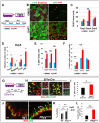
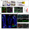
Notch signaling restricts the pool of p63+ airway progenitors in vitro and in vivo. (A) Experimental design. Adult airway progenitors cultured under submerged condition (Sub) and subsequently in air-liquid interface (ALI) treated with DMSO (control, blue) or DAPT (γ-secretase inhibitor; red) from day –3 to ALI day 8. (B) Increased number of multiciliated cells (acetylated α-tubulin (α-tub) and abolishment of secretory cells (Scgb3a2+). Expansion of basal cells (p63+ Krt5+) in DAPT compared with control. (C) Relative number of Ki67+ cells per total DAPI-labeled cells (%) at each time point. (D) Relative number of cells labeled with each markers at ALI day 8 (% total). (E) Relative number of p63+ cells per total DAPI-labeled cells (%), or (F) Ki67+p63+ cells per total p63+ cells at each time point. (G) Experimental design summary: Ef1α-Cre lentivirus-mediated disruption of Notch signaling in adult wild-type or Rbpjf/f airway progenitors. (H) Immunofluorescence for Cre, p63 or DAPI at ALI day 6: there is an increase in the population of p63-Cre double-labeled cells (arrowheads). (I) Relative number of p63+ cells in Cre-labeled cells at ALI day 6. (J) Immunofluorescence for p63 and Krt5 in E18.5 wild-type and Rbpjcnull tracheal epithelium; arrowheads indicate expanded basal cell population. Morphometric analysis of (K) p63+ or Krt5+ cells, and (L) p63+Ki67+ cells per p63+ cells in E18.5 wild-type and Rbpjcnull mice. Data are mean±s.e.m. from five or six fields per sample, n=3. Student's t-test *P<0.05 or **P<0.01. Scale bars: 10 µm.
Fig. 2.
Fig. 2.
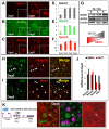
Notch3 is selectively activated in p63-negative undifferentiated airway progenitors. (A-C,G) At ALI day 0, nuclear signals are prominent for Notch3 but not for Notch2 or Notch1, which appear only later during differentiation. (A-C) Immunofluorescence of ALI day 0 and day 8 cells using antibodies against Notch1 (cleaved NICD1: Val1744) (A), the C-terminus of Notch2 (B) and the C-terminus of Notch3 (C) proteins. (D-F) Real-time RT-PCR of Notch1, Notch2 and Notch3 mRNA at ALI days 0, 2, 4, 8 (data are mean±s.e.m., n=3 per group). (G) Western blot confirming predominant activation of Notch3 at ALI day 0 and Notch1 at day 8; β-actin is used as an internal control. (H,I) Immunofluorescence at day 0 showing that Notch3 is activated in the nuclei of p63-negative cells and Hes1-expressing cells. (J) Real-time PCR of Notch1, Notch 2, Notch3 and Hes1 in ALI day 0 cultures treated with DMSO (control) or DAPT for 72 h showing that only Notch3 and Hes1 are differentially expressed between controls and DAPT at this stage. (K) Lineage analysis of adult Krt5-CreERT2; R26-tdTomato mice treated with tamoxifen (Tm, 10 mg/kg i.p.) 10 days prior to the sacrifice; airway progenitors cultured for 7 days to confluence; immunofluorescence for Notch3, Td Tomato and p63 showing Notch3 signals (arrows) in lineage-labeled tdTomato+ cells at ALI day 0. Scale bar: 10 µm.
Fig. 3.
Fig. 3.
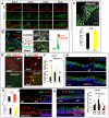
Endogenous Notch3 activation identifies parabasal cells and restricts the pool of p63+ cells in vivo and in vitro. (A) Immunofluorescence of Notch3 and p63 in developing and adult tracheal epithelium; nuclear Notch3 and p63 overlapping extensively at E14.5 (dashed line, yellow), signals becoming more segregated later (>E16.5). (B) Nuclear Notch3+ cells occupy a parabasal position and express Krt8 in developing (left, E18.5) and adult (right, ALI Day 0) airways. Diagram represents confocal image depicting luminal (top) and basal (bottom) views of airway epithelium. There is enrichment of p63+ in basal layer and Notch3+Krt8+ in parabasal layers; horizontal lines indicate the comparison of E18.5 and adult (ALI Day 0) epithelium. (C) Notch3 inactivation expands the p63+ pool in the embryonic (E14.5) airways of Notch3−/− mice. (D,E) Immunofluorescent staining of ALI cultures at day 0 showing significant increase in the p63+ population and in Ki67+p63+ double-labeled cells (arrowheads) in Notch3−/− cultures compared with wild-type cultures. (F) Increased cell proliferation in cultures of Notch3−/− no longer seen after ALI day 4. (G,H) Adult Notch3−/− airways showing expansion of the population of basal (p63+) and luminal (Krt8+) cells, (I) increased pseudostratification and (J) the appearance of Krt5+p63− parabasal cells (arrows) not seen in wild type (asterisks). In the inset in J, arrowhead indicates Krt5 and DAPI in parabasal cells. (K) Notch3−/− airways have increased number of Krt5+ single and Krt5+Krt8+ double-labeled cells and decreased number of Krt8+Krt5− cells. Data are mean±s.e.m., analysis of six fields (n=3 per group; *P<0.05, **P<0.01, Student's t-test). Scale bars: 8 µm.
Fig. 4.
Fig. 4.
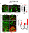
Constitutive activation of Notch3 restricts the number and precociously differentiated p63+ cells. Diagram shows lentiviral gene transduction of control (Ctr-GFP) or NICD3-HA in airway epithelial progenitors from wild-type and Notch3−/− mice. (A) Immunofluorescence/confocal analysis at day 0 showing major reduction in the p63+ cell population in Notch3−/− transduced with NICD3 (DAPI, GFP, HA show transduction efficiency): no co-expression of p63 and HA (circled areas indicate distinct nuclear signals) in contrast to the extensive double labeling in day 0 Ctr-GFP cultures (circled area indicates cytoplasmic GFP and nuclear p63). (C,D) NICD3 transduction induces precocious Scgb3a2 expression and the secretory cell phenotype in Notch3−/− and wild-type cultures at day 0. (E) By ALI day 8, Notch3−/− cultures transduced with NICD3 showed extensive areas of Scgb3a2+HA+ double labeling, suggesting prominent induction of the secretory phenotype (circled areas) compared with Ctr-GFP cultures. (B,F) Morphometric analysis of the percentage of p63 (B) or Scgb3a2 (F) cells double-labeled with HA (NICD3-HA lentivirus) or GFP (Ctr-GFP lentivirus) in ALI cultures from Notch3−/− or wild-type airway progenitors. Data are mean±s.e.m. of the percentage labeling in six fields at day 0 or day 8, n=3 per group; **P<0.01, Student's t-test. Scale bars: 8 µm.
Fig. 5.
Fig. 5.
Jag1 and Jag2 have partially overlapping functions in restricting basal cell expansion. (A) Expression pattern of Jag1 and Jag2 by in situ hybridization and p63 by immunohistochemistry in developing airways. Stronger signals for Jag2 compared with Jag1 in E14.5-E15.5 proximal airways initially in all epithelial cells and then overlapping with p63 at E15.5 (double in situ hybridization/immunohistochemistry; aw, airway; bv, blood vessels). Diagram summarizes temporal-spatial patterns from the panels above and data not shown. Proximal-distal domains of expression in trachea (Tr), extrapulmonary and intrapulmonary proximal airways of the lung (Lu). Solid and dotted bars represent strong and weak signals, respectively, as revealed by in situ hybridization or immunohistochemistry. (B) Jag1 and Jag2 are expressed in adult p63+ airway progenitors in culture (ALI day 2). (C) p63 immunofluorescence and morphometric analysis (% labeling) showing expansion of basal cells in the E14.5 trachea and extrapulmonary airways of Jag1cnull mice. (D) Double p63-Scgb1a1 immunofluorescence in E18.5 wild-type Jag1cnull and Jag2cnull tracheas showing local (squares) expansion of p63+ cells in Jag2cnull mice (arrows) not present in wild type or Jag1cnull. (E) In E18.5 Jag2cnull airways, expansion of p63+ cells is accompanied by reduced thickness of the Krt8-labeled cell layer, and (F) suppression of the secretory (Scgb3a2+) phenotype with expansion of multiciliated cell population (β-tubulin+). Data are mean±s.e.m. of the percentage of p63+ cells/total cells, n=3 per group; **P<0.01, Student's t-test. Scale bars: 10 µm in A-C,E,F; 100 µm in D.
Fig. 6.
Fig. 6.
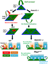
Summary of results and proposed model of Notch regulation of airway epithelial progenitor cell expansion and differentiation. Under homeostatic conditions, basal cells express Jag ligands but do not activate Notch signaling for self-renewal and continue to expand. Once the pool of p63+ basal cells is properly expanded, Notch3 becomes selectively expressed in the cells occupying a parabasal position and Notch3 signaling is activated by Jag2 and/or Jag1 from the adjacent basal cells. This pool of Notch3+ progenitor cells remains undifferentiated until later, when signaling by Notch1 and Notch2 is turned on for secretory-multiciliated cell differentiation. The balance of basal and parabasal cells is properly achieved by modulating endogenous levels of Notch3 activation selectively, but differentiation is not dependent on Notch3. The basal and multiciliated cell pools can be collectively expanded by inhibiting signaling by all Notch receptors (as in the Rbpjcnull mice). Diagrams summarize these conclusions in light of the Notch genetic models.

Similar articles

Cited by

References

    1. Blanpain C., Lowry W. E., Pasolli H. A. and Fuchs E. (2006). Canonical notch signaling functions as a commitment switch in the epidermal lineage. Genes Dev. 20, 3022-3035 10.1101/gad.1477606 - DOI - PMC - PubMed
    1. Bouras T., Pal B., Vaillant F., Harburg G., Asselin-Labat M.-L., Oakes S. R., Lindeman G. J. and Visvader J. E. (2008). Notch signaling regulates mammary stem cell function and luminal cell-fate commitment. Cell Stem Cell 3, 429-441 10.1016/j.stem.2008.08.001 - DOI - PubMed
    1. Breuer R., Zajicek G., Christensen T. G., Lucey E. C. and Snider G. L. (1990). Cell kinetics of normal adult hamster bronchial epithelium in the steady state. Am. J. Respir. Cell Mol. Biol. 2, 51-58 10.1165/ajrcmb/2.1.51 - DOI - PubMed
    1. Chen J. and Krasnow M. A. (2012). Integrin Beta 1 suppresses multilayering of a simple epithelium. PLoS One 7, e52886 10.1371/journal.pone.0052886 - DOI - PMC - PubMed
    1. Chiche A., Moumen M., Petit V., Jonkers J., Medina D., Deugnier M.-A., Faraldo M. M. and Glukhova M. a. (2013). Somatic loss of p53 leads to stem/progenitor cell amplification in both mammary epithelial compartments, basal and luminal. Stem Cells 31, 1857-1867 10.1002/stem.1429 - DOI - PubMed

Publication types

MeSH terms