. 2015 Mar 15;466(3):525-36.
doi: 10.1042/BJ20140698.

αvβ6 integrin is required for TGFβ1-mediated matrix metalloproteinase2 expression

Affiliations

αvβ6 integrin is required for TGFβ1-mediated matrix metalloproteinase2 expression

Anindita Dutta et al. Biochem J. .

Abstract

Transforming growth factor (TGF) β1 activity depends on a complex signalling cascade that controls expression of several genes. Among others, TGFβ1 regulates expression of matrix metalloproteinases (MMPs) through activation of Smads. In the present study, we demonstrate for the first time that the αvβ6 integrin interacts with TGFβ receptor II (TβRII) through the β6 cytoplasmic domain and promotes Smad3 activation in prostate cancer (PrCa) cells. Another related αv integrin, αvβ5, as well as the αvβ6/3 integrin, which contains a chimeric form of β6 with a β3 cytoplasmic domain, do not associate with TβRII and fail to show similar responses. We provide evidence that αvβ6 is required for up-regulation of MMP2 by TGFβ1 through a Smad3-mediated transcriptional programme in PrCa cells. The functional relevance of these results is underscored by the finding that αvβ6 modulates cell migration in an MMP2-dependent manner on an αvβ6-specific ligand, latency-associated peptide (LAP)-TGFβ. Overall, these mechanistic studies establish that expression of a single integrin, αvβ6, is sufficient to promote activation of Smad3, regulation of MMP2 levels and consequent catalytic activity, as well as cell migration. Our study describes a new TGFβ1-αvβ6-MMP2 signalling pathway that, given TGFβ1 pro-metastatic activity, may have profound implications for PrCa therapy.


Conflict of interest statement

The other authors do not have any conflict of interests.

Figures

Figure 1
Figure 1. αvβ6 is required for TGFβ1 induction of MMP2
(A) RWPE cells (left panels) transiently transfected with 100 nM β1C siRNA, β6 siRNA or non- transfected, were serum starved for 24 h, before being stimulated with 10 ng/ml TGFβ1 for 48 h. Cell lysates were analyzed by IB and probed with 2A1 Ab to β6 or an Ab to MMP2. ERK was used as a loading control. PC3-high cells (right panels) stably transfected with β5-shRNA (shβ5) or β6-shRNA (shβ6) were serum starved for 24 h; then stimulated with 20 or 40 ng/ml TGFβ1 for 48 h. Cell lysates were analyzed by 10% SDS-PAGE, under non-reducing conditions and probed with 2A1 Ab to β6, or under reducing conditions and probed with an Ab to MMP2. ERK was used as a loading control. (B) FACS analysis of β6 (solid line) in RWPE and PC3-high cells is shown. β1 (broken line) was used as positive control for each cell line. Dotted-lines represent staining with an isotype negative control Ab. (C) PC3-high cells, stably transfected with β5- shRNA (shβ5) or β6-shRNA (shβ6) were serum starved for 24 h; then stimulated with 20 ng/ml TGFβ1 for 48 h. Cell lysates were analyzed by 10% SDS-PAGE, under reducing conditions and probed with MT1-MMP Ab. FAK was used as a loading control. (D) MMP2 activity in culture supernatants of shβ5- or shβ6-PC3-high cells (left panels) and αvβ5- or αvβ6-PC3-zero (right panels) cells was analyzed by gelatin zymography (Zg). Cells were either stimulated by TGFβ1 (20 ng/ml) or left resting for 48 h. Serum-free culture medium from BPH1 cells was used as a control for MMP activity. Fibronectin (FN) protein levels, analyzed by IB, were used as a loading control.
Figure 2
Figure 2. TGFβ1 upregulates MMP2 in αvβ6+ - cells
(A-B) PC3-high cells were serum starved for 24 h; then stimulated with increasing concentrations of TGFβ1 for 48 h. PC3-high cell lysates were analyzed by IB and probed with 2A1 Ab to β6 (10% SDS-PAGE under non-reducing conditions) (A) or with an Ab to β5 (left panel; 10% SDS-PAGE under reducing conditions), to β3 (middle panel; 10% SDS-PAGE under reducing conditions) or to αv (right panel; 7.5% SDS-PAGE under non-reducing conditions) (B). ERK was used as a loading control. (C) Cell lysates from 10, 20 ng/ml TGFβ1 (48 h) stimulated or unstimulated PC3-zero cells were analyzed by 10% SDS-PAGE and probed with 2A1 Ab to β6 (under non-reducing conditions), an Ab to β5 or to β3 (under reducing conditions). ERK was used as loading control. (D) PC3-high cells stably transfected with β5-shRNA (shβ5) or β6-shRNA (shβ6) were serum starved for 24 h; then stimulated with 20 ng/ml TGFβ1 for 48 h. Cell lysates were analyzed by 10% SDS-PAGE, under reducing conditions and probed with TIMP2 Ab. FAK was used as a loading control. (E) PC3-high (αvβ6+; left panel) and PC3-zero (αvβ6-; right panel) cells were serum starved for 24 h; then stimulated with 10, 20, 40 ng/ml TGFβ1 for 48 h or unstimulated (-). MMP2 activity in serum-free culture supernatants was analyzed by gelatin zymography (Zg). Serum-free culture medium from BPH1 cells was used as a control for MMP activity. Fibronectin (FN) protein levels, analyzed by IB, were used as loading controls.
Figure 3
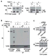
Figure 3. αvβ6 specifically interacts with TβRII
(A) αvβ6, αvβ3 or αvβ5 integrins were immunoprecipitated from 20 ng/ml TGFβ1 stimulated (48 h) PC3-high cell lysates and the immunoprecipitates were analyzed by 10% SDS-PAGE (under non-reducing conditions, left panels; or under reducing conditions, right panels) in order to detect TβRII. Mouse or rabbit IgG was used as a negative control (Neg.Ctr.) Ab. β6 (left panel), integrin expression was analyzed in the immunoprecipitates by IB using 2A1 Ab to β6. In the right panels an Ab to β3 or β5 was used. αvβ6 and TβRII expression was also detected in the cell lysate (left panel). (B) TβRII was immunoprecipitated from TGFβ1 stimulated PC3-high cell lysates using two different Abs against TβRII (1 and 2) and analyzed by 10% SDS-PAGE, under non-reducing conditions followed by IB to detect β6. (C) PC3-high (top panels), RWPE and PC3-zero (bottom panels) cells were transiently transfected with β1C or TβRII siRNA and the cell lysates were analyzed by 10% SDS-PAGE under non-reducing conditions and probed with rabbit Ab against TβRII. LNCaP cell lysate was used as a negative control. ERK was used as a loading control.
Figure 4
Figure 4. Association of TβRII with β6 through the β6 cytoplasmic domain induces MMP2
(A) PC3-zero cells were transfected as follows: pcDNA, β6 extracellular and β3 cytoplasmic (β63) or β3 extracellular and β6 cytoplasmic (β36) chimera. Non-transfected PC3-zero cells are designated as Parental. Expression of either extracellular (left panel) or cytoplasmic (right panel) domain of β6 in Parental and PC3-zero transfectants was analyzed by IB. 2A1 Ab (10% SDS-PAGE under non-reducing conditions; left panel) was used to detect β6 extracellular domain or C-19 Ab (10% SDS-PAGE under reducing conditions; right panel) to detect β6 cytoplasmic domain. ERK was used as a loading control. (B) PC3-zero cells, transfected with pcDNA, β63 integrin chimera or β36 integrin chimera, were stimulated with 20 ng/ml TGFβ1 (48 h) or unstimulated. αvβ6 integrin was immunoprecipitated from lysates using either C-19 (which recognizes β6 cytoplasmic domain (cyto); left panel) or 10D5 (specific for β6 extracellular domain (extra); right panel) Ab. Goat IgG (left panel) or mouse IgG (right panel) was used as a negative control (Neg.Ctr.) Ab. The immunoprecipitates were analyzed by IB in order to detect TβRII. β6 integrin expression was also analyzed in the immunoprecipitates by using C-19 (10% SDS-PAGE under reducing conditions; left panel) or 2A1 (10% SDS-PAGE under non-reducing conditions; right panel) as described above. (C) PC3-zero cells were transfected with pcDNA, β63 integrin chimera, β36 integrin chimera or were non-transfected (Parental). Cells were serum starved for 24 h before being incubated with 20 ng/ml TGFβ1 for 48 h. Cell lysates were analyzed by 10% SDS-PAGE under reducing conditions and probed with an Ab to MMP2. AKT was used as a loading control.
Figure 5
Figure 5. αvβ6 supports TGFβ1-induced Smad3 phosphorylation
(A) MMP2 (left) and MMP9 (right) mRNA levels were analyzed by qRT-PCR in Parental, shβ5- and shβ6-PC3-high cells. These cells were serum starved for 24 h, followed by incubation with or without 20 ng/ml TGFβ1 for 48 h. MMP2 and MMP9 mRNA expression levels were normalized to GAPDH. *, P=0.003. (B–C) PC3-high cells were transiently transfected with the following siRNAs: 100 nM β1C, Smad2, Smad3 and starved for 24 h in serum-free medium before being incubated with 20 ng/ml TGFβ1 for 48 h. Cells were analyzed for MMP2 mRNA levels using total RNA by qRT-PCR; expression levels were normalized to GAPDH (B). *, P=0.014. Cells were also lysed (C) and analyzed by 10% SDS-PAGE under reducing conditions, followed by IB using an Ab to Smad2 or Smad3. ERK was used as loading control. (D) shβ5-, shβ6- and Parental PC3-high cells (left panels) were serum starved for 24 h, incubated with 20 ng/ml TGFβ1 for 0, 5, 10 or 30 min. Similarly, αvβ5 or αvβ6-transfected and Parental PC3-zero cells (right panels) were also serum starved for 24 h before being incubated with 20 ng/ml TGFβ1 for 0 or 10 min. Proteins in cell lysates were separated by 10% SDS-PAGE under reducing conditions and probed with Abs to phospho- Smad3 or Smad3.
Figure 6
Figure 6. Smad3 phosphorylation is required for TGFβ1 induction of MMP2 mRNA
(A–B) PC3- high cells were serum starved for 24 h, pre-treated with 3, 10, 30 μM SIS3 or same volume of DMSO (vehicle) for 1 h before being incubated with 20 ng/ml TGFβ1 for 0 or 10 min. Cell lysates were analyzed by 10% SDS-PAGE under reducing conditions and probed with phospho-Smad3 Ab. Smad3 was used as loading control (A). MMP2 mRNA levels were analyzed by qRT-PCR (B). MMP2 mRNA expression levels were normalized to GAPDH. *, P=0.00078. **, P=0.00079. (C) αvβ6 stably transfected PC3-zero cells (αvβ6-PC3-zero) were pre-treated with 10 μM SIS3 or the same volume of DMSO (vehicle) for 1 h and incubated with 20 ng/ml TGFβ1 for 0 and 10 min. MMP2 mRNA levels were analyzed by qRT-PCR (C). MMP2 mRNA expression levels were normalized to GAPDH. *, P=0.0087. **, P=0.0079.
Figure 7
Figure 7. MMP2 promotes cell migration in αvβ6-expressing cells
Migration assays were performed using TGFβ1 pre-stimulated cells seeded on BSA, type I Collagen or LAP-TGFβ1-coated transwell chambers. Cells were allowed to migrate on different matrix ligands for 6 hr in the presence of TGFβ1. The differences in cell migration between shβ5- and shβ6-PC3-high cells (A) as well as between αvβ6-Ctr.shRNA-PC3-zero and αvβ6-shMMP2-PC3-zero (B) on LAP-TGFβ1 are statistically significant. *, P=0.004 and **, P=0.002.
Figure 8
Figure 8. αvβ6 increases MMP2 levels through Smad3 activation
Our model shows that the αvβ6 integrin, through its cytoplasmic domain, interacts with TβR, initiates the Smad3-mediated downstream signaling cascade. As a consequence, tumor cells produce MMP2, which is released in the extracellular matrix (left). On the other hand, other related αv-containing integrins, αvβx, as well as the αvβ6/3 integrin, which contains a chimeric form of β6 with a β3 cytoplasmic domain, fail to show similar responses due to the lack of association with TβRII (right).

Similar articles

Cited by

References

    1. Fornaro M, Manes T, Languino LR. Integrins and prostate cancer metastases. Cancer Metastasis Rev. 2001;20:321–331. - PubMed
    1. Edlund M, Miyamoto T, Sikes RA, Ogle R, Laurie GW, Farach-Carson MC, Otey CA, Zhau HE, Chung LW. Integrin expression and usage by prostate cancer cell lines on laminin substrata. Cell Growth Differ. 2001;12:99–107. - PubMed
    1. Goel HL, Li J, Kogan S, Languino LR. Integrins in prostate cancer progression. Endocr Relat Cancer. 2008;15:657–664. - PMC - PubMed
    1. Qin J, Vinogradova O, Plow EF. Integrin bidirectional signaling: a molecular view. PLoS Biol. 2004;2:0726–0729. - PMC - PubMed
    1. Felding-Habermann B. Integrin adhesion receptors in tumor metastasis. Clin Exp Metastasis. 2003;20:203–213. - PubMed

Publication types

MeSH terms