. 2014 Oct;6(4):10-26.

Mechanisms of activation of voltage-gated potassium channels

Affiliations

Mechanisms of activation of voltage-gated potassium channels

A V Grizel et al. Acta Naturae. 2014 Oct.

Abstract

Voltage-gated potassium ion channels (Kv) play an important role in a variety of cellular processes, including the functioning of excitable cells, regulation of apoptosis, cell growth and differentiation, the release of neurotransmitters and hormones, maintenance of cardiac activity, etc. Failure in the functioning of Kv channels leads to severe genetic disorders and the development of tumors, including malignant ones. Understanding the mechanisms underlying Kv channels functioning is a key factor in determining the cause of the diseases associated with mutations in the channels, and in the search for new drugs. The mechanism of activation of the channels is a topic of ongoing debate, and a consensus on the issue has not yet been reached. This review discusses the key stages in studying the mechanisms of functioning of Kv channels and describes the basic models of their activation known to date.

Keywords: activation; modeling; potassium ion channels; structure.


Figures
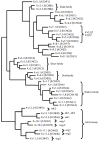
Fig. 1
Fig. 1
The phylogenetic tree of Kv channels based on the alignment of amino acid sequences. Braces combine channels belonging to the same family. Names are given according to the system of the International Union of Pure and Applied Chemistry (IUPAC) (alternative names according to the Gene Nomenclature Committee of the Human Genome Organization [2] are given in brackets)
Fig. 2
Fig. 2
Structure of Kv channels. A. Scheme of a single α-subunit of the Kv channel. Transmembrane segments S1–S6 and pore-forming P-loop are marked. Charged Arg of the membrane voltage sensor S4 are marked with “+” signs. PD –pore domain. B. Crystal structure of a single α-subunit of the Kv1.2 channel [21]. S1–6 segments, cytoplasmic domain T1, linker connecting the transmembrane portion with the T1 domain (T1–1), as well as N- and C-termini are marked. Charged Arg residues of the membrane voltage sensor S4 are indicated by blue circles. C. Crystal structure of the Kv1.2 channel in a complex with the β-subunit (marked as β, grey colored) (modified from [21]). TM –transmembrane region. D. Gate of the Kv2.1 channel. Only two opposite subunits of Kvα are shown for clarity reasons. The S6 helix is shown in purple, the blue color denotes a highly conserved portion of S6T –PXP helix (Pro-Val-Pro in Kv2.1), a key component of the lower gate. Green spheres mark K+ ions in the selectivity filter (P-loop), which represents the upper gate of the channel
Fig. 3
Fig. 3
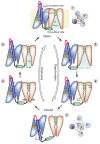
A. Scheme of the conformational transitions in Kv channels: C – closed channel; O – open channel; I – inactivated channel. B. N-type inactivation. The inactivation peptide enters the pore and physically blocks the transfer of ions after the activation of the channel. C. C-type inactivation. The selectivity filter acts as the second gate and closes, preventing the penetration of ions. The channels completely return to the closed conformation when the potential drops to the resting potential level (modified from [35])
Fig. 4
Fig. 4
A. Schematic arrangement of S4 helix residues in the Kv10 and Kv2.1 channels. The distribution of residues with high impact (HI) and low impact (LI) on the opening/closing process is shown. Three parallel stripes along the S4 helix (HI charged residues, HI hydrophobic residues, LI hydrophobic residues) are continuous for both channels and form a three-step coil [59]. B. Kv channel activation scheme according to the SHM model [59] – screw rotation and motion of S4 helix (white cylinder) in a fixed gate channel (GC)
Fig. 5
Fig. 5
Various models of Kv channels activation. All channels and their parts are shown in lateral orientation: the extracellular space is at the top, and the cytoplasm is at the bottom. A. Scheme of the paddle model (PM) of Kv activation. The movement of the paddles (blue ovals) is shown. Red “+” signs mark Arg in the S4 helix. B. PM based on the crystal structure of KvAP [92] in open and closed conformation. Paddle S3b–4 is shown in red. S1–4 helices are marked. The channel is shown as a frontal section. C. Sliding-helix model (SHM). The changes in the VSD domain of the Shaker channel are shown. Movable S4 segments and the S4–5 loop are purple-colored. Positively charged side chains of the S4 helix and negatively charged side chains of the S1–3 helices interacting with each other are colored blue and red, respectively [83]. D. SHM. The full-sized channels are given in closed and open conformations. S1–6 helices are numbered. Helices of the VSD domain are shown in different colors. Helices of the pore domain (S5–6) are purple-colored [83] E. Scheme of S4 helix movement (grey cylinder) during the activation of the Kv channel, according to the transport model (TM), showing how depolarization changes the availability of Arg residues (shown as blue circles) from the inner and outer aqueous cavities [48]. F. TM of Kv channels activation –closed and open conformations of the Shaker channel are shown. Transmembrane helices are color-coded: S1 –white, S2 –yellow, S3 –red, S4 –blue; pore domain is shown in green; Arg in S4 are shown in purple [48]
Fig. 6
Fig. 6
Comparison of Kv1.2 channel models [53] in the activated (open) state (left) and in the rested (closed) state (right). All transmembrane helices are shown as cylinders, except for S3 and S4 shown as spirals. Only one VSD domain is shown. S1 and S2 helices are shown in grey; the S4– S5 linker is purple-colored. The positions of Cα carbon atoms of Arg in the S4 helix are marked as R1 and R4 and highlighted in blue. The amino acid residue E226 of the S2 helix is marked as E1; E236 of the S2 helix, as E2; and residue D259 of the S3 helix, as D; these amino acids are highlighted in red. A. Lateral view. B. View from the extracellular space
Fig. 7
Fig. 7
Comparison of Kv1.2 VSD domain models in the open (left) [21] and closed (right) conformations according to the consensus model (CM). The S1 helix is shown in grey; S2,in yellow; S3, in red; and S4, in blue. Cα atoms of the R294 residue move in the vertical direction by 7-10 A. The values of the root mean square fluctuations (RMSF) reflect the variation in the vertical z coordinate calculated for a Cα atom. The blue spheres with lateral radicals represent the basic charged amino acid residues of the S4 helix (R1–R4) that interact with amino acid residues in other helices (their side chains are marked) [55]
Fig. 8
Fig. 8
A. The charge transfer center (CTC) is highly conserved among VSD-containing proteins. The alignment of the sequences of the chimeric channel Kv1.2/2.1 (GI: 160877792), Shaker (GI: 13432103), human channel Nav1.1 (GI: 115583677), human channel Cav1.1 (GI: 110349767), human channel Hv1 (GI: 91992155), and VSP (GI: 76253898) is shown. Only CTC-forming portions of the S2 and S3 segments are given. The highly conserved residues forming the site are marked: F – green; E and D – red. F corresponds to Phe233 in the chimeric channel Kv1.2/2.1. B. The five-stage model of Kv channel activation with four steps of VSD movement. At each stage, different, positively charged residues of the S4 helix (R1– R5, indicated by numbers) consistently occupy the CTC (shown as a circle). When all four sensors reach stage 5, the pore opens [114]
Fig. 9
Fig. 9
Five key intermediate stages of the Kv1.2 channel VSD domain, according to the model of charge transfer (MCT): initial upper position – α; three intermediate positions – β, γ, δ; and lower closed position – ε. The basic residues of the S4 helix are shown as blue sticks; amino acid residues and lipid PO4 - group that form salt bridges with R1–R5 are indicated by red sticks and yellow spheres, respectively. The highly conserved residue F233 of the S2 helix is shown as blue spheres [115]
Fig. 10
Fig. 10
Model of Kv deactivation (MKD). Intermediate stages that the VSD domain of the Shaker channel passes during deactivation [116]. A. Molecular models of the VSD domain: O – open channel, C1-C2 – intermediate states, C3 – closed conformation, C4 – deep closed state, which occurs under special conditions. At each stage, the side chain of one of the Arg residues of the S4 helix (blue sticks) passes through the CTC (F290, green stick), while the Arg side chains located close to the CTC form salt bridges with the negatively charged residues of the S1-S3 helices (red sticks; E247 in S1 and E283 in S2 above F290; and E293 in S2 and 316D in S3 below F290). At all the stages, the portion of the S4 helix situated opposite F290 transits into a 310 helix (purple), but at the C4 stage this portion is relaxed into an α-helix (yellow). Thus, the portion of the 310 helix slides along the S4 segment without energy consumption, which prevents the rotation of this segment during the activation/deactivation of the channel. B. Schematic, demonstrating the movements of the S4 helix. Color coding as in Fig. 10A
Fig. 11
Fig. 11
Mechanistic model of Kv activation/ deactivation (MMd) [56]. Effect of the hyperpolarization potential on the activated channel (1) initiates the inward movement of the S4 helix and weakens the bond between VSD and the pore domains. As a result, a depletion of ion transport in the pore cavity (2) and subsequent hydrophobic collapse of the pore occur. Closure of the upper (Ile402 in Kv1.2) and the lower gates [PVP motif; Leu331 (S5)–Pro405 (S6)] stops the ion current (3). The S4 helix continues to move inwardly; as soon as the S4 movement stops, the S4–S5 linker lowers completely and VSD domains are removed from the pore; the channel transits into the closed state (4). The impact of the depolarization potential on the closed channel leads to the movement of the S4 helix in an outward direction. The lower gate destabilizes when all four segments of the S4 and S4-S5 linker rise (5) and all VSD domains approach the pore again; the transition 4 => 5 represents the rate-limiting step of channel activation. Fluctuation of the lower gate causes opening of the pore and its partial rehydration. This allows potassium ions to enter into the pore and to initiate the channel conductance (6); the transition 5 => 6 is potential-independent. The presence of ions promotes complete rehydration of the pore leading to the complete opening of the upper and lower gates and returning the channel to the open state (1). Distribution of VSD domains (circles) relative to the pore domain (squares) is shown schematically (view from the extracellular side) [56]

Similar articles

Cited by

References

    1. Yu F.H., Yarov-Yarovoy V., Gutman G.A., Catterall W.A.. Pharmacol. Rev. 2005;57:387–395. - PubMed
    1. Gutman G.A., Chandy K.G., Grissmer S., Lazdunski M., McKinnon D., Pardo L.A., Robertson G.A., Rudy B., Sanguinetti M.C., Stühmer W., Wang X.. Pharmacol. Rev. 2005;57:473–508. - PubMed
    1. Hoshi T., Zagotta W.N., Aldrich R.W.. Science. 1990;250:533–538. - PubMed
    1. Yellen G.. Nature. 2002;419:35–42. - PubMed
    1. Bosma M.M., Hille B.. Endocrinology. 1992;130:3411–3420. - PubMed

LinkOut - more resources