. 2015 Jan 8;57(1):150-64.
doi: 10.1016/j.molcel.2014.12.001. Epub 2014 Dec 31.

Ubiquitin-SUMO circuitry controls activated fanconi anemia ID complex dosage in response to DNA damage

Affiliations

Ubiquitin-SUMO circuitry controls activated fanconi anemia ID complex dosage in response to DNA damage

Ian Gibbs-Seymour et al. Mol Cell. .

Abstract

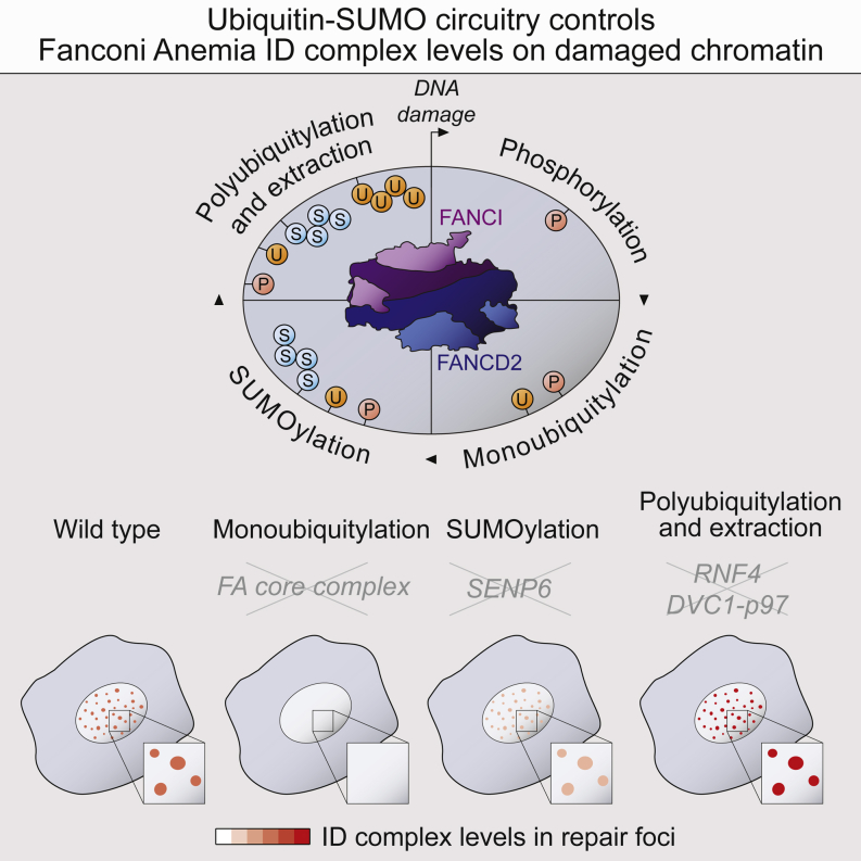
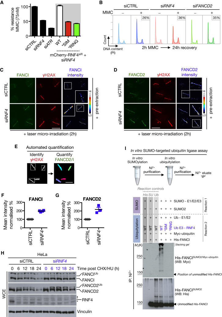
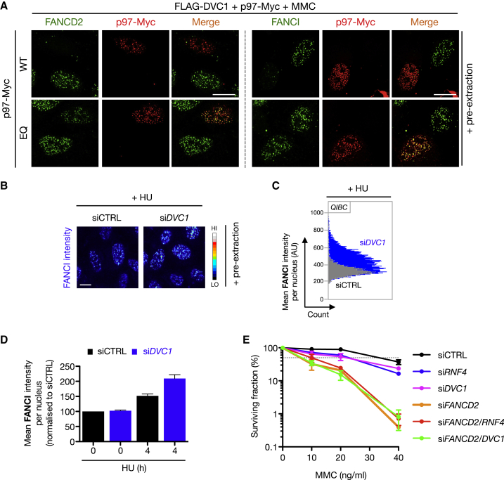
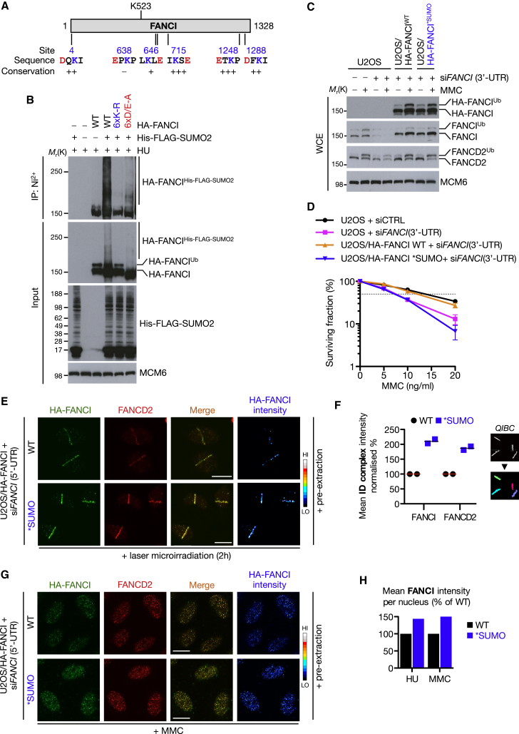
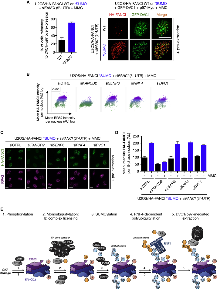
We show that central components of the Fanconi anemia (FA) DNA repair pathway, the tumor suppressor proteins FANCI and FANCD2 (the ID complex), are SUMOylated in response to replication fork stalling. The ID complex is SUMOylated in a manner that depends on the ATR kinase, the FA ubiquitin ligase core complex, and the SUMO E3 ligases PIAS1/PIAS4 and is antagonized by the SUMO protease SENP6. SUMOylation of the ID complex drives substrate selectivity by triggering its polyubiquitylation by the SUMO-targeted ubiquitin ligase RNF4 to promote its removal from sites of DNA damage via the DVC1-p97 ubiquitin segregase complex. Deregulation of ID complex SUMOylation compromises cell survival following replication stress. Our results uncover a regulatory role for SUMOylation in the FA pathway, and we propose that ubiquitin-SUMO signaling circuitry is a mechanism that contributes to the balance of activated ID complex dosage at sites of DNA damage.


Figures

None
Graphical abstract
Figure 1
Figure 1
The FANCI/FANCD2 (ID) Complex Is SUMOylated after DNA Damage (A) Stable HeLa/FLAG-SUMO2 cells were treated with doxycycline (DOX) for 24 hr to induce FLAG-SUMO2 expression. Cells were then lysed under denaturing conditions, subjected to FLAG immunoprecipitation (IP), and analyzed by immunoblotting using the indicated antibodies. WCE, whole-cell extract; CIS, cisplatin. MCM6 is used as a loading control. (B) HeLa cells were treated with HU for 24 hr and lysed under denaturing conditions, before IP with anti-FANCI antibody or preimmune serum (IgG). Immunopurified material was analyzed by immunoblotting with the indicated antibodies. (C) Same as (B) except FANCD2 antibody was used for the IP. (D) HeLa/FLAG-SUMO1 cells were induced with DOX and then subjected to MMC for various durations and processed as in (A). (E) HeLa/FLAG-SUMO2 cells treated with FANCI or FANCD2 siRNA and induced with DOX were subjected to MMC or HU for a further 24 hr and processed as in (A). (F) HeLa/FLAG-SUMO2 cells were biochemically fractionated, diluted in denaturing buffer, and processed as in (A). See also Figure S1.
Figure 2
Figure 2
ID complex SUMOylation Requires ATR, the FA Core Complex, and the SUMO E3 Ligases PIAS1 and PIAS4 (A) HeLa/FLAG-SUMO1 cells induced with DOX were transfected with siRNA against the SUMO E3 ligase PIAS4 and then subjected to MMC, HU, or APH for a further 24 hr. Cells were then lysed under denaturing conditions, subjected to FLAG IP, and analyzed by immunoblotting using the indicated antibodies. (B) Same as (A), using HeLa/FLAG-SUMO2 cells and PIAS1 siRNA. (C) U2OS cells transfected with Strep-HA-PIAS4 or empty vector (−) were subjected to replication stress using indicated agents for 24 hr. Strep-HA-PIAS4 complexes were purified with Strep-Tactin Sepharose and analyzed by immunoblotting. (D) Recombinant His-FANCI was SUMOylated in vitro and analyzed by immunoblotting with anti-His and anti-FANCI antibodies. , denotes unmodified His-FANCI band after reblotting. (E) U2OS cells transfected with control (CTRL) or PIAS1 siRNA were treated with MMC, pre-extracted, and then subjected to in situ proximity ligation assay using the indicated combination of antibodies. Scale bar represents 10 μm. (F) HeLa/FLAG-SUMO1/2 cells induced with DOX were transfected with FANCM siRNA and subjected to MMC for 24 hr. Cells were then processed as in (A). (G) FANCD2-deficient human fibroblasts (PD20) complemented with empty vector (EV), FANCD2 WT, or K561R mutant were treated with HU for 24 hr before lysis under denaturing conditions. FANCD2 was immunopurified using anti-FANCD2 antibody. Bound material was analyzed by immunoblotting with indicated antibodies. See also Figure S2.
Figure 3
Figure 3
SENP6 Antagonizes ID Complex SUMOylation (A) Schematic of QIBC methodology used to analyze ID complex chromatin loading. (B) U2OS cells were transfected with the indicated siRNAs against known SUMO proteases, treated with MMC (0.3 μM) for 24 hr, and then pre-extracted in situ to isolate chromatin bound proteins. Immunostained cells were processed for QIBC as outlined in (A). (C) Examples from (B) of FANCD2 chromatin-bound levels using QIBC. Scale bar represents 10 μm. (D) Quantification of mean FANCI chromatin-bound intensity using the same approach as (B). Data represent mean ± SEM from two independent experiments. p < 0.05. (E) Same as (D), but using FANCD2 antibody. (F) U2OS cells cotransfected with GFP-FANCI and HA-SENP6CI were treated with MMC (0.3 μM), fixed 4 hr later, and immunostained with HA antibody. Scale bar represents 10 μm. (G) Same as (F) except cells were subjected to GFP IP followed by immunoblotting with indicated antibodies. (H) HeLa/FLAG-SUMO2 cells induced with DOX were treated with SENP6 siRNA and subjected to MMC or HU for 24 hr. Cell lysates were subjected to FLAG IP under denaturing conditions before immunoblotting with indicated antibodies. (I) U2OS cells transfected with indicated siRNAs were treated with MMC and processed for QIBC. Cells were stained with FANCI antibody and chromatin-bound FANCI was quantified. Data represent mean ± SEM from two independent experiments. (J) Same as (I) but using FANCD2 antibody. See also Figure S3.
Figure 4
Figure 4
RNF4 Is a Regulator of the FA Pathway and Polyubiquitylates the SUMOylated ID Complex (A) Cellular fitness of U2OS cells transfected with control (CTRL), ATR, or RNF4 siRNA was assessed using the multicolor competition assay (MCA) (Smogorzewska et al., 2007). Stable U2OS/mCherry-RNF4 siRNA-resistant (siR) cell lines were used for RNF4 allele complementation analysis. Data represent mean ± SEM from three independent experiments (B) HeLa cells were transfected with the indicated siRNAs, exposed to a pulse of MMC (50 ng/ml) for 2 hr, and then allowed to recover for 24 hr before fixation and analysis by flow cytometry. The proportion of cells in G2/M phase is indicated. (C) U2OS cells transfected with control (CTRL) or RNF4 siRNA were subjected to laser microirradiation, pre-extracted and fixed 2 hr later, and immunostained with FANCDI and γH2AX antibodies. Scale bar represents 10 μm. (D) Same as (C) except with FANCD2 antibody. (E) QIBC strategy to analyze data generated from laser microirradiation experiments in an automated unbiased manner. (F) Quantification of normalized mean FANCI intensities at sites of laser microirradiation. Each data point represents the quantification of 75–150 cells from three independent experiments. (G) Same as (F) but for FANCD2. (H) HeLa cells transfected with control (CTRL) or RNF4 siRNA were treated with cycloheximide (CHX) and HU for the indicated times. Protein extracts were analyzed by immunoblotting with indicated antibodies. , denotes cross-reactive bands. (I) His-FANCI was SUMOylated in vitro as in Figure 2D, purified on Ni2+ agarose and then subjected to in vitro ubiquitylation by RNF4. The Ni2+ beads were then washed extensively and analyzed by immunoblotting with indicated antibodies. See also Figure S4.
Figure 5
Figure 5
The DVC1-p97 Complex Promotes Extraction of the ID Complex from Sites of DNA Damage (A) U2OS cells cotransfected with FLAG-DVC1 and p97-Myc WT or ATPase-dead (EQ) were treated with MMC (0.3 μM) for 24 hr and then pre-extracted, fixed, and immunostained with indicated antibodies. Scale bar represents 10 μm. (B) U2OS cells were transfected with control (CTRL) or DVC1 siRNA, treated with HU for 24 hr, pre-extracted, fixed, immunostained, and analyzed by QIBC. A representative image is shown for chromatin-bound FANCI. (C) Representative plot from QIBC analysis from (B). (D) Quantification of data from (B). Data represent mean ± SEM from two biologically independent experiments. (E) Colony formation assay using HeLa cells transfected with indicated siRNAs and subjected to various doses of MMC for 24 hr. Data represent mean ± SEM from two independent experiments using technical triplicates per datapoint. See also Figure S5.
Figure 6
Figure 6
FANCI SUMOylation Regulates Activated ID Complex Dosage at Sites of DNA Damage (A) Location and conservation of potential SUMOylation sites in FANCI. Modified lysine residues in consensus SUMOylation motifs are shown in blue with the acidic residue in red. (B) HeLa cells transfected with HA-FANCI wild-type (WT) or HA-FANCI SUMO-site mutants (6xK-R or 6xD/E-A) together with His-FLAG-SUMO2 were subjected to HU treatment for 24 hr. SUMO conjugates were purified under denaturing conditions using Ni2+ agarose and analyzed by immunoblotting with HA antibody. (C) Indicated cell lines transfected with control or FANCI (3′UTR) siRNA were treated with MMC for 24 hr and whole-cell extracts were analyzed by immunoblotting with indicated antibodies. (D) Clonogenic survival of indicated U2OS cell lines depleted of endogenous FANCI using the 3′-UTR siRNA where indicated and treated with various doses of MMC. Data represent mean ± SEM from three independent experiments using technical triplicates per datapoint. (E) U2OS/HA-FANCI WT or SUMO cells transfected with FANCI (5′-UTR) siRNA were subjected to laser microirradiation, pre-extracted, and fixed after 2 hr and then immunostained with HA and FANCD2 antibodies. Scale bar represents 10 μm. (F) QIBC analysis of normalized mean HA-FANCI and FANCD2 intensities from (E). Data represent mean ± SEM from two independent experiments. (G) U2OS/HA-FANCI WT or SUMO cells transfected with FANCI (5′-UTR) siRNA were treated with MMC (0.3 μM) for 24 hr and immunostained as in (E). Scale bar represents 10 μm. (H) U2OS/HA-FANCI cell lines treated as in (G) or with HU were analyzed by QIBC. Mean intensity of chromatin loaded HA-FANCI was normalized to HA-FANCI WT. See also Figure S6.
Figure 7
Figure 7
SUMOylation-Deficient FANCI Is Insensitive to Regulation by RNF4, SENP6, and DVC1 (A) U2OS/HA-FANCI WT or SUMO cells transfected with FANCI (5′-UTR) siRNA were transfected with GFP-DVC1 and p97-Myc plasmids and then treated with MMC (0.3 μM) for 4 hr, pre-extracted, fixed, and immunostained with HA antibody. Scale bar represents 10 μm. Data represent mean ± SEM from two independent experiments. For each data point, 200–400 cells were quantified. (B) U2OS/HA-FANCI SUMO cells were depleted of endogenous FANCI together with the indicated factors and processed for QIBC. (C) Representative images from data shown in (B). (D) Quantification of QIBC data from (B). Data represent mean ± SEM from two independent experiments. (E) Model of ubiquitin-SUMO circuitry in regulation of the ID complex (see Discussion for details). Modification locations do not represent actual sites. Question marks denote potential, but presently unknown, mechanism of regulation. P, phosphorylation; U, ubiquitin; S1, SUMO1; and S2, SUMO2. See also Figure S7.

Similar articles

Cited by

References

    1. Adamo A., Collis S.J., Adelman C.A., Silva N., Horejsi Z., Ward J.D., Martinez-Perez E., Boulton S.J., La Volpe A. Preventing nonhomologous end joining suppresses DNA repair defects of Fanconi anemia. Mol. Cell. 2010;39:25–35. - PubMed
    1. Akkari Y.M., Bateman R.L., Reifsteck C.A., D’Andrea A.D., Olson S.B., Grompe M. The 4N cell cycle delay in Fanconi anemia reflects growth arrest in late S phase. Mol. Genet. Metab. 2001;74:403–412. - PubMed
    1. Barysch S.V., Dittner C., Flotho A., Becker J., Melchior F. Identification and analysis of endogenous SUMO1 and SUMO2/3 targets in mammalian cells and tissues using monoclonal antibodies. Nat. Protoc. 2014;9:896–909. - PubMed
    1. Bergink S., Ammon T., Kern M., Schermelleh L., Leonhardt H., Jentsch S. Role of Cdc48/p97 as a SUMO-targeted segregase curbing Rad51-Rad52 interaction. Nat. Cell Biol. 2013;15:526–532. - PubMed
    1. Ciccia A., Elledge S.J. The DNA damage response: making it safe to play with knives. Mol. Cell. 2010;40:179–204. - PMC - PubMed

Publication types

MeSH terms

Substances