Review
. 2015 Jan 1;593(1):97-110.
doi: 10.1113/jphysiol.2014.277921. Epub 2014 Oct 21.

Retour aux sources: defining the structural basis of glutamate receptor activation

Affiliations
Review

Retour aux sources: defining the structural basis of glutamate receptor activation

G Brent Dawe et al. J Physiol. .

Abstract

Ionotropic glutamate receptors (iGluRs) are the major excitatory neurotransmitter receptor in the vertebrate CNS and, as a result, their activation properties lie at the heart of much of the neuronal network activity observed in the developing and adult brain. iGluRs have also been implicated in many nervous system disorders associated with postnatal development (e.g. autism, schizophrenia), cerebral insult (e.g. stroke, epilepsy), and disorders of the ageing brain (e.g. Alzheimer's disease, Parkinsonism). In view of this, an emphasis has been placed on understanding how iGluRs activate and desensitize in functional and structural terms. Early structural models of iGluRs suggested that the strength of the agonist response was primarily governed by the degree of closure induced in the ligand-binding domain (LBD). However, recent studies have suggested a more nuanced role for the LBD with current evidence identifying the iGluR LBD interface as a "hotspot" regulating agonist behaviour. Such ideas remain to be consolidated with recently solved structures of full-length iGluRs to account for the global changes that underlie channel activation and desensitization.


Figures

Figure 1
Figure 1. A cyclic gating model recreates AMPAR responses to willardiine series agonists
A, simulated response of AMPARs to l-Glu, generated from a five-state cyclic gating model (Patneau & Mayer, 1991). R, RA, Rd, and O represent resting, agonist-bound, desensitized, and open-channel states of the receptor, respectively. The rate constants for state transitions with l-Glu are adapted from values in another study (Vyklicky et al. 1991). B and C, simulated responses of AMPARs to KA (B), as well as to the willardiine series agonists, S-5-fluorowillardiine (s-FW), S-5-bromowillardiine (s-BrW), and S-5-iodowillardiine (s-IW) (C). As the halogen substituent increases in size, the equilibrium to peak current ratio recorded from AMPAR/KARs in hippocampal neurons also increases. To recapitulate this effect, the rate constant k–2 was increased, while k–1 and k–3 were adjusted to maintain microscopic reversibility. The rate constants are adapted from published values (Patneau et al. 1992), except for the s-BrW simulation, in which values were assigned to reproduce experimental observations.
Figure 2
Figure 2. Agonist efficacy correlates with closure of the GluA2 ligand binding cleft
A, side view of the GluA2 LBD with apo (protein data bank (PDB) 1FTO) and l-Glu-bound (PDB 1FTJ) structures overlaid (left). The residue P632, found at the base of the D2 domain, is emphasized to illustrate how this region lifts up and separates upon agonist binding. Visualization of a single subunit highlights how the cleft between D1 and D2 is narrowed in the l-Glu-bound structure (right). B and C, in the presence of the allosteric modulator cyclothiazide (CTZ) to attenuate desensitization, agonist responsiveness correlates to the degree of closure between D1 and D2 at the ligand binding cleft. For example, willardiine series agonists with smaller halogen substituents were more efficacious and produced a greater degree of cleft closure. (Jin et al. 2003).
Figure 3
Figure 3. Early structural model of iGluR activation and desensitization
The binding of agonist molecules (red) to the LBD dimer permits subsequent rearrangement to one of two conformations: activated or desensitized. The activated state occurs when the two D2 lobes (dark blue) are lifted apart, generating forces on the transmembrane domains (grey bars) to open the channel pore. Alternatively, the desensitized state stems from the separation of the two D1 domains (light blue) at the dimer interface, relaxing the LBD such that the energy provided by ligand binding cannot open the pore. The structural states are arranged according to the cyclic gating model described in Fig. 1, although to better account for the complex functional behaviour of iGluRs, more advanced kinetic models (Robert & Howe, 2003) have been developed, into which the four agonist binding sites are incorporated (bottom right). Moreover, is not clear to what extent movements of LBD dimers occur in the absence of bound ligands. The ATD has also been excluded for simplicity.
Figure 4
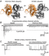
Figure 4. Crosslinking of the GluK2 LBD dimer interface yields non-decaying current responses
A, side views of the LBD dimer interface of GluK2 Y521C/L783C (PDB 2I0C), in its entirety (left) and close-up (right), detailing the inter-protomer disulphide bond (yellow). B, activation profiles of wild-type GluK2 and Y521C/L783C receptors in response to 10 mm l-Glu (holding potential –60 mV). Adapted from Daniels et al. (2013) with permission.
Figure 5
Figure 5. Cysteine crosslinking of the GluK2 LBD dimer interface disrupts activation
A, side views of the LBD dimer interface of GluK2 Y521C/L783C (left, PDB 2I0C) and GluA2 S729C (right, PDB 2I3W) in front of the corresponding l-Glu-bound wild-type receptors (transparent, PDB 3G3F and 1FTJ). For both mutants the distance across the interface between the D1 domains is increased. B, activation profiles of wild-type GluK2 and Y521C/L783C receptors at the single-channel level with a typical unitary response highlighted (black) for both receptors. C, simulations of GluK2 unitary channel activity in the presence (left) or absence (right) of desensitization, generated from a cyclic gating model (Bowie et al. 1998). When a single channel is simulated with desensitized states removed, the open channel probability is much greater than observed experimentally for the double cysteine mutant. Responses were recorded or simulated using 10 mm l-Glu and a holding potential of −100 mV, and experimental data were filtered at 1 kHz. Adapted from Daniels et al. (2013) with permission.
Figure 6
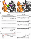
Figure 6. Occupancy of the GluK2 cation binding pocket sustains activation
A, side views of the LBD dimer interface of wild-type GluK2 (left, PDB 3G3F) and the D776K mutant (right, PDB 2XXX). The former includes two allosteric sodium ions (purple) and a chloride ion (green) bound at the apex of the interface, while the latter possesses a charged amino group (blue) on residue 776 tethering into the electronegative pocket (red) normally occupied by sodium. B, representative single-channel responses of GluK2 (left) and D776K (right) to 10 mm l-Glu (holding potential −60 mV, filtered at 1 kHz). C, averaged responses of individual sweeps taken from the same patch recordings as shown in B, which mimic the phenotype exhibited by a large population of receptors. For wild-type GluK2 several individual responses (grey) are overlaid behind the average response, while a fit of the current decay (red) suggests the unitary events are representative of those that contribute to macroscopic decay kinetics, which occur over a similar time course. (Dawe et al. 2013).
Figure 7
Figure 7. Structural rearrangements of full-length iGluRs during activation and desensitization
A, full-length GluA2 receptor bound by the competitive antagonist ZK200775 (top, PDB 3KG2) or KA and the allosteric modulator R,R-2b (bottom, PDB 4U1W), which potentiates GluA2 current responses in equilibrium conditions (Kaae et al. 2007). Both structures retain a twofold axis of symmetry, with the A/C and B/D subunits having distinct arrangements. The latter structure is thought to represent a ‘pre-open’ state of the receptor during the activation process. B, in contrast to the unliganded, apo state of GluA2 (left, PDB 4U2P), the binding of an agonist with positive modulator (centre, PDB 4U1W) causes separation between A/C subunits at the level of the LBD–TM 3 linker, generating forces that could open the pore. Addition of the con-ikot-ikot snail toxin further increases the B/D distance (right, PDB 4U5D). C, the tetrameric LBD of GluA2 bound by l-Glu and the allosteric modulator LY451646 (left, PDB 4UQK), a potentiator of AMPAR equilibrium currents (Gates et al. 2001), believed to be in an activated state. In contrast, the LBD of GluK2 bound by 2S,4R-4-methylglutamate (right, PDB 4UQQ) is believed to be in a desensitized state, characterized by large horizontal rotation of the B/D subunits.

Similar articles

Cited by

References

    1. Adams MD. Oxender DL. Bacterial periplasmic binding protein tertiary structures. J Biol Chem. 1989;264:15739–15742. - PubMed
    1. Akaike N. Concentration clamp technique. In: Walz W, editor; Boulton A, Baker G, editors. Neuromethods, Patch-Clamp Applications and Protocols. Humana Press Inc; 1995. pp. 141–154.
    1. Armstrong N. Gouaux E. Mechanisms for activation and antagonism of an AMPA-sensitive glutamate receptor: crystal structures of the GluR2 ligand binding core. Neuron. 2000;28:165–181. - PubMed
    1. Armstrong N, Jasti J, Beich-Frandsen M. Gouaux E. Measurement of conformational changes accompanying desensitization in an ionotropic glutamate receptor. Cell. 2006;127:85–97. - PubMed
    1. Armstrong N, Mayer M. Gouaux E. Tuning activation of the AMPA-sensitive GluR2 ion channel by genetic adjustment of agonist-induced conformational changes. Proc Natl Acad Sci U S A. 2003;100:5736–5741. - PMC - PubMed

Publication types

Substances

LinkOut - more resources