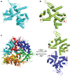
Molecular mechanism of mitochondrial calcium uptake
- PMID: 25548802
- PMCID: PMC11113575
- DOI: 10.1007/s00018-014-1810-1
Molecular mechanism of mitochondrial calcium uptake
Abstract
Mitochondrial calcium uptake plays a critical role in various cellular functions. After half a century of extensive studies, the molecular components and important regulators of the mitochondrial calcium uptake complex have been identified. However, the mechanism by which these protein molecules interact with one another and coordinate to regulate calcium passage through mitochondrial membranes remains elusive. Here, we summarize recent progress in the structural and functional characterization of these important protein molecules, which are involved in mitochondrial calcium uptake. In particular, we focus on the current understanding of the molecular mechanism underlying calcium through two mitochondrial membranes. Additionally, we provide a new perspective for future directions in investigation and molecular intervention.
Figures


Similar articles
-
The regulation of OXPHOS by extramitochondrial calcium.Biochim Biophys Acta. 2010 Jun-Jul;1797(6-7):1018-27. doi: 10.1016/j.bbabio.2010.02.005. Epub 2010 Feb 6. Biochim Biophys Acta. 2010. PMID: 20144582 Review.
-
Small-Molecule Modulators of Mitochondrial Channels as Chemotherapeutic Agents.Cell Physiol Biochem. 2019;53(S1):11-43. doi: 10.33594/000000192. Cell Physiol Biochem. 2019. PMID: 31834993 Review.
-
From the Identification to the Dissection of the Physiological Role of the Mitochondrial Calcium Uniporter: An Ongoing Story.Biomolecules. 2021 May 23;11(6):786. doi: 10.3390/biom11060786. Biomolecules. 2021. PMID: 34071006 Free PMC article. Review.
-
A Calcium Guard in the Outer Membrane: Is VDAC a Regulated Gatekeeper of Mitochondrial Calcium Uptake?Int J Mol Sci. 2021 Jan 19;22(2):946. doi: 10.3390/ijms22020946. Int J Mol Sci. 2021. PMID: 33477936 Free PMC article. Review.
-
SLC25A23 augments mitochondrial Ca²⁺ uptake, interacts with MCU, and induces oxidative stress-mediated cell death.Mol Biol Cell. 2014 Mar;25(6):936-47. doi: 10.1091/mbc.E13-08-0502. Epub 2014 Jan 15. Mol Biol Cell. 2014. PMID: 24430870 Free PMC article.
Cited by
-
Endoplasmic reticulum & mitochondrial calcium homeostasis: The interplay with viruses.Mitochondrion. 2021 May;58:227-242. doi: 10.1016/j.mito.2021.03.008. Epub 2021 Mar 26. Mitochondrion. 2021. PMID: 33775873 Free PMC article.
-
UCP2 modulates single-channel properties of a MCU-dependent Ca(2+) inward current in mitochondria.Pflugers Arch. 2015 Dec;467(12):2509-18. doi: 10.1007/s00424-015-1727-z. Epub 2015 Aug 16. Pflugers Arch. 2015. PMID: 26275882 Free PMC article.
-
PRMT1-mediated methylation of MICU1 determines the UCP2/3 dependency of mitochondrial Ca(2+) uptake in immortalized cells.Nat Commun. 2016 Sep 19;7:12897. doi: 10.1038/ncomms12897. Nat Commun. 2016. PMID: 27642082 Free PMC article.
-
Gestation age-associated dynamics of mitochondrial calcium uniporter subunits expression in feto-maternal complex at term and preterm delivery.Sci Rep. 2019 Apr 2;9(1):5501. doi: 10.1038/s41598-019-41996-3. Sci Rep. 2019. PMID: 30940880 Free PMC article.
-
MIRO GTPases in Mitochondrial Transport, Homeostasis and Pathology.Cells. 2015 Dec 31;5(1):1. doi: 10.3390/cells5010001. Cells. 2015. PMID: 26729171 Free PMC article. Review.
References
Publication types
MeSH terms
Substances
LinkOut - more resources
Full Text Sources
Other Literature Sources

