. 2015 Apr;27(4):756-69.
doi: 10.1016/j.cellsig.2014.12.009. Epub 2014 Dec 27.

Dimerization of cAMP phosphodiesterase-4 (PDE4) in living cells requires interfaces located in both the UCR1 and catalytic unit domains

Affiliations

Dimerization of cAMP phosphodiesterase-4 (PDE4) in living cells requires interfaces located in both the UCR1 and catalytic unit domains

Graeme B Bolger et al. Cell Signal. 2015 Apr.

Abstract

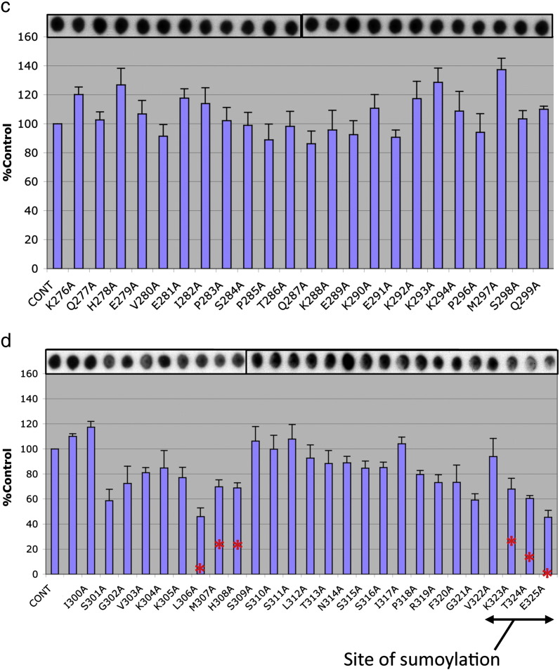
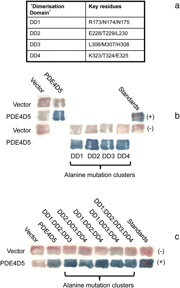
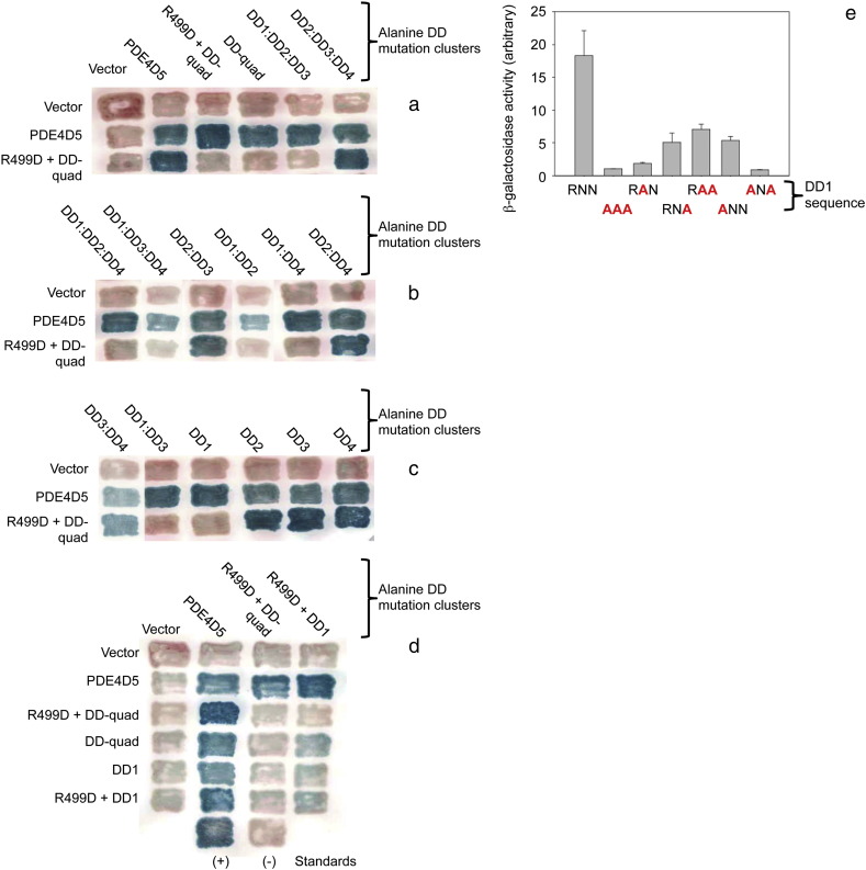
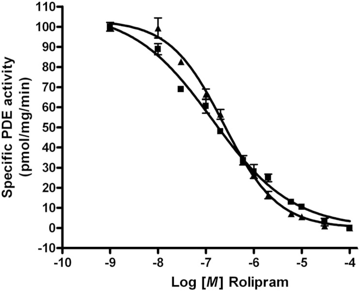
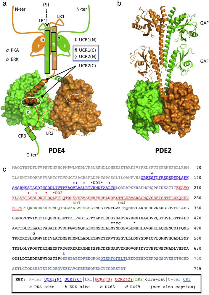
PDE4 family cAMP phosphodiesterases play a pivotal role in determining compartmentalised cAMP signalling through targeted cAMP breakdown. Expressing the widely found PDE4D5 isoform, as both bait and prey in a yeast 2-hybrid system, we demonstrated interaction consistent with the notion that long PDE4 isoforms form dimers. Four potential dimerization sites were uncovered using a scanning peptide array approach, where a recombinant purified PDE4D5 fusion protein was used to probe a 25-mer library of overlapping peptides covering the entire PDE4D5 sequence. Key residues involved in PDE4D5 dimerization were defined using a site-directed mutagenesis programme directed by an alanine scanning peptide array approach. Critical residues stabilising PDE4D5 dimerization were defined within the regulatory UCR1 region found in long, but not short, PDE4 isoforms, namely the Arg(173), Asn(174) and Asn(175) (DD1) cluster. Disruption of the DD1 cluster was not sufficient, in itself, to destabilise PDE4D5 homodimers. Instead, disruption of an additional interface, located on the PDE4 catalytic unit, was also required to convert PDE4D5 into a monomeric form. This second dimerization site on the conserved PDE4 catalytic unit is dependent upon a critical ion pair interaction. This involves Asp(463) and Arg(499) in PDE4D5, which interact in a trans fashion involving the two PDE4D5 molecules participating in the homodimer. PDE4 long isoforms adopt a dimeric state in living cells that is underpinned by two key contributory interactions, one involving the UCR modules and one involving an interface on the core catalytic domain. We propose that short forms do not adopt a dimeric configuration because, in the absence of the UCR1 module, residual engagement of the remaining core catalytic domain interface provides insufficient free energy to drive dimerization. The functioning of PDE4 long and short forms is thus poised to be inherently distinct due to this difference in quaternary structure.

Keywords: Dimerisation; Dimerization; PDE4; Phosphodiesterase; Rolipram; cAMP; cyclic AMP.


Figures

Fig. 1
Fig. 1
PDE4 dimerizes in living cells. Yeast 2-hybrid experiments using PDE4D5 as both ‘bait’ and ‘prey’. PDE4D5 was expressed unmutated (“wild-type”) or with the indicated point mutations. PDE4D5 was expressed unmutated (“wild-type”) or with the indicated deletion or point mutations. All patches in each column use identical ‘prey’ and all patches in each row use identical ‘bait’, as described in Materials and methods. Controls are vectors alone and standards are RasV12–Raf, as done before by us . Positive interactions, assessed with a filter β-galactosidase assay, produce blue patches, while negative interactions produce pink patches. (a) Full-length PDE4D5 (amino acids 1 to 749; “long”), or PDE4D5 lacking its unique N-terminal domain and UCR1, but containing UCR2 and the catalytic region (amino acids 206 to 747 of PDE4D5; “short”), was expressed as either a fusion to LexA (rows) or to GAL4 (columns) and the various mutants tested for interaction. (b) The EELD:AALA mutant reflects that ablating the UCR1:UCR2 interaction while the VFLL:AAAA mutation reflects that used to ablate PDE4D3 dimerization . This shows data typical of experiments performed at least 3 times.
Fig. 2
Fig. 2
Delineation of PDE4D5 dimerization sites by peptide array. An immobilized library of 25-mer peptides sequentially shifted by 5 amino acids along the entire sequence of PDE4D5 was probed with either purified PDE4D5-GST or GST alone. Dark spots represent areas of interaction between PDE4D5-GST and the PDE4D5 peptide array. Areas of interaction are identified by a colour code where red represents UCR1, blue represents UCR2 and green represents sequences within the catalytic unit. No interaction was detected between GST alone and the PDE4D5 peptide array (data not shown).
Fig. 3
Fig. 3
Alanine scanning pinpoints amino acids that are important for PDE4D5 dimerization. Alanine scanning analysis (where each of the indicated residues is replaced with alanine or aspartate if residue is an alanine) was undertaken on three PDE4D5 self-association domains highlighted in Fig. 2. Arrays were overlayed with PDE4D5-GST or GST alone. Dark spots represent areas of interaction between PDE4D5-GST and the PDE4D5 peptide array. Cont = control spot chosen from data depicted in Fig. 2 and where all 25 amino acids correspond to the native sequence. Data represents the mean plus S.E. from three independent experiments. No interaction was detected between GST alone and the PDE4D5 peptide array (data not shown).
Fig. 3
Fig. 3
Alanine scanning pinpoints amino acids that are important for PDE4D5 dimerization. Alanine scanning analysis (where each of the indicated residues is replaced with alanine or aspartate if residue is an alanine) was undertaken on three PDE4D5 self-association domains highlighted in Fig. 2. Arrays were overlayed with PDE4D5-GST or GST alone. Dark spots represent areas of interaction between PDE4D5-GST and the PDE4D5 peptide array. Cont = control spot chosen from data depicted in Fig. 2 and where all 25 amino acids correspond to the native sequence. Data represents the mean plus S.E. from three independent experiments. No interaction was detected between GST alone and the PDE4D5 peptide array (data not shown).
Fig. 4
Fig. 4
Characterization of ‘Dimerization Domain’ (DD) mutants. (a) Amino acid co-ordinates for the 4 DD regions on the PDE4D5 protein used throughout this study. (b) Mutations in individual DD domains do not ablate the interaction. The amino acids in each individual DD domain were mutated to alanine and tested for their effect on PDE4D5 dimerization in 2-hybrid assays, as in Fig. 1. (c) Mutations in combinations of DD domains do not ablate the interaction. Combinations of DD domain mutants were tested for their effect on PDE4D5 dimerization. This shows data typical of experiments performed at least 3 times.
Fig. 5
Fig. 5
Structure of dimeric PDE4 core catalytic domain assembly and the effect of the R449D mutation on dimerization. (a) (i) The dimeric PDE4D core catalytic domain with bound cilomilast inhibitor (blue surface) is shown (PDB: 1XOM; [73]). Assembly is centred on a C2 symmetry axis (asterisk) at the junction of helices-9 and -10; residues contributing to the focal interaction are highly organised through a network of hydrogen bonds. Helix-11 makes a single point of contact in the dimer through R499 in a salt bridge to H-loop residue, D463. Key residues contributing to the dimerization interface are only partially conserved in other PDE families. In contrast to the highly organised H-loop structure in PDE4, the corresponding region (dotted circle) in PDE2 and PDE5 is conformationally mobile and dynamically folds into the catalytic pocket (blue arrow) to regulate catalytic activity (see Discussion). (ii) Surface rendition derived from (i) highlighting the core catalytic domain dimerization interface on one subunit (orange) and contact residues from the second subunit (green stick). (b) Mutations in PDE4D5 were tested for their ability to ablate dimerization: The ‘DD-quad’ mutant is a combination of all four DD domain mutations where each are described individually in Fig. 4a. The R449D-PDE4D5 mutant and the combined R499D + DD-quad-PDE4D5 mutant were also studied. Two-hybrid assays were used as in Fig. 1. The data show that neither the R499D-PDE4D5 mutant nor the DD-quad-PDE4D5 mutant ablated the interaction, but that the combined R499D + DD-quad-PDE4D5 mutant markedly ablated the interaction. (c) Rescue of the charge effect of the R499D-PDE4D5 mutant by the D463R-PDE4D5 mutant. The D463R-PDE4D5 mutant was tested singly and in combination with the DD-quad-PDE4D5 mutant. The data show that the D463R-PDE4D5 mutant effectively rescued the ablation of the interaction seen with the combined R499D + DD-quad-PDE4D5 mutant. This shows data typical of experiments performed at least 3 times.
Fig. 5
Fig. 5
Structure of dimeric PDE4 core catalytic domain assembly and the effect of the R449D mutation on dimerization. (a) (i) The dimeric PDE4D core catalytic domain with bound cilomilast inhibitor (blue surface) is shown (PDB: 1XOM; [73]). Assembly is centred on a C2 symmetry axis (asterisk) at the junction of helices-9 and -10; residues contributing to the focal interaction are highly organised through a network of hydrogen bonds. Helix-11 makes a single point of contact in the dimer through R499 in a salt bridge to H-loop residue, D463. Key residues contributing to the dimerization interface are only partially conserved in other PDE families. In contrast to the highly organised H-loop structure in PDE4, the corresponding region (dotted circle) in PDE2 and PDE5 is conformationally mobile and dynamically folds into the catalytic pocket (blue arrow) to regulate catalytic activity (see Discussion). (ii) Surface rendition derived from (i) highlighting the core catalytic domain dimerization interface on one subunit (orange) and contact residues from the second subunit (green stick). (b) Mutations in PDE4D5 were tested for their ability to ablate dimerization: The ‘DD-quad’ mutant is a combination of all four DD domain mutations where each are described individually in Fig. 4a. The R449D-PDE4D5 mutant and the combined R499D + DD-quad-PDE4D5 mutant were also studied. Two-hybrid assays were used as in Fig. 1. The data show that neither the R499D-PDE4D5 mutant nor the DD-quad-PDE4D5 mutant ablated the interaction, but that the combined R499D + DD-quad-PDE4D5 mutant markedly ablated the interaction. (c) Rescue of the charge effect of the R499D-PDE4D5 mutant by the D463R-PDE4D5 mutant. The D463R-PDE4D5 mutant was tested singly and in combination with the DD-quad-PDE4D5 mutant. The data show that the D463R-PDE4D5 mutant effectively rescued the ablation of the interaction seen with the combined R499D + DD-quad-PDE4D5 mutant. This shows data typical of experiments performed at least 3 times.
Fig. 6
Fig. 6
Key effects of DD1 region mutants on dimerization. (a,b,c) The effects of various combinations of DD domain mutants were studied in filter β-galactosidase assays for their effect on their interaction with R499D + DD-quad-PDE4D5 mutant. Two-hybrid assays were used as in Fig. 1. The data show that combinations containing mutations in DD1 ablated the interaction, while mutations in other combinations of DD domains had minimal effect. (d) Comparison of the DD1-PDE4D5 mutants in cis and trans with the R499D-PDE4D5 mutant. The R499D, DD-quad, and DD1 PDE4D5 mutants when singly and in combination, were tested against the same array of mutants. The data show that the DD1-PDE4D5 mutant blocks the interaction when present in either half of the dimer (i.e., regardless of whether it is present in the ‘prey’ or ‘bait’ construct.) (e) Quantitative analysis of DD1 region individual amino acid mutants: Individual amino acids in PDE4D5-DD1 (sequence RNN, Fig. 4a) were mutated to alanine and tested in a quantitative β-galactosidase assay for their effect on their interaction with R499D + DD-quad-PDE4D5. The data show that restoration of one or two amino acids to wild-type increases the interaction, at least in some cases, but never back to wild-type. This suggests either that (1) All three amino acids in the RNN motif contribute directly to the interaction; or that (2) Even single amino acid mutations in this area cause sufficient localized distortion of the PDE4D5 structure that they attenuate the interaction. This shows data typical of experiments performed at least 3 times.
Fig. 7
Fig. 7
PDE4D5 dimerization in mammalian cells. The indicated Flag and vsv-tagged PDE4D5 wild-type and mutant constructs were co-expressed in HEK293 cells. Cells were then disrupted and lysates taken for either immunoblotting with both anti-Flag and anti-vsv antisera or immunoprecipitated with anti-vsv antisera prior to immunoblotting with both anti-Flag and anti-vsv antisera. The data shown are representative of experiments performed at least three times using different transfected cell preparations.
Fig. 8
Fig. 8
Dimeric and monomeric PDE4D5 are similarly inhibited by rolipram. Lysates of HEK293 cells transfected to express either wild-type- (dimeric) or DD1-R499D- (monomeric) PDE4D5 were used to assess the ability of increasing concentrations of rolipram to inhibit their activity using 1 μM cAMP as substrate. Data shows means ± SD for a single experiment with triplicate assays. Data from replicates of such experiments was used to determine IC50 values of 1.4 ± 0.2 × 10− 7 M for dimeric wild-type PDE4D5 and 2.3 ± 0.3 × 10− 7 M for monomeric DD1-R499D-PDE4D5, with only four amino acids mutated (SD, n = 3 separate experiments).
Fig. 9
Fig. 9
PDE4 long form dimerization model and sequence key. (a) Cross-capped model for PDE4 long form assembly based on the 3G45 UCR2-capped core catalytic domain crystal structure of Burgin et al. . Sequence analysis fitted to a helical wheel model suggests that the UCR1(C) and UCR2(N) regions likely adopt helical 2° structure with extensive hydrophobic contact surfaces suitable for helix bundle assembly . Inter-helix structural transition [e.g. mediated by phosphorylation (PKA site a; [23–26])] is proposed to control stability of the UCR2(N) cap and thence gate substrate entry to the catalytic pocket (white oval). The stability of the UCR2 cap is also affected by additional phosphorylation (ERK site b; [28,29,70,71]) and competitive capping with a C-terminal regulatory sequence (CR3; [68]) may further influence enzyme activity. The definition of LR2 (Linker Region 2) was coined as that region of amino acids that joins UCR2 to the catalytic unit and is unique to each of the four PDE4 sub-families. (b) Structure of the PDE2 dimer (PDB: 3IBJ, ; see Discussion). (c) PDE4D5 sequence key, highlighting domain boundaries and residues implicated in PDE4 long form dimer assembly. Residues marked: and • are proposed to contribute to extended hydrophobic contact surfaces along helical 2° structure motifs for UCR1(C) and UCR2(N) . Quadruple alanine mutagenesis of the VFLL set (•) reportedly ablates PDE4D3 dimerization and in the present work also compromises PDE4D5 dimerization. Sequences identified as possible dimerization domains in the peptide array studies of the present work are identified (DD1, 173-RNN-175; DD2, 228-ETL-230; DD3, 306-LMH-308; DD4, 323-KTE-325). PKA and ERK phosphorylation sites are marked (a and b respectively). Focal residues contributing to the core catalytic domain dimerization interface are marked (*, c, d), where c and d are respectively D463 and R499 and form an important ion pairing that underpins the stability of this interface (cf.Fig. 5a).

Similar articles

Cited by

References

    1. Houslay M.D. Trends Biochem. Sci. 2010;35:91–100. - PubMed
    1. Lugnier C. Pharmacol. Ther. 2006;109:366–398. - PubMed
    1. Maurice D.H., Ke H., Ahmad F., Wang Y., Chung J., Manganiello V.C. Nat. Rev. Drug Discov. 2014;13:290–314. - PMC - PubMed
    1. Rogne M., Tasken K. Horm. Metab. Res. Horm. Metab. Res. 2014;46(12):833–840. - PubMed
    1. Zaccolo M. Br. J. Pharmacol. 2009;158:50–60. - PMC - PubMed

Publication types

MeSH terms