. 2014 Dec 29;9(12):e115974.
doi: 10.1371/journal.pone.0115974. eCollection 2014.

Conditional knockout of prolyl hydroxylase domain protein 2 attenuates high fat-diet-induced cardiac dysfunction in mice

Affiliations

Conditional knockout of prolyl hydroxylase domain protein 2 attenuates high fat-diet-induced cardiac dysfunction in mice

Heng Zeng et al. PLoS One. .

Abstract

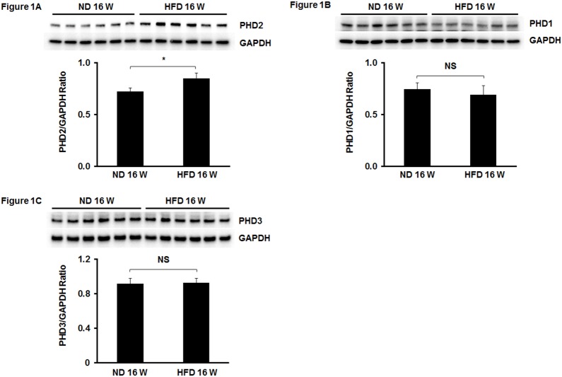
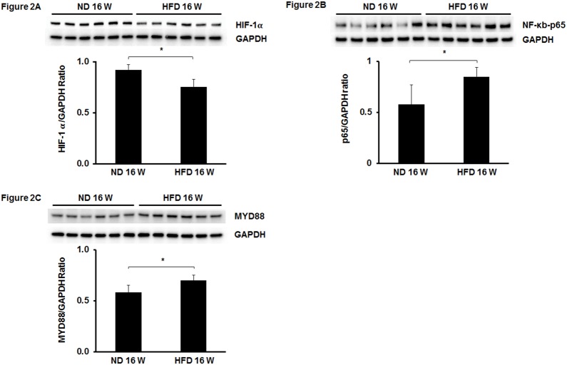
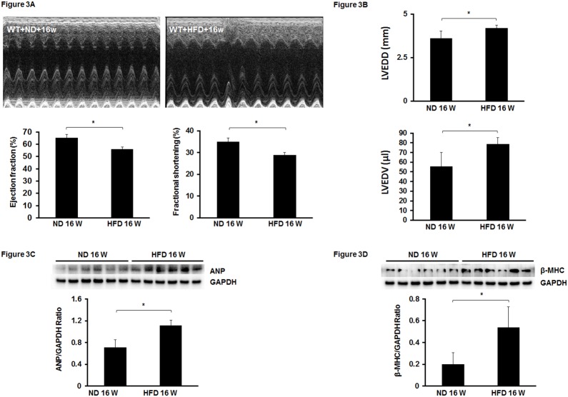
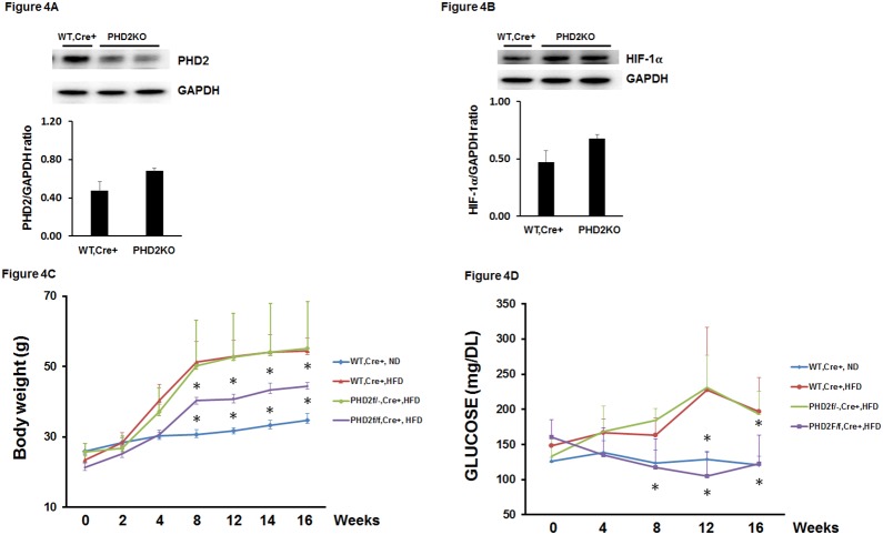
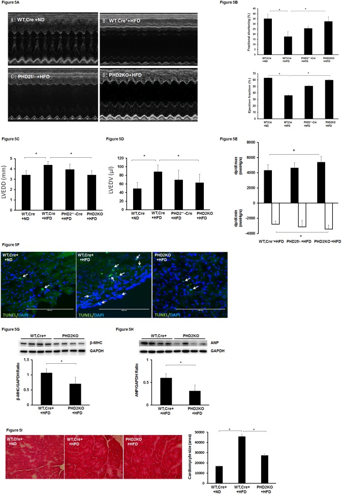
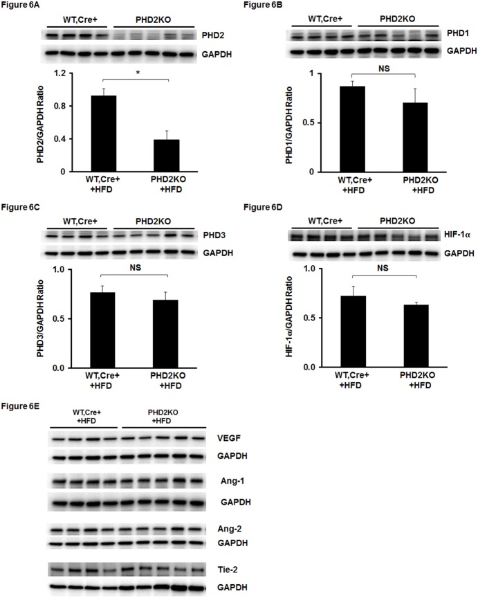
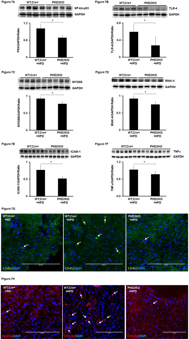
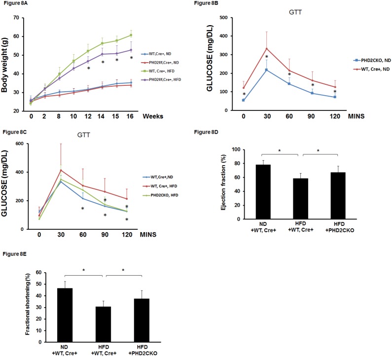
Oxygen sensor prolyl hydroxylases (PHDs) play important roles in the regulation of HIF-α and cell metabolisms. This study was designed to investigate the direct role of PHD2 in high fat-diet (HFD)-induced cardiac dysfunction. In HFD fed mice, PHD2 expression was increased without significant changes in PHD1 and PHD3 levels in the heart. This was accompanied by a significant upregulation of myeloid differentiation factor 88 (MYD88) and NF-κB. To explore the role of PHD2 in HFD-induced cardiac dysfunction, PHD2 conditional knockout mice were fed a HFD for 16 weeks. Intriguingly, knockout of PHD2 significantly reduced MYD88 and NF-κb expression in HFD mouse hearts. Moreover, knockout of PHD2 inhibited TNFα and ICAM-1 expression, and reduced cell apoptosis and macrophage infiltration in HFD mice. This was accompanied by a significant improvement of cardiac function. Most importantly, conditional knockout of PHD2 at late stage in HFD mice significantly improved glucose tolerance and reversed cardiac dysfunction. Our studies demonstrate that PHD2 activity is a critical contributor to the HFD-induced cardiac dysfunction. Inhibition of PHD2 attenuates HFD-induced cardiac dysfunction by a mechanism involving suppression of MYD88/NF-κb pathway and inflammation.


Conflict of interest statement

Competing Interests: The authors have declared that no competing interests exist.

Figures

Figure 1
Figure 1. Expression of PHDs in the hears of HFD mice.
(A) Western blot analysis demonstrating that PHD2 expression was significantly upregulated in the hearts of HFD mice compared to normal chow diet (ND) mice (n = 6 mice, *p<0.05). (B and C) Western blot analysis of PHD1 and PHD3 expression showing that there was no significantly difference in PHD1 and PHD3 expression between HFD and normal diet (ND) mice (n = 6 mice, p>0.05). NS =  Not significant.
Figure 2
Figure 2. Expression of HIF-1α, NF-κb and MyD88 in the hears of HFD mice.
(A and B) Western blot analysis revealing that HIF-1α expression was significantly decreased in the hearts of HFD mice compared to normal diet (ND) mice (n = 6 mice, *p<0.05). (B) NF-κb expression was significantly upregulated in the hearts of HFD mice compared to normal diet (ND) mice (n = 6 mice, *p<0.05). (C) MyD88 expression was significantly increased in the hearts of HFD mice compared to normal diet (ND) mice (n = 6 mice, *p<0.05).
Figure 3
Figure 3. Feeding with HFD for 16 weeks leads to cardiac dysfunction in mice.
(A) Upper panel: Representative images of M mode of echocardiography. Low panel: Feeding mice with HFD for 16 weeks significantly impaired cardiac performance indicated by decreases in EF and FS levels in HFD mice compared to ND mice (n = 5–7 mice, *p<0.05). (B) Left ventricular end diastolic diameter (LVEDD) and left ventricular end diastolic volume (LVEDV) were significantly elevated in HFD mice compared to ND mice at 16 weeks (n = 5–7 mice, *p<0.05). (C and D) Expression of cardiac hypertrophic markers ANP and β-MHC in HFD and ND mice. The levels of ANP and β-MHC were significantly higher in the hearts of HFD mice than that of ND mice at 16 weeks (n = 6 mice, *p<0.05).
Figure 4
Figure 4. Knockout of PHD2 prevents HFD-induced increases in body weight and glucose levels in mice.
(A and B) Western blot analysis confirming that treated PHD2f/f-Cre+ mice with tamoxifen for 7 days reduced PHD2 expression in the hearts. The expression of HIF-1α was increased in the hearts of PHD2f/f-Cre+ mice treated with tamoxifen for 7 days compared to wild type (WT, Cre+) mice treated with tamoxifen for 7 days (n = 2 mice). (C and D) Pretreatment of WT, Cre+ mice with tamoxifen for 7 days then fed with HFD for 16 weeks led to a gradual increase in body weight and elevation of fasting glucose levels compared to WT, Cre+ mice fed with normal chow diet (ND) (n = 10 mice, *p<0.05). Pretreatment of PHD2f/f-Cre+ mice with tamoxifen for 7 days then fed with HFD for 16 weeks significantly reduced body weight growth and fasting glucose levels compared to WT, Cre+ mice fed with HFD (n = 10 mice, *p<0.05). Pretreatment of PHD2f/−Cre+ mice with tamoxifen for 7 days then fed with HFD for 16 weeks did not alter body weight growth and fasting glucose levels compared to WT, Cre+ mice fed with HFD (n = 10 mice, p>0.05).
Figure 5
Figure 5. Knockout of PHD2 improves HFD-induced impairment of cardiac function in mice.
(A) Representative images of M mode of echocardiography. (B) Pretreatment of WT, Cre+ mice with tamoxifen for 7 days then fed with HFD for 16 weeks resulted in a significant reduction of FS% and EF% compared to WT, Cre+ mice fed with ND (n = 7 mice, *p<0.05). Pretreatment of PHD2f/f-Cre+ mice with tamoxifen for 7 days then fed with HFD for 16 weeks significantly increased FS% and EF% levels compared to WT, Cre+ mice fed with HFD (n = 7 mice, *p<0.05). Pretreatment of PHD2f/−Cre+ mice with tamoxifen for 7 days then fed with HFD for 16 weeks had little effects on FS% and EF% levels compared to WT, Cre+ mice fed with HFD (n = 6 mice, p>0.05). (C and D) Left ventricular end diastolic diameter (LVEDD) and left ventricular end diastolic volume (LVEDV) were significantly elevated in WT, Cre+ + HFD mice compared to WT, Cre+ + ND mice at 16 weeks (n = 5–7 mice, *p<0.05). Knockout of PHD2 (PHD2KO) significantly reduced HFD-induced elevation of LVEDD and LVEDV (n = 7 mice, *p<0.05). (E) Cardiac dp/dt max and dp/dt min levels were significantly improved in PHD2KO + HFD mice compared to WT, Cre+ + HFD mice (n = 6 mice, *p<0.05). (F) Apoptotic cells were stained with apoptotic marker TUNEL (green) and nuclei were counterstained with DAPI (blue). Immunohistochemical analysis of apoptotic cells showing apoptotic cells (TUNEL positive cells, white arrow) were increased in the hearts of WT, Cre+ + HFD mice compared to that of WT, Cre+ + ND mice. Knockout of PHD2 dramatic reduced the number of TUNEL positive cells in the hearts of HFD mice. (G and H) The levels of ANP and β-MHC were significantly reduced in the hearts of PHD2KO + HFD mice compared to WT, Cre+ + HFD mice at 16 weeks (n = 4–5 mice, *p<0.05). (I) Cardiomyocytes were stained with H&E. Cardiomyocyte size (area) was significantly reduced in the hearts of PHD2KO + HFD mice compared to WT, Cre+ + HFD mice at 16 weeks (40X, n = 3 mice, *p<0.05).
Figure 6
Figure 6. Knockout of PHD2 does not rescue HFD-induced reduction of HIF-1α expression in HFD mice.
(A) Western blot analysis confirming that PHD2 expression was significantly reduced in PHD2f/f-Cre+ mice treated with tamoxifen for 7 days and fed with HFD for 16 weeks compared to WT, Cre+ + HFD mice (n = 4–5 mice, *p<0.05). (B–F) Western blot analysis showing that expression of PHD1 and PHD3 was not significantly altered in PHD2KO + HFD mice compared to WT, Cre+ + HFD mice (n = 4–5 mice, NS). The expression of HIF-1α did not changed in the hearts of PHD2KO + HFD mice compared to WT, Cre+ + HFD mice (n = 4–5 mice, NS). VEGF and angiopoietins/Tie-2 system were not altered in PHD2KO + HFD mice compared to WT, Cre+ + HFD mice (n = 4–5 mice, NS).
Figure 7
Figure 7. Knockout of PHD2 reduces HFD-induced NF-κb activation and macrophage infiltration in HFD mice.
(A) NF-κb expression was significantly decreased in the hearts of PHD2KO+HFD mice compared to WT, Cre+ + HFD mice at 16 weeks (n = 4 mice, *p<0.05). (B) TLR4 expression was significantly reduced in the hearts of PHD2KO+HFD mice compared to WT, Cre+ + HFD mice at 16 weeks (n = 4–5 mice, *p<0.05). (C) MyD88 expression was significantly suppressed in the hearts of PHD2KO+HFD mice compared to WT, Cre+ + HFD mice at 16 weeks (n = 4–5 mice, *p<0.05). (D) IRAK-4 expression was significantly inhibited in the hearts of PHD2KO+HFD mice compared to WT, Cre+ + HFD mice at 16 weeks (n = 4 mice, *p<0.05). (E and F) The expression of ICAM-1 and TNF-α was significantly reduced in the hearts of PHD2KO+HFD mice compared to WT, Cre+ + HFD mice at 16 weeks (n = 6 mice, *p<0.05). (G and H) Inflammatory cell infiltrations were stained with macrophage markers CD45 (green) and CD11b (Red). Nuclei were counterstained with DAPI (blue). Immunohistochemical analysis of macrophage infiltrations (CD45 and CD11b positive cells) showing the number of macrophage (white arrow) was increased in the hearts of WT, Cre+ + HFD mice compared to that of WT, Cre+ + ND mice. Knockout of PHD2 dramatic reduced the number of macrophage in the hearts of HFD mice.
Figure 8
Figure 8. Conditional knockout of PHD2 in HFD mice improves glucose tolerance and cardiac function.
(A) Conditional knockout of PHD2 in HFD mice significantly reduced body weight growth compared to WT, Cre+−HFD mice (n = 10 mice, *p<0.05). (B) Conditional knockout of PHD2 in normal chow diet mice significantly improved glucose tolerance compared to WT, Cre+ mice fed with ND (n = 7–10 mice, *p<0.05). (C) Conditional knockout of PHD2 in HFD mice significantly enhanced glucose tolerance compared to WT, Cre+−HFD mice (n = 10 mice, *p<0.05). (D and E) Conditional knockout of PHD2 in HFD mice significantly increased EF% and FS% compared to WT, Cre+−HFD mice (n = 5–7 mice, *p<0.05).

Similar articles

Cited by

References

    1. Semenza GL (2007) Hypoxia-inducible factor 1 (HIF-1) pathway. Sci STKE 2007:cm8 doi:10.1126/stke.4072007cm8. - DOI - PubMed
    1. Willam C, Maxwell PH, Nichols L, Lygate C, Tian YM, et al. (2006) HIF prolyl hydroxylases in the rat; organ distribution and changes in expression following hypoxia and coronary artery ligation. J Mol Cell Cardiol 41:68–77 doi:10.1016/j.yjmcc.2006. - DOI - PubMed
    1. Appelhoff RJ, Tian YM, Raval RR, Turley H, Harris AL, et al. (2004) Differential function of the prolyl hydroxylases PHD1, PHD2, and PHD3 in the regulation of hypoxia-inducible factor. J Biol Chem 279:38458–38465 doi:10.1074/jbc.M406026200. - DOI - PubMed
    1. Fong GH, Takeda K (2008) Role and regulation of prolyl hydroxylase domain proteins. Cell Death Differ 15:635–641 doi:10.1038/cdd.2008. - DOI - PubMed
    1. Nwogu JI, Geenen D, Bean M, Brenner MC, Huang X, et al. (2001) Inhibition of collagen synthesis with prolyl 4-hydroxylase inhibitor improves left ventricular function and alters the pattern of left ventricular dilatation after myocardial infarction. Circulation 104:2216–2221 doi:10.1161/hc4301.097193. - DOI - PubMed

Publication types

MeSH terms

Substances

LinkOut - more resources