. 2015 May;33(5):1390-404.
doi: 10.1002/stem.1926.

Signalling Through Retinoic Acid Receptors is Required for Reprogramming of Both Mouse Embryonic Fibroblast Cells and Epiblast Stem Cells to Induced Pluripotent Stem Cells

Affiliations

Signalling Through Retinoic Acid Receptors is Required for Reprogramming of Both Mouse Embryonic Fibroblast Cells and Epiblast Stem Cells to Induced Pluripotent Stem Cells

Jian Yang et al. Stem Cells. 2015 May.

Abstract

We previously demonstrated that coexpressing retinoic acid (RA) receptor gamma and liver receptor homolog-1 (LRH1 or NR5A2) with OCT4, MYC, KLF4, and SOX2 (4F) rapidly reprograms mouse embryonic fibroblast cells (MEFs) into induced pluripotent stem cells (iPSCs). Here, we further explore the role of RA in reprogramming and report that the six factors (6F) efficiently and directly reprogram MEFs into integration-free iPSCs in defined medium (N2B27) in the absence of feeder cells. Through genetic and chemical approaches, we find that RA signalling is essential, in a highly dose-sensitive manner, for MEF reprogramming. The removal of exogenous RA from N2B27, the inhibition of endogenous RA synthesis or the expression of a dominant-negative form of RARA severely impedes reprogramming. By contrast, supplementing N2B27 with various retinoids substantially boosts reprogramming. In addition, when coexpressed with LRH1, RA receptors (RARs) can promote reprogramming in the absence of both exogenous and endogenously synthesized RA. Remarkably, the reprogramming of epiblast stem cells into embryonic stem cell-like cells also requires low levels of RA, which can modulate Wnt signalling through physical interactions of RARs with β-catenin. These results highlight the important functions of RA signalling in reprogramming somatic cells and primed stem cells to naïve pluripotency. Stem Cells 2015;33:1390-1404.

Keywords: Epiblast stem cells; Induced pluripotent stem cells; Liver receptor homolog-1; Reprogramming; Retinoic acid receptor gamma; Retinoic acid receptors; β-Catenin.


Figures

Figure 1
Figure 1
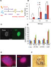
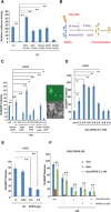
Reprogramming MEFs to iPSCs in N2B27 by 4F and 6F. (A): Diagram of the reprogramming strategy. After transfection, MEFs were recovered in M10 before being subsequently cultured in N2B27/LIF. Colonies were picked and cultured in N2B27/2i/LIF for characterization. (B): Effects of increased doses of reprogramming factors. GFP+ colonies from Rex1‐GFP MEFs were scored on day 18. *, p < .05; **, p < .01. (C): The morphology of a Rex1‐GFP+ 6F‐iPSC colony in 2i/LIF on day 10. Scale bar = 200 µm. (D): Quantitative real‐time PCR analysis of pluripotent gene expression in 4F‐ and 6‐iPSCs. The expression levels are shown relative to Gapdh and normalized to ESCs. (E): Immunostaining of 6F‐iPSCs for SSEA‐1 and Nanog. Scale bar = 100 µm. (F): Chimeric mice with a contribution (dark fur) from 6F‐iPSCs. The experiments were repeated at least three times, and error bars show standard deviations from the mean of triplicate measurements in one representative experiment. Abbreviations: DAPI, 4′,6‐diamidino‐2‐phenylindole; ESC, embryonic stem cell; GFP, green fluorescent protein; iPSC, induced pluripotent stem cell; LIF, leukemia inhibitory factor; MEF, mouse embryonic fibroblast cell.
Figure 2
Figure 2
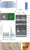
Efficient reprogramming of Rex1‐GFP MEFs to integration‐free iPSCs in N2B27/leukaemia inhibitory factor by 6F. (A): AP+ colonies of expressing 4F or 6F from the episomal vectors. Left panel: AP+ colonies. Right panel: colony numbers. **, p < .01. (B): A 6F GFP+ colony from the episomal vectors. Scale bar = 200 µm. (C): Characterization of iPSCs for loss of the episomal vectors. Whole cell lysis was used to detect residual episome or genome‐integrated episomal vectors (top four panels). Actb was used as a genomic DNA polymerase chain reaction (PCR) control. The bottom four panels show RT‐PCR‐amplified transcripts of exogenous factors. Actb expression was used as the control. E‐iPSCs were produced using episomal vectors. PB‐iPSCs were reprogrammed using piggyBac vectors. (D): Quantitative real‐time PCR analysis of pluripotent gene expression in iPSCs from 6F‐episomal vectors. The expression is shown relative to Gapdh and normalized to ESCs. (E): Teratomas derived from 6F E‐iPSCs. (F): Chimeric mice from iPSC injection into host blastocysts. Experiments were repeated at least three times, and the error bars show standard deviations from the mean of triplicate determinations of one representative experiment. Abbreviations: AP, alkaline phosphatase; ESC, embryonic stem cell; GFP, green fluorescent protein; iPSC, induced pluripotent stem cell; MEF, mouse embryonic fibroblast cell.
Figure 3
Figure 3
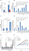
Retinoic acid (RA) signalling promotes mouse embryonic fibroblast cell (MEF) reprogramming in the defined condition. (A): The effect of withdrawing VA (retinol) from N2B27/leukemia inhibitory factor (LIF) on 4F and 6F reprogramming. *, p < .05; **, p < .01. (B): Reprogramming MEFs in medium containing retinoids. *, p < .05; **, p < .01. (C): The retinoic acid receptor gamma agonist CD437 (25.0 nM) increases AP+ colony numbers by 4F plus LRH1 in −VA medium. (D): RA signalling activated by various retinoids in a luciferase assay. (E): Quantitative real‐time PCR analysis of gene expression in MEFs cultured in N2B27/LIF in the presence of retinoids for 2 days. These genes include pluripotency‐related genes (c‐Myc, Klf4), Rars (α, β, γ) and genes involved in retinol metabolism (Adh1, Raldh2, Cyp26a1, and Stra6). The expression levels are shown relative to Gapdh and normalized to embryonic stem cells. (F): Proliferation of MEFs in retinoids. MEFs (1 × 10⁵) were plated in N2B27/LIF with or without vitamin A or ATRA. Cells were counted daily for 6 days. Δ, p < .05; ΔΔ, p < .01: ATRA 100.0 nM compared with −VA; *, p < .05; **, p < .01: ATRA 1.0 µM compared with −VA. In the MEF reprogramming experiments, AP+ colonies were scored on day 18 after transfection. The experiments were repeated at least three times, and the error bars shown standard deviations from the mean of triplicate determinations in one representative experiment. Abbreviations: AP, alkaline phosphatase; ATRA, all‐trans retinoic acid; 9cRA, 9‐cis‐retinoic acid; LRH1, liver receptor homolog‐1; VA, vitamin A.
Figure 4
Figure 4
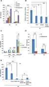
Reprogramming mouse embryonic fibroblast cells (MEFs) by 6F is both retinoic acid (RA) receptor ligand‐dependent and ligand‐independent. (A): MEFs were reprogrammed by 4F alone, 4F plus either RARG or LRH1, or 4F plus both RARG and LRH1 (6F) in +VA and −VA medium. Significantly more AP+ colonies are obtained by 6F, even in −VA. (B): Blocking RA signalling by citral (50.0 μM) or CD2665 (1.0 μM) as measured in a luciferase assay. (C): Blocking 4F‐ or 6F‐mediated MEF reprogramming by citral (50.0 μM). Adding ATRA rescues reprogramming. Note that 6F produces substantially more AP+ colonies in the presence of citral compared with the other conditions. (D): The blocking of MEF reprogramming by RARA‐DN. (E): The effects of blocking RA signalling by either citral (50.0 μM) or RARA‐DN on MEF reprogramming. Note that adding citral and expressing RARA‐DN almost completely blocked reprogramming. Adding ATRA at 10.0 nM could partially rescue the block. In the MEF reprogramming experiments, AP+ colonies were scored on day 18 after transfection. *, p < .05; **, p < .01. The experiments were repeated at least three times, and the error bars show standard deviations from the mean of triplicate determinations in one representative experiment. Abbreviations: AP, alkaline phosphatase; ATRA, all‐trans retinoic acid; LRH1, liver receptor homolog‐1; RARA‐DN, dominant‐negative form of RARA; RARG, retinoic acid receptor gamma; VA, vitamin A.
Figure 5
Figure 5
Retinoic acid (RA) signalling in EpiSC reprogramming. (A): RA signalling as measured by a luciferase assay in EpiSCs cultured in N2B27/activin/FGF2 with VA and other retinoids. The activities are shown relative to that of Renilla luciferase and normalized to that of EpiSCs in −VA. (B): A diagram of EpiSC reprogramming in 2i/LIF. (C): The effects of retinoids on reprogramming Oct4‐GFP reporter EpiSCs to induced pluripotent stem cells by LRH1. *, p < .05; **, p < .01. Scale bar = 200 µm. (D): RA signalling at various stages of EpiSC reprogramming. **, p < .01. (E): The effects of expressing RARG (under the CMV early enhancer/chicken β actin promoter) on reprogramming EpiSCs by LRH1. Note that even small amounts of Rarg PB transposon have deleterious effects. *, p < .05; **, p < .01. (F): Blocking LRH1‐mediated EpiSC reprogramming by ligand depletion (citral), RA receptor inhibition (CD2665) or expression of RARA‐DN. Reprogramming was partially restored by 0.1 nM ATRA. Oct4‐GFP+ colonies were scored on day 9 after transfection. *, p < .05; **, p < .01. In all the experiments, Oct4‐GFP+ colonies were scored on day 9 after transfection. The experiments were repeated at least three times, and the error bars show standard deviations from the mean of triplicate determinations in one representative experiment. Abbreviations: ATRA, all‐trans retinoic acid; 9cRA, 9‐cis‐retinoic acid; EpiSCs, epiblast stem cells; FGF2, fibroblast growth factor 2; LIF, leukemia inhibitory factor; LRH1, liver receptor homolog‐1; RARA‐DN, dominant‐negative form of RARA; RARG, retinoic acid receptor gamma; VA, vitamin A.
Figure 6
Figure 6
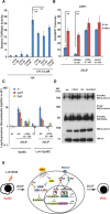
Retinoic acid (RA) signalling modulates the Wnt pathway. (A): Wnt signalling as measured by a TOPflash assay. EpiSCs were cultured in N2B27‐VA or supplemented with ATRA, CD437, or CD2665 with or without CH for 24 hours before the assay. The activities are shown relative to Renilla luciferase and normalized to EpiSC in N2B27‐VA. **, p < .01. (B): The attenuation of Wnt signalling by IWR‐1 improves EpiSC reprogramming. After Lrh1 transfection, Oct4‐GFPEpiSCs were cultured in 2i/LIF medium without VA and supplemented with ATRA, CD437, or CD2665 with or without IWR‐1 (2.0 µM) for 4 days. The cells were subsequently cultured in 2i/LIF‐VA for another 4 days. **, p < .01. (C): The activation of RA signalling downregulates CH‐induced expression of Wnt target genes. EpiSCs or Lrh1 transfected EpiSCs were cultured in 2i/LIF‐VA and 2i/LIF‐VA supplemented with ATRA for 24 hours for quantitative real‐time PCR analysis. The expression is shown relative to Gapdh and normalized to EpiSCs in N2B27/AF‐VA. (D): The interaction of RARG and β‐catenin. Nuclear extracts of EpiSCs cultured in N2B27‐VA with CD437 (5.0 nM) or CH (3.0 µM) were immunoprecipitated with an anti‐RARG antibody. The pull‐down proteins were blotted with an anti‐β‐catenin antibody and an anti‐RARG antibody, sequentially. Rabbit IgG was the negative control, and Histone H3 was the loading control. Western blotting of β‐catenin in EpiSCs prior to immunoprecipitation was also performed. In all experiments, iPSC colonies were scored on day 9 after transfection. The experiments were repeated at least three times, and the error bars show standard deviations from the mean of triplicate determinations in one representative experiment. (E): A proposed model of the interaction between Wnt and RA signalling in EpiSCs reprogramming. Abbreviations: AF, activin/fibroblast growth factor 2; APC, Adenomatosis Polyposis Coli; ATRA, all‐trans retinoic acid; CH, CHIR9902; 9cRA, 9‐cis‐retinoic acid; DVL, dishevelled; EpiSCs, epiblast stem cells; GSK, glycogen synthase kinase; iPSC, induced pluripotent stem cell; IP, immunoprecipitation; LEF, lymphoid enhancing factor; LIF, leukemia inhibitory factor; LRH1, liver receptor homolog‐1; MW, molecular weight; RAR, RA receptor; RARE, retinoic acid response element; RARG, retinoic acid receptor gamma; RXR, retinoid X receptor; TCF, T‐cell factor; VA, vitamin A; WB, Western blot.

Similar articles

Cited by

References

    1. Takahashi K, Yamanaka S. Induction of pluripotent stem cells from mouse embryonic and adult fibroblast cultures by defined factors. Cell 2006;126:663–676. - PubMed
    1. Hamanaka S, Yamaguchi T, Kobayashi T et al. Generation of germline‐competent rat induced pluripotent stem cells. PLoS One 2011;6:15. - PMC - PubMed
    1. Takahashi K, Tanabe K, Ohnuki M et al. Induction of pluripotent stem cells from adult human fibroblasts by defined factors. Cell 2007;131:861–872. - PubMed
    1. Yu J, Vodyanik MA, Smuga‐Otto K et al. Induced pluripotent stem cell lines derived from human somatic cells. Science 2007;318:1917–1920. - PubMed
    1. Huangfu D, Maehr R, Guo W et al. Induction of pluripotent stem cells by defined factors is greatly improved by small‐molecule compounds. Nat Biotechnol 2008;26:795–797. - PMC - PubMed

Publication types