. 2015 Jan 20;6(2):1101-14.
doi: 10.18632/oncotarget.2728.

MicroRNA-130a associates with ribosomal protein L11 to suppress c-Myc expression in response to UV irradiation

Affiliations

MicroRNA-130a associates with ribosomal protein L11 to suppress c-Myc expression in response to UV irradiation

Yuhuang Li et al. Oncotarget. .

Abstract

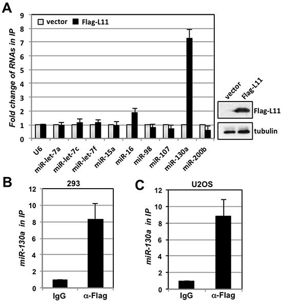
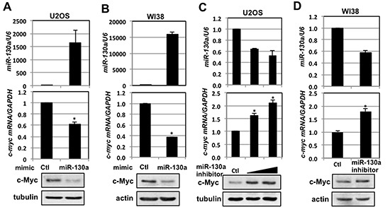
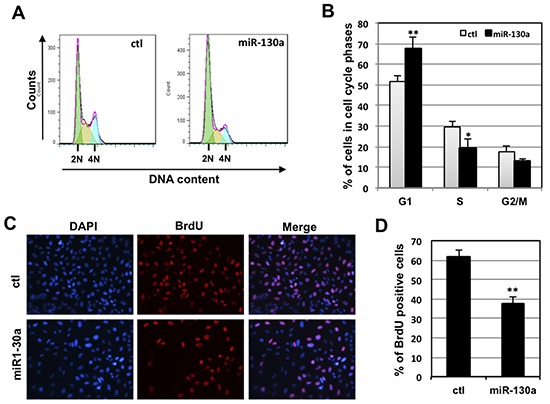
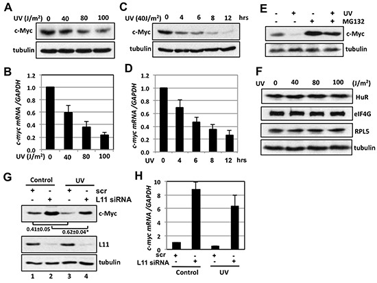
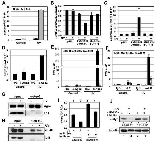
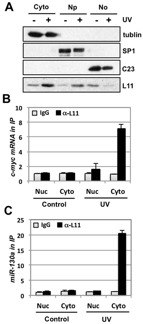
The oncoprotein c-Myc is essential for cell growth and proliferation while its deregulated overexpression is associated with most human cancers. Thus tightly regulated levels and activity of c-Myc are critical for maintaining normal cell homeostasis. c-Myc is down-regulated in response to several types of stress, including UV-induced DNA damage. Yet, mechanism underlying UV-induced c-Myc reduction is not completely understood. Here we report that L11 promotes miR-130a targeting of c-myc mRNA to repress c-Myc expression in response to UV irradiation. miR-130a targets the 3'-untranslated region (UTR) of c-myc mRNA. Overexpression of miR-130a promotes the Ago2 binding to c-myc mRNA, significantly reduces the levels of both c-Myc protein and mRNA and inhibits cell proliferation. UV treatment markedly promotes the binding of L11 to miR-130a, c-myc mRNA as well as Ago2 in cells. Inhibiting miR-130a significantly suppresses UV-mediated c-Myc reduction. We further show that L11 is relocalized from the nucleolus to the cytoplasm where it associates with c-myc mRNA upon UV treatment. Together, these results reveal a novel mechanism underlying c-Myc down-regulation in response to UV-mediated DNA damage, wherein L11 promotes miR-130a-loaded miRISC to target c-myc mRNA.


Figures

Figure 1
Figure 1. L11 associates with miR-130a
(A) Identification of miR-130a as a L11-associated miRNA. 293 cells transfected with control or Flag-L11 were subjected to RNA-IP using anti-Flag antibody followed by detection of indicated miRNAs using RT-qPCR. The expression of Flag-L11 is shown in the right panel. (B–C) L11 associates with miR-130a in cells. Lysates from 293 (B) and U2OS (C) cells transfected with Flag-L11 were immunoprecipitated with control mouse IgG or anti-Flag antibody, followed by RT-qPCR detection of miR-130a.
Figure 2
Figure 2. miR-130a regulates c-Myc levels
(A–B) Overexpression of miR-130a decreases c-Myc levels. U2OS (A) or WI38 (B) cells transfected with control or miR-130a mimics were assayed for the relative expression of miR-130a normalized with U6 snRNA (top panels), c-myc mRNA normalized with GAPDH mRNA (middle panels) by RT-qPCR, and c-Myc protein levels (bottom panels) by IB. *p < 0.01, compared with control transfected cells. (C–D) Inhibition of miR-130a increases c-Myc levels. U2OS (C) or WI38 (D) cells transfected with control or miR-130a hairpin inhibitors were assayed for the relative expression of c-myc mRNA normalized with GAPDH mRNA (middle panels) by RT-qPCR and c-Myc protein expression (bottom panels) by IB. *p < 0.01, compared with control transfected cells.
Figure 3
Figure 3. miR-130a targets c-myc mRNA through its 3′-UTR
(A) Overexpression of miR-130a reduces the activity of luciferase reporter with c-myc 3′-UTR. 293 cells transfected with control pGL3 or pGL3-myc-3′UTR in the presence of β-gal plasmid together with control or miR-130a mimic as indicated were assayed for the relative luciferase activity normalized to β-gal expression. *p < 0.01, compared with cells transfected with pGL3-myc-3′UTR and control miRNA mimic. (B) Three putative miR-130a binding sites (BS-1, BS-2 and BS-3) in the c-myc 3′-UTR predicted by RNA22 program. (C) Schematic diagram of the pGL3-myc-3′UTR vectors. The first nucleotide after stop codon is indicated as “1”. (D) miR-130a regulates c-Myc via BS-1. 293 cells transfected with control or miR-130a mimic together with the indicated pGL3 or pGL3-myc-3′UTR vectors were assayed for the relative luciferase activity normalized to β-gal expression. *p < 0.01, compared with cells transfected with control miRNA mimic and corresponding luciferase reporters. (E–F) Ago2 associates with miR-130a at c-myc mRNA. U2OS cells transfected with control or miR-130a mimic were subjected to RNA-IP using control IgG or anti-Ago2 antibody, followed by RT-qPCR detection of c-myc and GAPDH mRNA (E) as well as U6 and miR-130a (F).
Figure 4
Figure 4. miR-130a inhibits cell proliferation
(A–B) Overexpression of miR-130a inhibits cell cycle progression. U2OS cells were transfected with control or miR-130a mimic for 48 hours. The cells were trypsinized, stained with PI, and analyzed by flow cytometry. The histograms of PI staining from one representative experiment indicating the G1 (2N DNA), G2/M (4N DNA) and S (between G1 and G2/M phases) phases are shown in panel (A). The mean percentages of cells in different cell cycle phases from three independent experiments are shown in panel (B). *p < 0.05; **p < 0.01, compared with control transfected cells. (C–D) BrdU incorporation assays. U2OS cells were transfected with control or miR-130a mimic as above. At 48 hour post-transfection, the cells were incubated with BrdU for another 10 hours. The cells were fixed and stained with anti-BrdU antibodies (red) and DAPI (blue) (C). The average of BrdU-positive cells is shown in (D). **p < 0.01, compared with control transfected cells.
Figure 5
Figure 5. L11 is involved in UV-induced c-Myc downregulation
(A–D) UV irradiation decreases c-Myc levels. U2OS cells were exposed to different dosages of UV (A) (B) or 40 J/m2 UV for different times (C) (D). The cells were assayed for the expression of c-Myc protein by IB (A) (C) and c-myc mRNA by RT-qPCR (B) (D). (E) MG132 treatment partially rescued the c-Myc reduction by UV treatment. U2OS cells treated with 40 J/m2 UV were cultured in the presence or absence of 40 μM MG132 for 6 hours followed by IB. (F) UV treatment does not reduce the levels of HuR, eIF4G and ribosomal protein L5 (RPL5). U2OS cells were treated with different dosages of UV for 6 hours and assayed by IB. (G–H) Knockdown of L11 abolished the c-Myc reduction by UV treatment. U2OS cells transfected with scrambled or L11 siRNA were treated with 40 J/m2 UV for 6 hours. The cells were subjected to IB detection of c-Myc protein (G) and RT-qPCR detection of c-myc mRNA (H). *p < 0.01, compared to scrambled RNA transfected cells.
Figure 6
Figure 6. L11 recruits miR-130a-loaded miRISC to c-myc mRNA in response to UV irradiation
(A) UV treatment increases the L11 binding to c-myc mRNA. U2OS cells treated without or with UV were subjected to RNA-IP using control IgG or anti-L11 antibodies, followed by RT-qPCR assays. (B–C) L11 inhibition of c-Myc in response to UV requires its binding to the c-myc 3′-UTR. 293 cells transfected with pGL3, pGL3-myc 3′-UTR-FL, or pGL3-myc 3′-UTR-F1 were treated with or without UV. The cells were then assayed for the relative luciferase activity normalized to β-gal expression (B) and subjected to RNA-IP using anti-L11 antibodies, followed by RT-qPCR detection of the luciferase mRNA. (D) UV treatment increases Ago2 binding to c-myc mRNA. U2OS cells treated with or without UV were subjected to RNA-IP using control IgG or anti-Ago2 antibodies, followed by RT-qPCR detection of c-myc mRNA (D). (E–F) UV treatment increases the binding of L11 and Ago2 to miR-130a, and, to a less extent, to miR-24. U2OS cells treated with or without UV were subjected to RNA-IP using control IgG or anti-Ago2 (E) or anti-L11 (F) antibodies, followed by RT-qPCR detection of miR-130a, miR-24 and the control U6 RNA. (G–H) UV treatment increases the L11 binding to Ago2, but not eIF4G. U2OS cells treated with or without UV were subjected to co-IP with anti-Ago2 (G) and anti-eIF4G (H) antibodies followed by IB. (I–J) Inhibiting miR-130a abolishes c-Myc reduction by UV treatment. U2OS cells transfected with control or miR-130a inhibitor were treated with or without UV. The cells were assayed for the expression of c-myc mRNA by RT-qPCR (I) and c-Myc protein by IB (J). *p < 0.05, compared the ratio of lane 4 to lane 3 with the ratio of lane 2 to lane 1. In all above assays, cells were treated with 40 J/m2 UV and harvested at 6 hours post-treatment.
Figure 7
Figure 7. UV irradiation promotes L11 interaction with miR-130a and c-myc mRNA in the cytoplasm
(A) UV treatment releases L11 from the nucleolus into the nucleoplasm and the cytoplasm. U2OS cells treated with or without 40 J/m2 UV for 6 hours were subjected to isolation of cytoplasm (Cyto), nucleoplasm (Np), and the nucleolus (No) fractions, followed by IB detection of indicated proteins. Tubulin, SP1, and nucleolin (C23) were used as cytoplasm, nucleoplasm and nucleolar markers, respectively. (B–C) UV treatment increases L11 binding to miR-130a and c-myc mRNA in the cytoplasm. The cytoplasmic and the nuclear (Nuc) fractions isolated from U2OS cells treated with or without 40 J/m2 UV for 6 hours were immunoprecipitated with anti-L11 antibodies or control rabbit IgG, followed by RT-qPCR detection of c-myc mRNA (B) and miR-130a (C).

Similar articles

Cited by

References

    1. Dang CV. MYC on the path to cancer. Cell. 2012;149:22–35. - PMC - PubMed
    1. Luscher B, Vervoorts J. Regulation of gene transcription by the oncoprotein MYC. Gene. 2012;494:145–160. - PubMed
    1. Meyer N, Penn LZ. Reflecting on 25 years with MYC. Nature reviews. Cancer. 2008;8:976–990. - PubMed
    1. Nesbit CE, Tersak JM, Prochownik EV. MYC oncogenes and human neoplastic disease. Oncog ene. 1999;18:3004–3016. - PubMed
    1. Liu J, Levens D. Making myc. Current topics in microbiology and immunology. 2006;302:1–32. - PubMed

Publication types

MeSH terms

LinkOut - more resources