. 2015 Feb 6;290(6):3563-75.
doi: 10.1074/jbc.M114.601872. Epub 2014 Dec 23.

Salinomycin and other polyether ionophores are a new class of antiscarring agent

Affiliations

Salinomycin and other polyether ionophores are a new class of antiscarring agent

Collynn F Woeller et al. J Biol Chem. .

Abstract

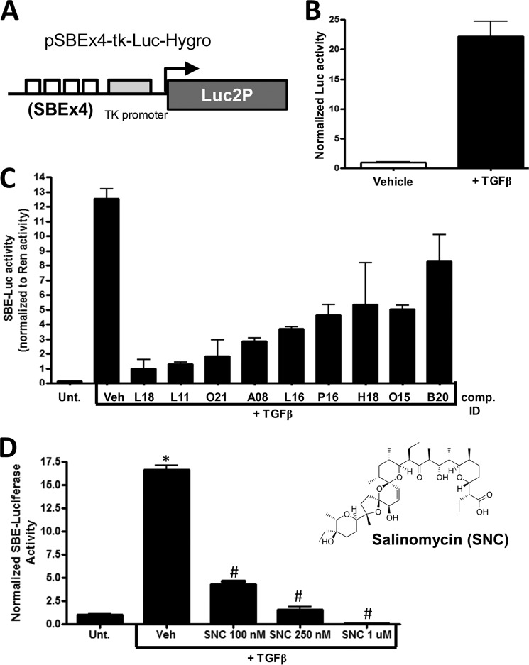
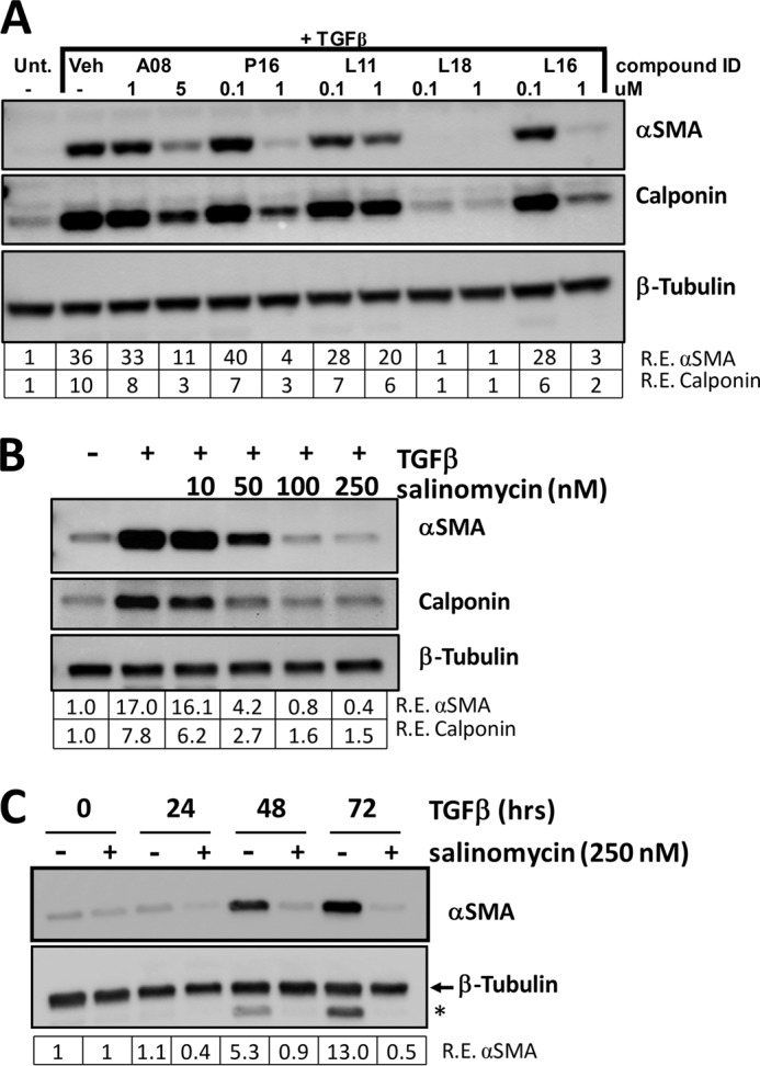
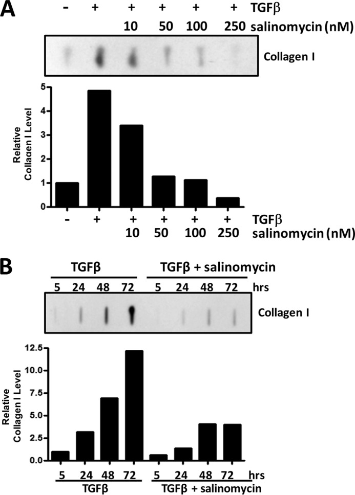
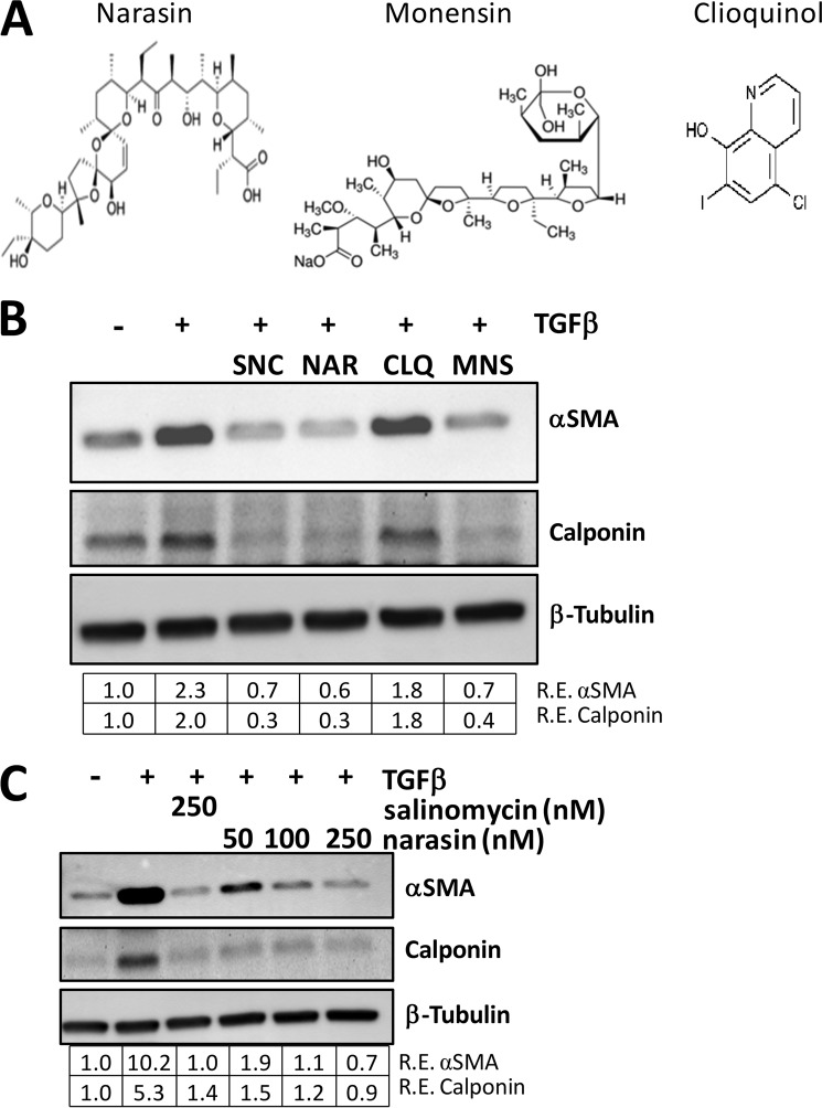
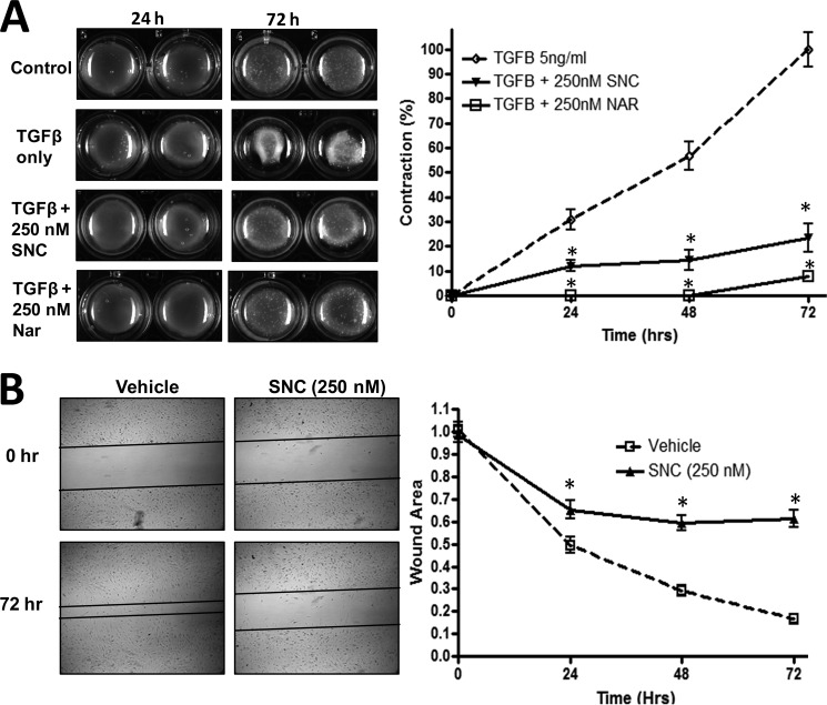
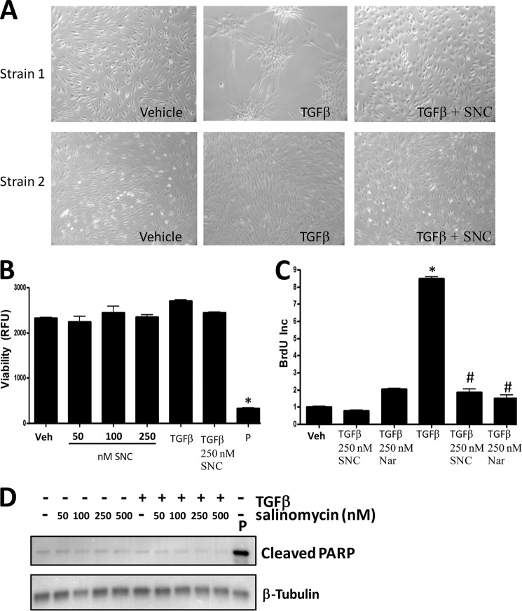
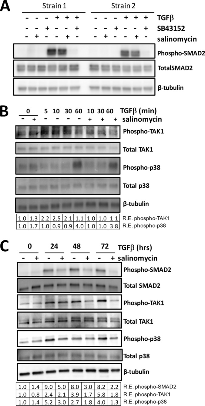
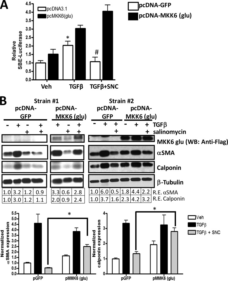
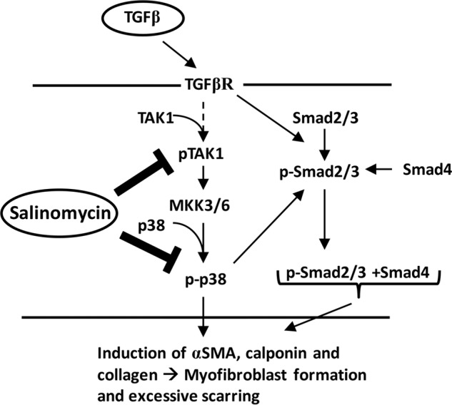
Although scarring is a component of wound healing, excessive scar formation is a debilitating condition that results in pain, loss of tissue function, and even death. Many tissues, including the lungs, heart, skin, and eyes, can develop excessive scar tissue as a result of tissue injury, chronic inflammation, or autoimmune disease. Unfortunately, there are few, if any, effective treatments to prevent excess scarring, and new treatment strategies are needed. Using HEK293FT cells stably transfected with a TGFβ-dependent luciferase reporter, we performed a small molecule screen to identify novel compounds with antiscarring activity. We discovered that the polyether ionophore salinomycin potently inhibited the formation of scar-forming myofibroblasts. Salinomycin (250 nm) blocked TGFβ-dependent expression of the cardinal myofibroblast products α smooth muscle actin, calponin, and collagen in primary human fibroblasts without causing cell death. Salinomycin blocked phosphorylation and activation of TAK1 and p38, two proteins fundamentally involved in signaling myofibroblast and scar formation. Expression of constitutively active mitogen activated kinase kinase 6, which activates p38 MAPK, attenuated the ability of salinomycin to block myofibroblast formation, demonstrating that salinomycin targets the p38 kinase pathway to disrupt TGFβ signaling. These data identify salinomycin and other polyether ionophores as novel potential antiscarring therapeutics.

Keywords: Collagen; Fibrosis; MKK6; Myofibroblast; Polyether Ionophores; SMAD Transcription Factor; Salinomycin; TGFβ; p38; p38 MAPK.


Figures

FIGURE 1.
FIGURE 1.
A small molecule screen identifies salinomycin as a potential antiscarring compound. A, the Smad-dependent reporter construct. Four tandem SBEs were inserted upstream of the minimal tk promoter. Downstream of the promoter is a destabilized version of the firefly luciferase gene (luc2P) present in the pLuc2P-Hygro plasmid that also harbors the hygromycin resistance gene. B, the reporter plasmid was introduced into the HEK293FT cell line, and individual colonies were selected with hygromycin (200 μg/ml) to develop a Smad/TGFβ-dependent luciferase reporter cell line. 1 ng/ml treatment of TGFβ for 24 h resulted in a robust increase of luciferase activity. C, the HEK293FT-luc reporter line was screened with the 2300-compound Spectrum collection of small molecules. Several hits from the initial screen that blocked TGFβ-induced luciferase activity were tested further in a Dual-Luciferase screen including a constitutive Renilla luciferase to normalize the SBE luciferase activity. Compound L18, which corresponded to salinomycin, a polyether ionophore antibiotic, inhibited the reporter construct more than 10-fold. Unt., untreated; Veh, vehicle; comp., compound. D, salinomycin (two-dimensional structure, right) exhibited a dose-dependent decrease in TGFβ-induced SBE-luciferase activity. 1 μm salinomycin reduced SBE luciferase activity to below baseline levels, whereas 100 nm salinomycin reduced SBE luciferase activity more than 3-fold. Experiments were repeated three times in triplicate, and similar results were observed in all tests. *, p < 0.01 versus untreated cells; #, p < 0.01 versus TGFβ with vehicle.
FIGURE 2.
FIGURE 2.
Salinomycin inhibits TGFβ-induced expression of myofibroblast markers in primary human fibroblasts. A, human fibroblasts were treated with two different concentrations of five small molecules from the Spectrum collection and treated with TGFβ (1 ng/ml) for 72 h. The cells were then lyses and analyzed by Western blot for the myofibroblast markers αSMA and calponin. β-tubulin was used as a loading control. Relative expression (R.E.) of αSMA and calponin (both normalized to β-tubulin) as determined by densitometry is shown below each lane. Salinomycin (compound ID L18) dramatically reduced expression of both myofibroblast markers to at or below untreated (Unt.) fibroblast expression levels. Veh, vehicle. B, to determine the minimal dose at which salinomycin effectively inhibited expression of αSMA and calponin, human fibroblasts were treated with salinomycin (10–250 nm) and TGFβ (1 ng/ml) for 72 h, and cells were isolated and analyzed as in A. 250 nm salinomycin inhibited expression of αSMA and calponin by more than 17- and 5-fold, respectively. C, to determine the time points at which salinomycin inhibits myofibroblast marker expression, human fibroblasts were treated with vehicle or 250 nm salinomycin in the presence of 1 ng/ml TGFβ, and then cells were harvested at 0, 24, 48, and 72 h. Cell extracts were collected and analyzed by Western blot for αSMA and β-tubulin. The tubulin band is indicated by the arrow, and the asterisk denotes the signal from αSMA expression. Salinomycin inhibits expression of αSMA at 24, 48, and 72 h. Experiments were repeated using at least three different primary human fibroblast strains, with similar results seen in all strains. A representative experiment is shown.
FIGURE 3.
FIGURE 3.
Salinomycin prevents TGFβ-induced collagen production in primary human fibroblasts. A, primary human fibroblasts were treated with salinomycin (10–250 nm) and TGFβ (1 ng/ml) for 72 h, and then cell culture supernatants were collected and analyzed for collagen I levels by slot blot analysis. Salinomycin reduced collagen I levels in a dose-dependent manner, with salinomycin (100 and 250 nm) reducing collagen production to baseline levels. B, fibroblasts were treated with 250 nm salinomycin and TGFβ (1 ng/ml), and cell culture supernatants were collected at 5, 24, 48, and 72 h to analyze production of collagen I. Salinomycin prevented collagen production at 24, 48, and 72 h. Experiments were performed in at least three primary human fibroblast strains, with representative strains shown.
FIGURE 4.
FIGURE 4.
The polyether ionophores salinomycin, narasin, and monensin inhibit myofibroblast formation. A, molecular structures of the ionophores narasin, monensin, and clioquinol. Narasin, a methylated derivative of salinomycin, and monensin are polyether ionophores, whereas clioquinol is an unrelated ionophore. B, human fibroblasts were treated with vehicle (DMSO), TGFβ, or TGFβ plus 250 nm of the indicated compounds for 72 h and then analyzed for myofibroblast markers by Western blot. Salinomycin (SNC), narasin (NAR), and monensin (MNS) all inhibited expression of αSMA and calponin, whereas clioquinol (CLQ) did not. Relative expression (R.E.) of αSMA and calponin (both normalized to β-tubulin) as determined by densitometry is shown below each lane. C, human fibroblasts were treated with 10–250 nm narasin or 250 nm salinomycin and TGFβ (1 ng/ml) for 72 h, and then cells were isolated and analyzed as in B. Salinomycin (250 nm) inhibited expression of αSMA and calponin 10- and 5-fold, respectively, whereas narasin exhibited a dose-dependent decrease in expression of the myofibroblast markers, where 250 nm narasin inhibited expression of αSMA and calponin by more than 10- and 5-fold, respectively. Experiments were repeated in two different strains, with representative results shown.
FIGURE 5.
FIGURE 5.
Salinomycin blocks TGFβ-induced myofibroblast contraction and migration. A, human fibroblasts were cultured in a collagen gel matrix as described in the text. After collagen gel formation, cells were treated with TGFβ (5 ng/ml) or TGFβ plus either salinomycin (SNC, 250 nm) or narasin (Nar, 250 nm) and allowed to contract for 72 h. Gel matrices were imaged at the 0, 24, 48, and 72 h time points. The gel area was quantified using ImageJ. TGFβ-induced contraction was set to 100%, and all other measurements were normalized to it. Representative images are shown, demonstrating that TGFβ readily induced myofibroblast contraction, whereas salinomycin and narasin inhibit contraction by more than 80% and 90%, respectively, 72 h post-TGFβ. *, p < 0.01 versus TGFβ-treated cells. B, human fibroblasts were cultured in a 12-well culture dish until they reached a confluent monolayer. A scratch wound was then induced in the cultures, and wells were washed to remove cells and debris. Cells were treated with TGFβ (5 ng/ml) or TGFβ plus salinomycin (250 nm) for 72 h to allow myofibroblast migration. Open areas were quantified using ImageJ software. Areas were normalized to time 0 (area = 1.0 at time 0). Images were captured at 0, 24, 48, and 72 h. After 72 h, TGFβ-treated cells had reduced the scratched area to less than 20% of the original area. Salinomycin treatment prevented myofibroblast migration 3- to 4-fold over TGFβ only. *, p < 0.01 versus TGFβ-treated cells. Experiments were repeated in triplicate, with two different strains and representative images shown.
FIGURE 6.
FIGURE 6.
Salinomycin does not affect viability or basal proliferation of human fibroblasts but does block TGFβ-induced proliferation. A, human fibroblasts were treated with vehicle (DMSO), TGFβ (1 ng/ml), or TGFβ plus 250 nm salinomycin (SNC) and cultured for 72 h. After culture, cell images were taken (original magnification ×200). Two different primary fibroblast strains at two different culture confluence states are shown. TGFβ induces morphology changes in human fibroblasts, indicative of myofibroblast formation. Cotreatment with salinomycin effectively blocked the morphological changes induced by TGFβ, and fibroblasts appeared similar to vehicle-treated fibroblasts. B, human fibroblasts were treated with vehicle (Veh, DMSO); 50, 100, or 250 nm salinomycin; TGFβ (1 ng/ml); and TGFβ plus 250 nm salinomycin or puromycin (P) (5 μg/ml) for 72 h in the presence of the redox-sensitive fluorescent dye Alamar Blue. After 72 h, fluorescence was measured to analyze cellular viability. RFU, relative fluorescent units. Puromycin, which served as a positive control, resulted in a total loss of cell viability, whereas salinomycin did not significantly affect cell viability at the tested doses either in the presence or absence of TGFβ. C, human fibroblasts were treated with vehicle (DMSO), 250 nm salinomycin, 250 nm narasin (Nar), TGFβ alone, or TGFβ plus salinomycin or narasin (250 nm) for 24 h before addition of BrdU. BrdU treatment was carried out for an additional 24 h, and then cells were fixed and stained for BrdU incorporation, which served as a measure of cell proliferation. Salinomycin and narasin did not affect basal fibroblast proliferation (first three columns). However, salinomycin and narasin significantly blocked TGFβ-induced proliferation (last three columns). Results are from a representative experiment performed in triplicate. *, p < 0.01 versus vehicle; #, p < 0.01 versus TGFβ treatment. D, primary human fibroblasts were treated with salinomycin (50–500 nm) in the presence or absence of TGFβ (1 ng/ml) for 72 h, and then cell extracts were collected and analyzed by Western blot for the apoptotic marker cleaved PARP. β-tubulin was used as a loading control. Cells were treated with puromycin as a positive control to induce apoptosis. As expected, puromycin induced cleaved PARP levels. However, salinomycin at the doses used (50–500 nm) did not induce PARP cleavage.
FIGURE 7.
FIGURE 7.
Salinomycin does not directly target TGFβ-induced SMAD2 phosphorylation but inhibits TGFβ-induced TAK1 and p38 phosphorylation. A, human fibroblasts were treated with vehicle, salinomycin, or SB-43152 with or without TGFβ. Cells were harvested at 1 h and analyzed for phospho-SMAD2, total SMAD2, and β-tubulin (loading control) by Western blot. Two representative strains are shown, demonstrating that, at 1 h of treatment, TGFβ induces phosphorylation of SMAD2 and SB-43152 completely ablates phospho-SMAD2, whereas salinomycin has no effect. B, fibroblasts were treated with TGFβ and/or salinomycin, and cells were harvested 0, 5, 10, 30, and 60 min after TGFβ treatment to analyze levels of phospho-TAK1, total TAK1, phospho-p38, and total p38 by Western blot. Salinomycin blocked phospho-TAK production 10 and 30 min after TGFβ treatment. R.E., relative expression. C, Human fibroblasts were treated as in A, except that cells were harvested at the 0, 24, 48, and 72 h time points to analyze levels of phospho-TAK1, totalTAK1, phospho-p38, total p38, phospho-SMAD2, and total SMAD2 by Western blot. The relative expression level of each phosphoprotein (normalized to β-tubulin) as determined by densitometry is shown below each lane. Salinomycin reduced TGFβ-induced phospho-Smad2 levels more than 2-fold at 48 h. Likewise, salinomycin also inhibited phospho-TAK1 levels at 24, 48, and 72 h. Salinomycin blocked p38 phosphorylation at the 24, 48, and 72 h time points, with an 80% reduction at 24 h and more than a 3-fold reduction at 72 h. Data are representative of at least three different experiments performed with different human fibroblasts strains.
FIGURE 8.
FIGURE 8.
Expression of constitutively active MKK6 attenuates the inhibitory effect of salinomycin on TGFβ. A, a Smad-luc reporter, a constitutive CMV-Renilla reporter, and either a control plasmid (pcDNA-GFP) or the pcDNA3.1 FLAG-MKK6(glu) plasmid were introduced into human fibroblasts by electroporation. After electroporation, cells were treated with vehicle (Veh, DMSO), TGFβ, or TGFβ plus 250 nm salinomycin (SNC). After 24 h, cells were lysed, and luciferase activity was measured. Smad-luc activity was normalized to Renilla activity. As expected, TGFβ induced SBE-luc activity over vehicle treatment in both control plasmid and MKK6(glu) plasmid electroporated samples. Salinomycin blocked TGFβ-induced Smad-luc activity in control plasmid samples. However, the presence of MKK6(glu) attenuated the effect of salinomycin on Smad-luc activity. *, p < 0.01 in vehicle versus TGFβ; #, p < 0.01 in TGFβ versus TGFβ plus salinomycin for control pGFP electroporated cells. The experiment was repeated in triplicate in two different fibroblast strains. B, a control plasmid or the pcDNA3.1 FLAG-MKK6(glu) plasmid, which harbors the cDNA for a constitutively active MKK6 protein, was introduced into human fibroblasts by electroporation. Cells were then treated with vehicle, TGFβ, or TGFβ plus 250 nm salinomycin for 72 h. In cells expressing the control pGFP plasmid, TGFβ-induced αSMA and calponin expression was blocked by salinomycin. However, in cells expressing the MKK6(glu) plasmid, salinomycin did not block αSMA and calponin expression. The experiment was performed in three different fibroblast strains, with a representative experiment shown. WB, Western blot; R.E., relative expression. C, Western blot densitometry of the experiment shown in B. *, p < 0.01 in pGFP versus pMKK6(glu).
FIGURE 9.
FIGURE 9.
Potential model for the salinomycin mode of action. Active TGFβ, present at high concentrations during wound healing, exuberant scarring, or chronic inflammation, binds to the TGFβ receptor (TGFβR). Activation of the TGFβ receptor triggers a range of cell signaling events, including the phosphorylation and activation of TAK1 and Smad2/3. TAK activates MKK3/6 by phosphorylation, leading to phosphorylation and activation of p38. Active p38 leads to a further increase in Smad2/3 phosphorylation and activation. These signaling pathways reinforce the myofibroblast program, leading to excessive expression of αSMA, calponin, and collagen to promote myofibroblast generation and scar formation. Salinomycin or other polyether ionophores can block the activation and phosphorylation of TAK1 and p38, thereby limiting the activation of Smad2/3 and blocking myofibroblast formation.

Similar articles

Cited by

References

    1. Friedlander M. (2007) Fibrosis and diseases of the eye. J. Clin. Invest. 117, 576–586 - PMC - PubMed
    1. Noble P. W., Barkauskas C. E., Jiang D. (2012) Pulmonary fibrosis: patterns and perpetrators. J. Clin. Invest. 122, 2756–2762 - PMC - PubMed
    1. Bahn R. S. (2010) Graves' ophthalmopathy. N. Engl. J. Med. 362, 726–738 - PMC - PubMed
    1. Hinz B. (2007) Formation and function of the myofibroblast during tissue repair. J. Invest. Dermatol. 127, 526–537 - PubMed
    1. Niessen F. B., Spauwen P. H., Schalkwijk J., Kon M. (1999) On the nature of hypertrophic scars and keloids: a review. Plast. Reconstr. Surg. 104, 1435–1458 - PubMed

Publication types

MeSH terms