. 2015 May;33(5):1419-33.
doi: 10.1002/stem.1944.

A Regulatory Network Involving β-Catenin, e-Cadherin, PI3k/Akt, and Slug Balances Self-Renewal and Differentiation of Human Pluripotent Stem Cells In Response to Wnt Signaling

Affiliations

A Regulatory Network Involving β-Catenin, e-Cadherin, PI3k/Akt, and Slug Balances Self-Renewal and Differentiation of Human Pluripotent Stem Cells In Response to Wnt Signaling

Tyng-Shyan Huang et al. Stem Cells. 2015 May.

Abstract

The mechanisms underlying disparate roles of the canonical Wnt signaling pathway in maintaining self-renewal or inducing differentiation and lineage specification in embryonic stem cells (ESCs) are not clear. In this study, we provide the first demonstration that self-renewal versus differentiation of human ESCs (hESCs) in response to Wnt signaling is predominantly determined by a two-layer regulatory circuit involving β-catenin, E-cadherin, PI3K/Akt, and Slug in a time-dependent manner. Short-term upregulation of β-catenin does not lead to the activation of T-cell factor (TCF)-eGFP Wnt reporter in hESCs. Instead, it enhances E-cadherin expression on the cell membrane, thereby enhancing hESC self-renewal through E-cadherin-associated PI3K/Akt signaling. Conversely, long-term Wnt activation or loss of E-cadherin intracellular β-catenin binding domain induces TCF-eGFP activity and promotes hESC differentiation through β-catenin-induced upregulation of Slug. Enhanced expression of Slug leads to a further reduction of E-cadherin that serves as a β-catenin "sink" sequestering free cytoplasmic β-catenin. The formation of such a framework reinforces hESCs to switch from a state of temporal self-renewal associated with short-term Wnt/β-catenin activation to definitive differentiation. Stem Cells 2015;33:1419-1433.

Keywords: Differentiation; E-cadherin; Human embryonic stem cell; Self-renewal; Slug; Wnt; β-Catenin.


Figures

Figure 1
Figure 1
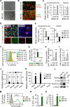
Short‐term GSK3 inhibition temporally promotes E‐cadherin expression and human embryonic stem cell (hESC) self‐renewal whereas long‐term inhibition generates opposite results. (A–L): hESCs were treated with either vehicle (Veh) or BIO for 6 hours. Twenty‐four hours after removal of BIO, hESCs exhibit thick and compact colonies (A, bright‐field images) that express higher levels of E‐cadherin (E‐cad), Nanog, and Oct3/4 (B and D, immunofluorescence staining; C and E, quantitative image analysis of fluorescence intensity, n > 100 from three independent experiments for each group; F and G, flow cytometry). They also show high cloning efficiency (H, clonogenic assays, n = 3). After removal of BIO and continual culture for 4 days, BIO treatment leads to a high cell yield (I). In contrast, long‐term BIO treatment leads to opposing results (D–G; J, Q‐PCR; L, Western blot), significantly enhancing the expression of mesoderm marker Mixl1 and Brachyury (K, Q‐PCR, HNF3β: endoderm, Pax6: ectoderm). Nuclei were counterstained with DAPI (D, blue). Scale bars = 100 µm (A), 10 µm (B), and 20 µm (D). (M–O): Representative flow cytometric histogram (M), percentage of live TCF‐eGFP‐positive cells (pregated on 7‐AAD‐negative live cells), and MFI after treatment with BIO for 6 hours or 4 days. All data are mean ± SD. **, p < .01 compared to vehicle control. Abbreviations: GFP, green fluorescent protein; MFI, median fluorescence intensity; TCF, T‐cell factor.
Figure 2
Figure 2
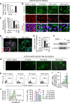
Dual function of β‐catenin in canonical Wnt signaling pathway is responsible for hESC self‐renewal and differentiation. (A): βS33Y‐Tet‐On hESCs were treated with doxycycline (DOX, 2 µg/ml) for 24 hours, followed by clonogenic assays (mean ± SD, n = 3, **, p < .01). (B–D): βS33Y‐Tet‐On hESCs were treated with different dosages of DOX for 4–6 days, followed by immunofluorescence staining of β‐catenin and Nanog (B), and quantification of mean pixel intensity of Nanog (C, n >100 cells/group). The nuclear translocation of β‐catenin was observed in 1,000 ng/ml of DOX group (D). Nuclei were counterstained with DAPI (blue). White arrows indicate cytoplasmic β‐catenin. Scale bars = 10 µm (insets in D: 1 µm). (E, F): Clonogenic assays (E, mean ± SD, n = 3, *, p < .05; **, p < .01) and Western blot analysis (F) for the same cells as described in (B)–(D). (G): TCF‐eGFP reporter activity remains invisible in the absence of DOX for 4–6 days but visible in the nuclear region in a DOX dose‐dependent manner, scale bars = 20 µm. (H, I): Flow cytometric analysis of 7xTCF‐eGFP hESCs as described in (G), pregated on single 7‐AAD‐negative live cells. The TCF‐eGFP reporter activity was measured by the percentage of GFP‐positive cells after treatment with different concentrations of DOX for 4–6 days (H). The expression of GFP‐positive cells is normalized to DOX‐untreated group that was arbitrarily set at 1 (I). (J): Flow cytometric analysis of the cells in (H) and (I) for E‐cadherin expression (E‐cad+) on the cell membrane indicates an inverse association with TCF‐eGFP activity as shown in (G)–(I), pregated on single 7‐AAD‐negative live cells. Data in (I) and (J) are means ± SD, n = 3, two independent experiments; **, p < .01 compared to DOX‐untreated group. Abbreviations: GFP, green fluorescent protein; hESC, human embryonic stem cell; TCF, T‐cell factor.
Figure 3
Figure 3
β‐Catenin enhances human embryonic stem cell (hESC) self‐renewal through upregulation of E‐cadherin. (A–C): Inducible over‐expression of E‐cadherin (E‐cad) in hESCs enhances E‐cadherin expression in cell‐cell junctions (A, immunofluorescence, left panel: enlarged images; DOX: 3 days), and increases the expression of E‐cadherin, Oct3/4, and Nanog (B, Q‐PCR, DOX: 24 hours) and cell density (C, bright‐field image and cell counting; low panel: enlarged images; DOX: 3 days). Scale bars = 10 µm (A), 20 µm (low panel of C), and 100 µm (upper panel of C); DOX, 2 µg/ml. (D, E): Reduced expression levels of E‐cadherin, Oct3/4, and Nanog (D, Q‐PCR) and diminished clonogenic capacity (E, clonogenic assays, n = 3, **, p < .01) in hESC 24 hours after E‐cadherin siRNA knockdown. H1, H9, or CA1 hESC lines were used in this figure.
Figure 4
Figure 4
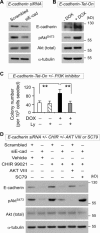
E‐cadherin associates with PI3K/Akt activation in response to short‐term Wnt/β‐catenin signaling‐enhanced human embryonic stem cell (hESC) self‐renewal. (A): Decreased phosphorylated AKT (pAKTS473) in hESCs after E‐cadherin knockdown for 48 hours (Western blot). (B): Increased phosphorylated AKT (pAKTS473) after E‐cadherin upregulation. E‐cadherin‐Tet‐On hESCs were treated with DOX (2 µg/ml) for 48 hours, followed by Western blot analysis. (C): Inhibition of PI3/Akt with LY294002 abrogates the enhanced clonogenic capacity induced by E‐cadherin upregulation. E‐cadherin‐Tet‐On hESCs were treated with or without DOX (2 µg/ml) in the presence or absence of LY294002 (5 µM) for 24 hours, followed by clonogenic assays (n = 3). (D): E‐cadherin knockdown abolishes short‐term GSK3 inhibition‐induced upregulation of E‐cadherin and phosphorylated Akt, while specific Akt activator SC79 counteracts the effect of E‐cadherin knockdown. After siRNA knockdown for 44 hours, hESCs (H9 and H1 lines) were treated with either CHIR99021 (6 µM) or vehicle (DMSO) for 6 hours, followed by Akt inhibitor VIII (6 µM), or Akt activator II SC79 (6 µg/ml), or vehicle for an additional 30 minutes. All data in this figure are mean ± SD and were generated from H1 and H9 lines. **, p < .01.
Figure 5
Figure 5
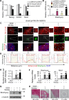
E‐cadherin sequesters β‐catenin to suppress Wnt‐induced hESC differentiation. (A): Q‐PCR analysis of hESCs cotransfected with βS33Y plasmids and either wild‐type E‐cadherin (WT‐Ecad) or EcadΔβ for 24 hours. pcDNA: control vector. (B): In comparison to control groups, co‐overexpression of EcadΔβ and βS33Y for 24 hours significantly increases Brachyury expression (Q‐PCR). (C): EcadΔβ‐Tet‐On hESCs were treated with or without doxycycline (DOX+, 2 µg/ml) or vehicle in the presence or absence of Wnt3a protein (100 ng/ml) for 4–6 days and analyzed by double‐immunostaining. Nuclei were counterstained with DAPI (blue). Scale bar = 20 µm. (D): Subcellular localization analysis of the expression of active β‐catenin and Brachyury as shown in (C). Plots of the intensity profile of active β‐catenin (red), Brachyury (green), and DAPI (blue) over a linear section of a whole cell (white lines in C) are representative of >100 cells/group analyzed. Data are expressed as AU versus length in microns. The arrowheads indicate the cell‐cell contact areas; the yellow area in the middle of each plot marks the nuclear region; mean ± SD in red on each plot represent the mean pixel intensity of the nuclear active β‐catenin from >100 cells/group. (E): Quantification of the ratio of active β‐catenin nuclear staining/DAPI intensity for the indicated groups in (C). Data are mean ± SD, four replicates from H9 line, **, p < .01 compared to DOXWnt3a group. (F, G): Doxycycline‐induced EcadΔβ over‐expression promotes hESC differentiation in response to Wnt3a treatment as revealed by significant upregulation of Brachyury and Mixl1 (F, Q‐PCR; G, Western blot). (H): Alkaline phosphatase (AP) staining and quantitative analyses. At day 4 of treatments as indicated, cells were dissociated and reseeded. AP‐positive colonies were counted when >50% of the cells within the colony were positive for AP staining. The percentages of AP+ colonies were quantitatively assessed and are shown at the bottom. The dashed line indicates the boundary of the colony. Scale bar = 100 µm. The results represent three replicates ± SD from H1 and H9 lines. *, p <.05; **, p <.01. Abbreviations: AU, arbitrary unit; hESC, human embryonic stem cell.
Figure 6
Figure 6
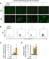
E‐cadherin sequesters β‐catenin and suppresses the functional interaction of β‐catenin and TCF in hESCs. Analysis of 7xTCF‐eGFP‐EcadΔβ‐Tet‐On hESCs after treatment with doxycycline (DOX+, 2 µg/ml) or vehicle in the presence or absence of Wnt3a protein (100 ng/ml) for the indicated time points. The cells were cultured on Matrigel‐coated plates. Media, DOX, and Wnt3a were changed daily. (A): TCF‐eGFP (green) expression pattern of hESCs in the absence or presence of DOX and/or Wnt3a for 6 hours or 4 days. Scale bar = 50 µm. (B–D): Representative flow cytometric plots (B), fold changes of TCF‐eGFP‐positive cells over DOXWnt3a control (C) and MFI (D) in the absence or presence DOX and/or Wnt3a for 4 days, pregated on single 7‐AAD‐negative live cells. Mean ± SD from three independent experiments. *, p < .05; **, p < .01. Abbreviations: GFP, green fluorescent protein; hESC, human embryonic stem cell; MFI, median fluorescence intensity; TCF, T‐cell factor.
Figure 7
Figure 7
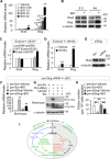
β‐Catenin‐induced Slug upregulation reinforces human embryonic stem cell (hESC) differentiation by downregulating E‐cadherin and upregulating Brachyury. (A, B): Long‐term but not short‐term BIO treatment enhances Slug expression in hESCs (A, Q‐PCR; B, Western blot). (C): hESCs cotransfected with EcadΔβ and βS33Y, but not with vectors (pcDNA) or with wild‐type E‐cadherin (WT‐Ecad) and βS33Y, enhances Slug expression (Q‐PCR). (D): Over‐expression of EcadΔβ together with short‐term Wnt3a treatment (100 ng/ml) promotes Slug expression in hESCs (Q‐PCR). (E): hESCs infected with pLKO‐siSlug plasmid (siSlug) for 2 days reduce Slug expression (Western blot). Scrambled: pLKO‐puro vector with a scrambled sequence that does not target any mRNA. (F–H): Slug knockdown (pre‐siSlug) followed by BIO treatment for 4 days results in a decrease of differentiation marker Brachyury (F, Q‐PCR; G, Western blot) and clonogenic capacity (H, clonogenic assays, n = 3) in hESCs. All data are mean ± SD, *, p <.05; **, p < .01. (I): The proposed working model (detailed in Supporting Information Fig. S7).

Similar articles

Cited by

References

    1. ten Berge D, Kurek D, Blauwkamp T et al. Embryonic stem cells require Wnt proteins to prevent differentiation to epiblast stem cells. Nat Cell Biol 2011;13:1070–1075. - PMC - PubMed
    1. Sato N, Meijer L, Skaltsounis L et al. Maintenance of pluripotency in human and mouse embryonic stem cells through activation of Wnt signaling by a pharmacological GSK‐3‐specific inhibitor. Nat Med 2004;10:55–63. - PubMed
    1. Singla DK, Schneider DJ, LeWinter MM et al. wnt3a but not wnt11 supports self‐renewal of embryonic stem cells. Biochem Biophys Res Commun 2006;345:789–795. - PubMed
    1. Miki T, Yasuda SY, Kahn M. Wnt/beta‐catenin signaling in embryonic stem cell self‐renewal and somatic cell reprogramming. Stem Cell Rev 2011;7:836–846. - PubMed
    1. Doble BW, Patel S, Wood GA et al. Functional redundancy of GSK‐3alpha and GSK‐3beta in Wnt/beta‐catenin signaling shown by using an allelic series of embryonic stem cell lines. Dev Cell 2007;12:957–971. - PMC - PubMed

Publication types

MeSH terms