. 2015 Jul;230(7):1594-606.
doi: 10.1002/jcp.24906.

Wild-type and specific mutant androgen receptor mediates transcription via 17β-estradiol in sex hormone-sensitive cancer cells

Affiliations

Wild-type and specific mutant androgen receptor mediates transcription via 17β-estradiol in sex hormone-sensitive cancer cells

Takao Susa et al. J Cell Physiol. 2015 Jul.

Abstract

We previously encountered regulatory processes wherein dihydrotestosterone (DHT) exerted its inhibitory effect on parathyroid hormone-related protein (PTHrP) gene repression through the estrogen receptor (ER)α, but not the androgen receptor (AR), in breast cancer MCF-7 cells. Here, we investigated whether such aberrant ligand-nuclear receptor (NR) interaction is present in prostate cancer LNCaP cells. First, we confirmed that LNCaP cells expressed large amounts of AR at negligible levels of ERα/β or progesterone receptor. Both suppression of PTHrP and activation of prostate-specific antigen genes were observed after independent administration of 17β-estradiol (E2), DHT, or R5020. Consistent with the notion that the LNCaP AR lost its ligand specificity due to a mutation (Thr-Ala877), experiments with siRNA targeting the respective NR revealed that the AR monopolized the role of the mediator of shared hormone-dependent regulation, which was invariably associated with nuclear translocation of this mutant AR. Microarray analysis of gene regulation by DHT, E2, or R5020 disclosed that more than half of the genes downstream of the AR (Thr-Ala877) overlapped in the LNCaP cells. Of particular interest, we realized that the AR (wild-type [wt]) and AR (Thr-Ala877) were equally responsible for the E2-AR interactions. Fluorescence microscopy experiments demonstrated that both EGFP-AR (wt) and EGFP-AR (Thr-Ala877) were exclusively localized within the nucleus after E2 or DHT treatment. Furthermore, reporter assays revealed that some other cancer cells exhibited aberrant E2-AR (wt) signaling similar to that in the LNCaP cells. We herein postulate the presence of entangled interactions between wt AR and E2 in certain hormone-sensitive cancer cells.


Figures

Figure 1
Figure 1
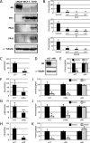
The androgen receptor (AR) exclusively mediates parathyroid hormone‐related protein gene repression by several steroid hormones in LNCaP cells. (A) Western blotting of AR, estrogen receptor (ER)α, progesterone receptor (PR)‐A and PR‐B proteins in LNCaP, MCF‐7, and T47D cells using AR (H‐280), ERα, and PR (reacted with both isoforms of PR‐A and PR‐B) antibodies. Anti‐α‐tubulin antibody was used as the loading control. (B) Parathyroid hormone‐related protein (PTHrP) mRNA in LNCaP cells treated with dihydrotestosterone (DHT), 17β‐estradiol (E2), or R5020 was analyzed by quantitative real‐time (qRT)‐PCR. The concentrations of the used hormones were 10−9, 10−8, and 10−7 M, respectively. Data are normalized to GAPDH mRNA levels and represented relative to the vehicle (%). * P < 0.01 compared with the vehicle. (C–E) Control siRNA (siCT) and AR siRNA (siAR) were introduced into the respective LNCaP cells. At 24 h after transfection, AR mRNA (C) and protein (D) were analyzed. (C) AR mRNA in the transfected LNCaP cells was analyzed by real‐time PCR. Data are normalized to GAPDH and represented as the ratio of AR mRNA in the siAR groups to that in the siCT groups (%). * P < 0.01 compared with siCT. (D) Western blotting with the AR antibody. Anti‐α‐tubulin antibody was used as the loading control. (E) Real‐time PCR assay of PTHrP mRNA. LNCaP cells with or without siAR were treated with 10−7 M DHT for 24 h. Black and gray bars indicate the effect of vehicle and DHT, respectively. Data are shown relative to the siCT of the vehicle (%). * P < 0.01 compared with each vehicle. (F–K) siCT and siRNAs for each NR (siERα, siERβ, and siPR) were introduced into the respective LNCaP cells. Data are normalized to GAPDH expression. (F–H) At 24 h after transfection, the mRNA expression of ERα (F), ERβ (G), and PR (H) in the LNCaP cells was analyzed by real‐time PCR. Data are presented as ratios relative to siCT (%). * P < 0.01 and ** P < 0.05 compared with the siCT lane. (I–K) At 24 h after transfection of each siRNA, LNCaP cells were treated with 10−7 M E2 (I, J) or R5020 (K) for another 24 h. Expression of the PTHrP gene was analyzed by real‐time PCR. Black and gray bars indicate the results after vehicle and steroid hormone treatment, respectively. Data are shown relative to the vehicle (%). * P < 0.01 compared with each vehicle. (B–C and E–K) Data are presented as means ± SD (n = 4).
Figure 2
Figure 2
AR as an exclusive mediator of prostate‐specific antigen activation by several steroid hormones in LNCaP cells. Levels of prostate‐specific antigen (PSA) mRNA in LNCaP cells treated with DHT (A, D), E2 (B, E, F), and R5020 (C, G) were analyzed by qRT‐PCR. The concentrations of the hormones were 10−9, 10−8, and 10−7 M for A–C, and 10−7 M for D‐G, respectively. (D–G) The effects of siCT and siNRs on PSA gene activation are shown. Data are normalized to GAPDH expression and presented as means ± SD (n = 4). Data are shown relative to the vehicle (%). * P < 0.01 and ** P < 0.05 compared with the vehicle.
Figure 3
Figure 3
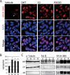
Nuclear translocation of the AR triggered by various steroid hormones in LNCaP cells. (A) Immunocytochemical analysis with AR antibody, AR (H‐280) was performed. At 24 h after treatment with each steroid hormone, LNCaP cells fixed by 4% paraformaldehyde were incubated with the anti‐AR antibody. Each hormone was added at a dose of 10−7 M. (Upper panel) AR visualized with the Alexa 546‐labeled secondary antibody (red). (Middle panel) Nuclear staining with DAPI (blue). (Lower panel) Merged images. Modest staining of the nuclear AR, even in the absence of hormones, is indicated by the arrow. Scale bar = 10 μm. (B) AR mRNA in LNCaP cells treated with DHT, E2, or R5020 was analyzed by quantitative real‐time PCR. The concentrations of the used hormones were 10−7 M, respectively. Data are normalized to GAPDH mRNA levels and represented relative to the vehicle (%). * P < 0.01 compared with the vehicle. (C) Western blotting of AR (H‐280) using fractionated LNCaP cells protein was performed. Anti‐α‐tubulin antibody was used as the loading control for whole protein (WP). Anti‐α‐tubulin antibody was used as the cell fraction control for cytoplasm protein (CP) and anti‐Pol II antibody were used as that for nuclear protein (NP), respectively. We also used anti‐Lamin A/C antibody as another NP fraction control (Supplemental Fig. 7C). V, D, E, and R indicated Vehicle, DHT, E2 and R5020, respectively.
Figure 4
Figure 4
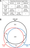
Comparison of DHT‐AR, E2‐AR, and R5020‐AR target gene profiles in LNCaP cells. Microarray analysis was performed to investigate the exhaustive target gene profiles of DHT, E2, and R5020 via the AR (Thr‐Ala877) in LNCaP cells. (A) The proportion of common target probes between each group is listed. The percentage and number of probes in each group is shown. (B) Venn diagram showing the distribution of the DHT‐AR (black), E2‐AR (red), and R5020‐AR (blue) target probes. The numbers outside the Venn diagram indicate the total number of each probe and those inside the Venn diagram indicate the number of probes in each group.
Figure 5
Figure 5
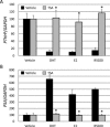
TSA commonly abandoned DHT, E2, and R5020 dependent repression of PTHrP as well as activation of PSA. The concentration of DHT, E2, and R5020 was 10−7 M. TSA was used concomitantly at the dose of 10−6 M for 24 h. The expression levels of PTHrP (A) and PSA mRNAs (B) in LNCaP cells were analyzed by quantitative real‐time PCR. Black and gray bars indicate treatment of Vehicle and TSA, respectively with each steroid hormone. * P < 0.01 compared with the vehicle.
Figure 6
Figure 6
Steroid hormone‐dependent promoter activation and nuclear translocation of the exogenously introduced AR (wild‐type) and AR (Thr‐Ala877) in LNCaP cells. (A) The plasmid pcDNA3.1‐based expression vectors, AR (wild‐type; wt) and AR (Thr‐Ala877), as well as an empty vector, were introduced into LNCaP cells together with the ARE × 3/pGL4‐TK reporter. At 24 h after transfection, the cells were treated with 10−9, 10−8, and 10−7 M of DHT, E2, and R5020, respectively for 24 h followed by luciferase assay. The blue, red, and green lines in each graph denote the response induced by the introduction of pcDNA3.1, AR (wt), and AR (Thr‐Ala877), respectively. Data are expressed as fold changes relative to the vehicle. * P < 0.01 compared with the vehicle. (B–E) At 24 h after transfection with enhanced green fluorescent protein (EGFP) (B), EGFP‐AR (Thr‐Ala877) (C), or EGFP‐AR (wt) (D and E), the indicated steroid hormones (10−7 M) were included for another 24 h. In E, 24 h before transfection with EGFP‐AR (wt), AR siRNA was introduced concurrently with cell seeding to knockdown the endogenous mutant AR (Thr‐Ala877). (Upper panels) EGFP (B), EGFP‐AR (Thr‐Ala877) (C), and EGFP‐AR (wt) (D and E) (green). (Middle panels) DAPI (blue). (Lower panels) Merged images. Note that the stained cells in each upper panel and the double stained cells in each lower panel originated from the DAPI‐positive cells shown in the middle panels, reflecting a transfectional efficiency of around 5%. Scale bar = 10 μm.
Figure 7
Figure 7
Steroid hormone‐dependent activation of the AR response element containing reporter activity in other prostate and breast cancer cells. Luciferase assay after introducing the empty pcDNA3.1, AR (wt) expression vector, or AR (Thr‐Ala877) expression vector together with the AR response element (ARE) × 3/pGL4‐TK vector in MDA‐MB‐453 (A), Rv22 (B), and MCF‐7 (C) cells. Cells were treated and assays were performed as described in Fig. 5. The black, gray, and white lines show the values for pcDNA3.1, AR (wt), and AR (Thr‐Ala877), respectively. Data are expressed as fold changes relative to the vehicle. * P < 0.01 and **P < 0.05 compared with pcDNA3.1 in each group, respectively.
Figure 8
Figure 8
Diverse interactions of the AR with the steroidal ligands in several cell lines. (Left) Previous reports by others postulated that the endogenous AR (Thr‐Ala877) upregulated or downregulated the AR target genes mediated not only by DHT, but also by E2 and R5020 in LNCaP cells. (Middle) In addition, we found that the extrinsic wild‐type AR overexpressed in the LNCaP cells responded to E2. Even in the Rv22 and MCF‐7 cells, the exogenous AR (wt) exhibited a skewed crosstalk with E2 in ARE‐containing reporter gene activity. (Right) In MDA‐MB‐453 cells, the endogenous AR (Glu‐His865), as well as the exogenous AR (wt), responded only to DHT, while the exogenous AR (Thr‐Ala877) maintained its responses to E2 and R5020. *Endo AR (Glu‐His865) regulates the endogenous FKBP51 gene. Exo AR (wt) and Exo AR (Thr‐Ala877) regulate the exogenous ARE‐containing reporter gene. Throughout the figure, “Exo” denotes “exogenously introduced” and “Endo” denotes “endogenously expressed”.

Similar articles

Cited by

References

    1. Ahlstrom M, Pekkinen M, Lamberg‐Allardt C. 2009. Dexamethasone downregulates the expression of parathyroid hormone‐related protein (PTHrP) in mesenchymal stem cells. Steroids 74:277–282. - PubMed
    1. Arriza JL, Weinberger C, Cerelli G, Glaser TM, Handelin BL, Housman DE, Evans RM. 1987. Cloning of human mineralocorticoid receptor complementary DNA: Structural and functional kinship with the glucocorticoid receptor. Science 237:268–275. - PubMed
    1. Asadi F, Kukreja S. 2005. Parathyroid hormone‐related protein in prostate cancer. Crit Rev Eukaryot Gene Expr 15:15–28. - PubMed
    1. Bektic J, Berger AP, Pfeil K, Dobler G, Bartsch G, Klocker H. 2004. Androgen receptor regulation by physiological concentrations of the isoflavonoid genistein in androgen‐dependent LNCaP cells is mediated by estrogen receptor beta. Eur Urol 45:245–251; discussion251. - PubMed
    1. Bolstad BM, Irizarry RA, Astrand M, Speed TP. 2003. A comparison of normalization methods for high density oligonucleotide array data based on variance and bias. Bioinformatics 19:185–193. - PubMed

Publication types

MeSH terms