. 2014 Dec 22:5:5780.
doi: 10.1038/ncomms6780.

Critical role of histone demethylase Jmjd3 in the regulation of CD4+ T-cell differentiation

Affiliations

Critical role of histone demethylase Jmjd3 in the regulation of CD4+ T-cell differentiation

Qingtian Li et al. Nat Commun. .

Abstract

Epigenetic factors have been implicated in the regulation of CD4(+) T-cell differentiation. Jmjd3 plays a role in many biological processes, but its in vivo function in T-cell differentiation remains unknown. Here we report that Jmjd3 ablation promotes CD4(+) T-cell differentiation into Th2 and Th17 cells in the small intestine and colon, and inhibits T-cell differentiation into Th1 cells under different cytokine-polarizing conditions and in a Th1-dependent colitis model. Jmjd3 deficiency also restrains the plasticity of the conversion of Th2, Th17 or Treg cells to Th1 cells. The skewing of T-cell differentiation is concomitant with changes in the expression of key transcription factors and cytokines. H3K27me3 and H3K4me3 levels in Jmjd3-deficient cells are correlated with altered gene expression through interactions with specific transcription factors. Our results identify Jmjd3 as an epigenetic factor in T-cell differentiation via changes in histone methylation and target gene expression.


Figures
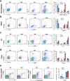
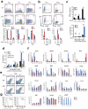
Figure 1
Figure 1. Jmjd3 deletion alters CD4+ T cell populations in different organs
Frequency of CD4+ T cell populations isolated from (a) small intestine, (b) spleen, (c) LN, and (d) colon of WT and Jmjd3 cKO mice (e) Frequency of thymic and splenic nTreg cell populations (CD25+CD4+GFP+) from WT and Jmjd3 cKO Foxp3-GFP reporter mice. Mean percentage of the indicated T cell populations ± SD shown as histograms (right panel)(representative of three independent experiments, n = 3 for each group, *p < 0.05 determined by Student's t-test).
Figure 2
Figure 2. Jmjd3 ablation alters the differentiation of naïve T cells into Th1, Th2, Th17, and Treg cell lineages in vitro
(a and b) Frequency of IFN-γ, IL-4, and IL-17-producing and Foxp3-expressing CD4+ T cells after exposure of WT and Jmjd3 cKO naïve CD4+ T cells to ThN, Th1, Th2, Th17, or Treg-inducing. Mean percentage of the indicated T cell populations ± SD shown as histograms (bottom panel). (c) IFN-γ (top panel) and IL-4 (bottom panel) production by WT and Jmjd3 cKO naïve CD4+ T cells stimulated with soluble anti-CD3 and anti-CD28 in the presence of T cell-depleted irradiated splenocytes. (d) IL-4 production by WT and Jmjd3 cKO naïve CD4+ T cells under Th2-stimulating cell culture conditions with exogenous addition of IFN-γ. (e) Frequency of Th2 cells after exposure to Th2-stimulating cell culture conditions plus exogenous IFN-γ. (f) Relative gene expression of T cell differentiation factors from WT and Jmjd3 cKO naïve CD4+ cells cultured under ThN, Th1, Th2, Th17, or Treg-inducing conditions. X axis, different cytokine condition; Y axis, relative expression at mRNA level. (g) Intracellular staining of T-bet and Gata3 in CD4+ T cells cultured under ThN conditions (representative of three independent experiments, n = 3 for each group,*p < 0.05, **p < 0.01 determined by Student's t-test).
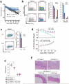
Figure 3
Figure 3. Jmjd3 ablation promotes Th2 differentiation and suppresses Th1 differentiation in vivo
(a) Naïve CD4+ T cells (CD4+CD25CD62L+CD44low) from WT and Jmjd3 cKO mice were FACS-purified and i.p. injected into irradiated Rag2−/−γc−/− mice (2 × 10⁶ cells/mouse). Body weight loss was monitored daily, indicating colitis progression (data are expressed as mean ± SD from three independent experiments, n = 5 mice in each group, *p < 0.05). (b) Percentages of splenic and colonic IL-4 and IFN-γ-producing CD4+ T cells from mice with colitis, one month following adoptive transfer of WT and Jmjd3 cKO naïve CD4+ T cells. Mean percentage of indicated T cell populations ± SD shown as histograms (right panel) (n = 3 mice per group, *p < 0.05). (c) WT and Jmjd3 cKO naïve CD4+ T cells (2 × 10⁶) were i.v. injected into irradiated Rag2−/−γc−/− mice. Ten days later, frequency of IL-4 and IFN-γ-producing T cells from isolated splenocytes. Mean percentage of the indicated cell populations ± SD as shown by histograms (right panel) (representative results from at least three independent experiments, n = 3 per group, *p < 0.05). (d) Naïve CD4+ T cells and nTreg cells (CD4+CD25+) from WT and Jmjd3 cKO mice were FACS-purified and i.p. injected into Rag2−/−γc−/− mice (1 × 10⁶ T cells + 0.2 × 10⁶ nTreg cells per mouse). Body weight loss was monitored daily for colitis progression and reported as the mean ± SD from three independent experiments (n = 5 mice in each group,*p < 0.05). (e) Colitis score for individual Rag2−/−γc−/− mice (points) receiving indicated T cells and Treg cells (n = 5 mice in each group, *p < 0.05 determined by Student's t-test). (f) H&E staining of colon from Rag2−/−γc−/− mice receiving indicated T cells. Scale bar = 200 μm.
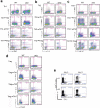
Figure 4
Figure 4. Jmjd3 ablation increases Th2, Th17, and Treg cell stability in vitro
(a) WT and Jmjd3 cKO naïve CD4+ T cells were differentiated under Th1-inducing conditions for 3 days, and then exposed to Th2, Th17 or Treg-inducing cell culture conditions for 3 days. Frequency of IFN-γ, Il-4, IL-17 and Foxp3-producing CD4+ T cells. (b) WT and Jmjd3 cKO naïve CD4+ T cells were differentiated under Th2 condition for 3 days, and then exposed to Th1, Th17 or Treg-inducing cell culture conditions for 3 days. Frequency of IFN-γ, IL-4, IL-17 and Foxp3-producing CD4+ T cells. (c) WT and Jmjd3 cKO naïve CD4+ T cells were differentiated under Th17 condition for 3 days, and then exposed to Th1, Th2 and Treg-inducing cell culture conditions for 3 days. Frequency of IFN-γ, IL-4, IL-17 and Foxp3-producing CD4+ T cells. (d) Naïve CD4+ T cells derived from WT and Jmjd3 cKO Foxp3-GFP reporter mice were differentiated under Treg conditions for 4 days. GFP+ cells were sorted and cultured under Th1, Th2 or Th17-inducing conditions for 2 days. Frequency of Foxp3-GFP+, IL-4, IL-17 and IFN-γ-producing CD4+ T cells. (e) WT and Jmjd3 cKO mice were i.v. injected with anti-CD3 to induce in vivo Th17 differentiation. Frequency of IL-17 and IFN-γ-producing CD4+ T cells from lymphocytes isolated from the small intestine, day 5 and day 8 after injection. (representative of three independent experiments, *p < 0.05 determined by Student's t-test).
Figure 5
Figure 5. Jmjd3 regulates CD44 and CXCR3 in CD4+ T cells
(a) T-bet protein expression in WT and Jmjd3 cKO CD44+ and CD44 CD4 SP thymocytes. (b) Percentage of CXCR3+ cells in CD4+ SP T cells from WT and Jmjd3 cKO mice (data expressed as mean + SD of three independent experiments, *p < 0.05, **p < 0.01). (c) Frequency of CD44 and CXCR3 in WT and Jmjd3 cKO CD4+ SP thymocytes. (d) Naïve CD4+ T cells (2 × 10⁶) derived from WT and Jmjd3 cKO mice were i.v. injected into irradiated Rag2−/−γc−/− mice (n = 3 for each group). Ten days later, frequency of CD44+CXCR3+-expressing T cells isolated from splenocytes. (representative of three independent experiments,*p < 0.05, **p < 0.01 determined by Student's t-test).
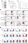
Figure 6
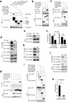
Figure 6. Jmjd3 regulates target gene expression by altering H3K4me3 and H3K27me3 levels
(a) Western blot of global H3K4me1/me2/me3 and H3K27me1/me2/me3 levels in thymic CD4+ SP T cells. (b) H3K4me3 and H3K27me3 in WT and Jmjd3 cKO CD4 SP T cells were analyzed by genome-wide ChIP-Seq. Venn diagram showing the numbers of genes with H3K27me3 and H3K4me3 modifications in Jmjd3 cKO cells compared with WT cells. (c) Average H3K27me3 profiles in WT and cKO samples at differentially methylated genes. TSS, transcription start site; TES, transcription end site. (d) T cell-related genes were classified into three groups based on H3K4me3 and H3K27me3 modifications in WT and Jmjd3 cKO CD4 SP thymocytes: group I, increased H3K27 and decreased H3K4; group II, unchanged H3K27 and decreased H3K4; and group III, unchanged H3K27 and H3K4. Red frames indicate the 2 kb region around the TSS. Scale bars represent 5kb region. (e) Validation of methylation changes in WT and cKO CD4 SP thymocytes by ChIP-qPCR (*p < 0.05 determined by Student's t-test).
Figure 7
Figure 7. Jmjd3 regulates Th1 and Th2 differentiation by facilitating the T-bet-RbBP5 and Smad3-Ash2L interaction
(a) 293T cells were cotransfected with HA-Jmjd3 and FLAG-tagged T-bet, GATA-3, Foxp3, and RORγT plasmids. Whole cell lysates (WCL) were immunoprecipitated with anti-T-bet, anti-GATA-3, anti-Foxp3, and anti-RORγT antibodies and immunoblotted with anti-FLAG antibody. (b) 293T cells were cotransfected with HA-Jmjd3 and FLAG-tagged Wdr5, Ash2L, or RbBP5. WCL were immunoprecipitated with anti-HA beads and immunoblotted with anti-FLAG antibody. (c) 293T cells were cotransfected with T-bet and FLAG-tagged Wdr5, Ash2L, Dpy30 or RbBP5. WCL were immunoprecipitated with anti-T-bet antibody and protein (A+G), and immunoblotted with anti-FLAG antibody. (d) Cell lysates were obtained from WT and Jmjd3 cKO Th1 cells and immunoprecipitated with anti-Jmjd3 antibody and protein (A+G). The immunoprecipitated product was immunoblotted with anti-Jmjd3, anti-T-bet, and anti-Ash2L antibodies. (e) WCL were obtained from WT and Jmjd3 cKO Th1 cells and immunoprecipitated with anti-T-bet antibody and protein (A+G). The immunoprecipitated product was immunoblotted with anti-RbBP5 antibody. (f) ChIP-qPCR analysis of T-bet binding to the promoter regions of Cxcr3 and Ifng genes in WT and Jmjd3 cKO Th1 cells. (g) WCL obtained from 293T cells cotransfected with HA-Jmjd3 and FLAG-Smad1, FLAG-Smad2, or FLAG-Smad3 plasmids were immunoprecipitated with anti-HA beads. The immunoprecipitated product was immunoblotted with anti-FLAG and anti-HA antibodies. (h) WCL and protein isolates from WT and Jmjd3cKO CD4+ T cells derived from thymus. (i) 293T cells were cotransfected with HA-tagged Smad3 and FLAG-tagged Wdr5, Ash2L, Dpy30 or RbBP5. WCL were immunoprecipitated with anti-FLAG beads and immunoblotted with anti-HA antibody. (j) Cell lysates were obtained from WT and Jmjd3 cKO Treg cells and immunoprecipitated with anti-Smad3 antibody. The immunoprecipitated product was immunoblotted with anti-Jmjd3, anti-Smad3 and anti-Ash2L antibodies. (k) ChIP-qPCR analysis of Smad3 binding to the promoter regions of Foxp3 gene in WT and Jmjd3 cKO Treg cells.
Figure 8
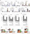
Figure 8. Jmjd3 functionally regulates gene expression and DNA-binding
(a) Ectopically expressed Jmjd3 rescues the gene expression in Jmjd3-deficient T cells. (b, c) Ectopically expressed Jmjd3 enhances the binding of the transcription factors, T-bet and Smad3, to their target genes (*p < 0.05). (d) Schematic diagram illustrating the proposed mechanistic role of Jmjd3 in the regulation of CD4+ T cell differentiation. Jmjd3 interacts with T-bet-RbBP5 and Ash2L to form a stable complex with transcription factors capable of binding to target gene promoters, thus allowing Jmjd3 to alter H3K327 methylation and Ash2L/RbBP5 to alter H3K4 levels to control target gene expression. Jmjd3 upregulates the expression of Th1 and Treg cell differentiation factors, including Foxp3, Ifng, Cd44, and Cxcr3, and downregulates the expression of the Th2 cell differentiation factors Rorc and Gata3. These changes in gene expression lead to the promotion of Th1 and Treg cell differentiation and the inhibition of Th2 and Th17 cell differentiation.

Similar articles

Cited by

References

    1. Weaver CT, Harrington LE, Mangan PR, Gavrieli M, Murphy KM. Th17: an effector CD4 T cell lineage with regulatory T cell ties. Immunity. 2006;24:677–88. - PubMed
    1. Zhu J, Yamane H, Paul WE. Differentiation of effector CD4 T cell populations (*). Annu Rev Immunol. 2010;28:445–89. - PMC - PubMed
    1. Harrington LE, et al. Interleukin 17-producing CD4+ effector T cells develop via a lineage distinct from the T helper type 1 and 2 lineages. Nat Immunol. 2005;6:1123–32. - PubMed
    1. Park H, et al. A distinct lineage of CD4 T cells regulates tissue inflammation by producing interleukin 17. Nat Immunol. 2005;6:1133–41. - PMC - PubMed
    1. Ivanov II, et al. The orphan nuclear receptor RORgammat directs the differentiation program of proinflammatory IL-17+ T helper cells. Cell. 2006;126:1121–33. - PubMed

Publication types

Substances