. 2015 Jun;17(6):876-92.
doi: 10.1111/cmi.12409. Epub 2015 Jan 26.

Host endoplasmic reticulum COPII proteins control cell-to-cell spread of the bacterial pathogen Listeria monocytogenes

Affiliations

Host endoplasmic reticulum COPII proteins control cell-to-cell spread of the bacterial pathogen Listeria monocytogenes

Antonella Gianfelice et al. Cell Microbiol. 2015 Jun.

Abstract

Listeria monocytogenes is a food-borne pathogen that uses actin-dependent motility to spread between human cells. Cell-to-cell spread involves the formation by motile bacteria of plasma membrane-derived structures termed 'protrusions'. In cultured enterocytes, the secreted Listeria protein InlC promotes protrusion formation by binding and inhibiting the human scaffolding protein Tuba. Here we demonstrate that protrusions are controlled by human COPII components that direct trafficking from the endoplasmic reticulum. Co-precipitation experiments indicated that the COPII proteins Sec31A and Sec13 interact directly with a Src homology 3 domain in Tuba. This interaction was antagonized by InlC. Depletion of Sec31A or Sec13 restored normal protrusion formation to a Listeria mutant lacking inlC, without affecting spread of wild-type bacteria. Genetic impairment of the COPII component Sar1 or treatment of cells with brefeldin A affected protrusions similarly to Sec31A or Sec13 depletion. These findings indicated that InlC relieves a host-mediated restriction of Listeria spread otherwise imposed by COPII. Inhibition of Sec31A, Sec13 or Sar1 or brefeldin A treatment also perturbed the structure of cell-cell junctions. Collectively, these findings demonstrate an important role for COPII in controlling Listeria spread. We propose that COPII may act by delivering host proteins that generate tension at cell junctions.


Figures

Figure 1
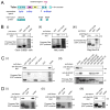
Figure 1. The Tuba SH36 domain associates with human Sec31A and other COPII components
A. Diagram depicting functional domains in Tuba and known mammalian or bacterial ligands of these domains. DH (Dbl Homology) and Bar (Bin/Amphiphysin/Rvs) domains are indicated. SH3 domains are colored blue. ‘Dyn2’ is an abbreviation for Dynamin 2. SH3 domains present in GST fusion proteins used for co-precipitation experiments are shown below Tuba. B. Association of Sec31A with GST-SH36. Lysates from Caco-2 BBE1 cells (i) HeLa cells (ii), or HeLa cells transfected with a plasmid expressing Flag-tagged human Sec31A (iii) were incubated with GST-SH36 or GST alone, followed by precipitation of the GST protein. The presence of Sec31A was detected by Western blotting of precipitates with anti-Sec31A or anti-Flag antibodies. Lysates not subjected to precipitation were used as a positive control for detection of Sec31A. Stripped membranes were stained with Ponceau Red to confirm precipitation of GST proteins. Images in (i) and (ii) are of spliced gels in which irrelevant lanes had been excised. C. Specificity of interaction of Sec31A with various SH3 domains. Lysates of Caco-2 BBE1 cells were incubated with GST-SH36, GST alone, GST fused to other SH3 domains in Tuba (i) or GST fused to SH3 domains in mammalian proteins apart from Tuba (ii). Co-precipitation experiments were performed as described in B. Precipitates were Western blotted with anti-Sec31A antibodies. In ii, membranes were stripped and reacted with anti-N-WASP antibodies. N-WASP detection confirmed activity of some SH3 domains, since several of these domains are known to interact with N-WASP. SH3 domains tested in ii include a domain from the p85 regulatory subunit of type IA PI 3-kinase (SH3.p85), two domains from the adaptor protein CrkII (SH3N.CrkII and SH3C.CrkII), and two domains from the endocytic protein intersectin-l (SH3A.int and SH3E.int). D. Association of GST-SH36 with the COPII components Sec13. Sec23A, and Sec24A. Caco-2 BBE1 cell lysates were used for co-precipitation experiments and Western blotted with anti-Sec13 (i), Sec23A (ii), or anti-Sec24A (iii) antibodies. Experiments in parts B–D were performed at least three times, with similar results.
Figure 2
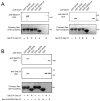
Figure 2. The Tuba SH36 domain interacts directly with the Sec31A-Sec13 complex
A. Binding of SH36 to wild-type Sec31A complexed with Sec13. Purified Sec31A-Sec13 complex was incubated with GST-SH36 and binding was assessed in co-precipitation assays as described in the Experimental Procedures. As negative controls, GST alone or GST fused to the SH35 domain of Tuba (GST-SH35) or the SH3E domain of intersectin-l (GST-SH3E.int) were used. Binding was detected by probing precipitates with antibodies against Sec31A (left panel) or anti-Sec13 (right panel). In the last lane, 100 ng of Sec31A-Sec13 protein was loaded as a control for antibody reactivity. B. Experiments with Sec31A deleted for its proline-rich domain (Sec31AΔPRD) complexed with Sec13. Co-precipitation experiments were performed with purified Sec31A-Sec13 or Sec31A-ΔPRD-Sec13 complexes and the indicated GST fusion proteins. The last two lanes contain 100 ng of purified Sec31A-Sec13 or Sec31AΔPRD-Sec13 complexes. Experiments in A and B were performed 3 times with similar results.
Figure 3
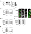
Figure 3. Sec31A controls spread of Listeria
A. RNAi-mediated depletion of Sec31A. Caco-2 BBE1 cells grown in transwells were either mock transfected in the absence of siRNA (−), or transfected with a control non-targeting siRNA (C) or an siRNA directed against Sec31A. Approximately 72 h after addition of siRNA, cells lysates were prepared and used for Western blotting analysis of Sec31A expression. A. Effect of Sec31A siRNA on target protein expression. A representative Western blot is in the left panel and quantification of Western blots from three experiments is presented in the right panel. B. Effect of Sec31A depletion on Listeria protrusion formation. After transfection with the indicated siRNA, Caco-2 BBE1 cells were infected with wild-type (wt) or ΔinlC strains of Listeria for 5.5 h prior to fixation and processing for confocal microscopy analysis. (i). Bar graph displaying mean relative protrusion formation values +/− SEM. (ii). Confocal microscopy images illustrating how protrusions were quantified. Protrusion efficiency is expressed as the proportion of total bacteria-associated F-actin structures in protrusions. These F-actin structures are defined as protrusions, actin tails, or symmetric actin. The top, middle, and bottom panels display human cells containing a Listeria protrusion, bacteria with F-actin tails, or bacteria decorated with symmetric F-actin, respectively. In the ‘merge’ image, EGFP-actin is green, total F-actin labeled with phalloidin is red, and bacteria are blue. Individual channels for EGFP-actin or F-actin (phalloidin labeling) are also shown. The regions in white boxes in the ‘merge’ images are expanded in the right panels. Protrusions (P) were identified as EGFP-positive actin tails projecting from transfected cells into neighboring EGFP-negative cells. Bacteria with actin tails (T) and symmetric F-actin (S) within the EGFP-positive cell body are shown. The scale bars indicate 5 micrometers. C. Bacterial-directed F-actin tail assembly in Sec31A-depleted cells. (i) The percentages of bacteria associated with F-actin (i) or the lengths of bacterial F-actin tails (ii) were quantified in the same samples used for protrusion analysis. D. Effect of Sec31A depletion on cell-to-cell spread of Listeria. Caco-2 BBE1 cells transfected with Sec31A or subjected to control conditions were infected with the indicated Listeria strains for 12 h, followed by processing for immunofluorescence and analysis by confocal microscopy. Spread was assessed by measuring surface areas of foci containing intracellular bacteria, as described in the Experimental Procedures. Mean relative surface areas +/− SEM of foci produced by wt or ΔinlC bacterial strains are presented. The data in A, B, and C are mean +/− SEM from three experiments, whereas results in D are from a single experiment, representative of three total experiments. *, P < 0.05 relative to control siRNA transfected cells infected with wt Listeria.
Figure 4
Figure 4. The COPII components Sec13 and Sar1 control Listeria protrusion formation
A. Effect of Sec13 depletion on Listeria protrusions. Caco-2 BBE1 cells were either mock transfected in the absence of siRNA (−), transfected with a control non-targeting siRNA (C), or transfected with an siRNA directed against Sec13. Approximately 72 h after addition of siRNA, cells lysates were prepared and used for analysis of Sec13 expression by Western blotting (i) or infected with wild-type (wt) or ΔinlC Listeria strains for 5.5 h followed by assessment of bacterial protrusions (ii). *, P < 0.05 relative to control siRNA transfected cells infected with wt Listeria. B. Impact of inhibition of Sar1 on Listeria protrusions. Caco-2 BBE1 cells were transfected with plasmids expressing an Ha-tagged dominant negative allele of human Sar1 (Sar1.T39N) or Ha-tagged luciferase as a control. About 72 h post-transfection, cells were infected with wt or ΔinlC Listeria strains and protrusion formation was quantified by confocal microscopy analysis. *, P < 0.05 relative to Ha-luciferase-expressing cells infected with wt bacteria. Data in A and B are each mean +/− SEM values from three experiments.
Figure 5
Figure 5. Effects of brefeldin A (BFA) on protein secretion and Listeria protrusion formation
Caco-2 BBE1 cells were transfected with a plasmid expressing Secreted Placental Alkaline Phosphatase (SEAP). Approximately 72 h later, cells were used for analysis of SEAP secretion or bacterial protrusion formation. A. SEAP activity. After treatment of Caco-2 BBE1 cells with the indicated concentrations of BFA (+) or with the vehicle DMSO for 1 h, levels of secreted (extracellular) SEAP were measured as described in the Experimental Procedures. *, P < 0.05 relative to cells treated with DMSO (−). B. Listeria protrusion formation. Caco-2 BBE1 cells were infected with wt or ΔinlC bacterial strains for 1 h followed by incubation for another 4.5 h in medium containing 5 μg/ml BFA (+) or DMSO (−). Samples of fixed and labeled cells were analyzed by confocal microscopy for quantification of bacterial protrusions. *, P < 0.05 relative to DMSO-treated cells (−) infected with wt Listeria. Data in A and B are mean +/− SEM from three experiments.
Figure 6
Figure 6. InlC displaces Sec31A from the Tuba SH36 domain
A. InlC-mediated inhibition in association of Sec31A with the SH36 domain. A constant amount of lysate from Caco-2 BBE1 cells was incubated with approximately 0.30 μM GST-SH36 and increasing concentrations (0–16.7 μM) of purified InlC. Sec31A in GST-SH36 precipitates was detected by Western blotting. The image is of a spliced gel in which irrelevant lanes had been excised. B. Control with the mutant protein InlC.F146A. (i). Caco-2 BBE1 cell lysates were incubated with approximately 0.30 μM GST-SH36 in the absence of competitor (−) or in the presence of 6.7 μM of InlC or InlC.F146A protein. GST alone (0.30 μM) was used as a negative control. Sec31A in precipitates was detected by Western blotting. Binding of InlC to GST-SH36 was confirmed by probing a stripped membrane with anti-InlC antibodies. (ii). Anti-InlC Western blot of purified wild-type and mutant InlC proteins. This experiment verified that the InlC.F146A protein does not have diminished reactivity to anti-InlC antibodies. Data in A and B are each representative of three experiments.
Figure 7
Figure 7. COPII components regulate apical junction morphology
A. Effect of depletion of Sec31A on cell junctions. (i). About 72 h after transfection with control siRNA or an siRNA targeting Sec31A, Caco-2 BBE1 cells were fixed and labeled for the tight junction protein ZO-1. Images of ZO-1 labeling were acquired using a confocal microscope. (ii). Linear indexes were quantified as described in the Experimental Procedures. B. Impact of Sec13 depletion on cell junctions. Transfection, imaging of cell junctions (i), and measurement of linear indexes (ii) was performed as described in A. C. Perturbation of cell junctions by a dominant negative allele of Sar1. Caco-2 BBE1 cells transfected with plasmids expressing Ha-tagged Sar1.T39N or luciferase for 72 h were fixed and labeled for Ha (red) and ZO1 (green). Images of ZO-1 labeling (i) and linear index values (ii) are shown. D. Effect of BFA on cell junctions. Caco-2 BBE1 cells were treated with 5 μg/ml BFA for 2 h, followed by processing for confocal microscopy. Images of ZO-1 labeling (i) and quantification of linear indexes (ii) are shown. Asterisks in A–D indicate cells with curved junctions. Scale bars represent 3 μm. Linear index data in A–D are mean+/− SEM values from three experiments. *, P < 0.05 relative to control siRNA (C) treated cells.

Similar articles

Cited by

References

    1. Basar T, Shen Y, Ireton K. Redundant roles for Met docking site tyrosines and the Gab1 pleckstrin homology domain in InlB-mediated entry of Listeria monocytogenes. Infect Immun. 2005;73:2061–2074. - PMC - PubMed
    1. Belov GA, Altan-Bonnet N, Kovtunovych G, Jackson CL, Lippincott-Schwartz J, Ehrenfeld E. Hijacking components of the cellular secretory pathway for replication of poliovirus RNA. J Virol. 2007;81:558–567. - PMC - PubMed
    1. Berger J, Hauber J, Hauber R, Geiger R, Cullen BR. Secreted placental alkaline phosphatase: a powerful new quantitative indicator of gene expression in eukaryotic cells. Gene. 1988;66:1–10. - PubMed
    1. Bergmann JE, Singer SJ. Immunoelectron microscopic studies of the intracellular transport of the membrane glycoprotein (G) of vesicular somatitis virus in infected Chinese hamster ovary cells. J Cell Biol. 1983;97:1777–1787. - PMC - PubMed
    1. Bryant DM, Datta A, Rodriguez-Fraticelli AE, Peranen J, Martin-Belmonte F, Mostov KE. A molecular network for de novo generation of the apical surface and lumen. Nat Cell Biol. 2010;12:1035–1045. - PMC - PubMed

Publication types

Substances