. 2014 Dec 19;4(1):86-94.
doi: 10.1242/bio.20148532.

Rab11a is required for apical protein localisation in the intestine

Affiliations

Rab11a is required for apical protein localisation in the intestine

Tomoaki Sobajima et al. Biol Open. .

Abstract

The small GTPase Rab11 plays an important role in the recycling of proteins to the plasma membrane as well as in polarised transport in epithelial cells and neurons. We generated conditional knockout mice deficient in Rab11a. Rab11a-deficient mice are embryonic lethal, and brain-specific Rab11a knockout mice show no overt abnormalities in brain architecture. In contrast, intestine-specific Rab11a knockout mice begin dying approximately 1 week after birth. Apical proteins in the intestines of knockout mice accumulate in the cytoplasm and mislocalise to the basolateral plasma membrane, whereas the localisation of basolateral proteins is unaffected. Shorter microvilli and microvillus inclusion bodies are also observed in the knockout mice. Elevation of a serum starvation marker was also observed, likely caused by the mislocalisation of apical proteins and reduced nutrient uptake. In addition, Rab8a is mislocalised in Rab11a knockout mice. Conversely, Rab11a is mislocalised in Rab8a knockout mice and in a microvillus atrophy patient, which has a mutation in the myosin Vb gene. Our data show an essential role for Rab11a in the localisation of apical proteins in the intestine and demonstrate functional relationships between Rab11a, Rab8a and myosin Vb in vivo.

Keywords: Apical membrane; Brain; Cell polarity; Intestine; Knockout mouse; Rab11a.


Conflict of interest statement

Competing interests: The authors have no competing interests to declare.

Figures

Fig. 1.
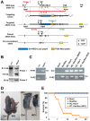
Fig. 1.. Generation and analysis of Rab11a knockout mice.
(A) Diagram of targeting strategies. Restriction maps of the Rab11a wild-type allele (+), the targeting vector containing an SA-IRES-LacZ-polyA sequence (blue horizontal bar) and PGK-Neo-polyA (orange horizontal bar), and the targeted allele (neo). Brown triangle, loxP site; green triangle, FRT site. Probe 1 hybridises with a 21.3-kb Apa I fragment from the wild-type allele and a 9.5-kb fragment from the targeted allele. Probe 2, the neomycin fragment in PGK-Neo-polyA, hybridises with a 19-kb Apa I fragment from the targeted allele. SA, splice acceptor; IRES, internal ribosomal entry site; PGK, phosphoglycerate kinase promoter; Neo, neomycin resistance gene cassette. (B) Southern blotting analysis of the targeted ES cell clones. Genomic DNA from parental embryonic stem cells (+/+) and targeted clones (neo/+) were digested with Apa I for hybridisation with the probes 1 and 2 in (A). (C) Genotyping PCR analysis using genomic DNA from control and knockout mouse tails. Primers ‘F+R1’, primers ‘F+R2’, and primers ‘Cre’ were used to detect the targeted allele (neo), the wild-type (+) and floxed (flox) alleles, and cre gene. (D) Control (Ctrl) and intestine-specific Rab11a knockout (IKO) mice at postnatal days 5 (P5) and 21 (P21). The IKO mice were smaller than control littermates at both ages. (E) Survival curves for control (n = 12–14) and IKO (n = 12) mice. IKO mice began dying 5 days after birth, and all mice were dead by postnatal day 25. NC, Nestin-Cre; VC, Villin-Cre.
Fig. 2.
Fig. 2.. Localisation of the apical protein DPPIV in the small intestine of Rab11a IKO mice at P5 and P21.
(A–D) Localisation of the apical protein DPPIV (green) and the lysosome marker Lamp2 (red) in control (Ctrl) and IKO mice at P5 (A,B) and P21 (C,D). Higher magnification views of the intestinal cells from Ctrl and IKO mice at P5 (B) and P21 (D). At P5 (A,B), intracellular DPPIV staining (green) is colocalised with Lamp2 (red) in IKO cells (arrows in B ‘IKO’), whereas DPPIV and Lamp2 (arrowheads in B ‘Ctrl’) did not colocalise in control cells. In addition, basolateral staining (arrowheads in B ‘IKO’) is more evident in IKO cells. At P21 (C,D), intracellular DPPIV staining is evident and the colocalisation of Lamp2 and DPPIV is greater in IKO mice (arrows in D). Scale bars: 20 µm (A,C), 10 µm (B,D).
Fig. 3.
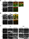
Fig. 3.. Localisation of apical and basolateral proteins in the small intestine of Rab11a IKO mice at P21.
(A) DPPIV and the TGN marker Golgin-97 (arrowheads) do not colocalise in intestinal epithelial cells from IKO mice at P21. High-magnification insets (white boxes) are shown to the right of these panels. (B) Localisation of the apical proteins AlP and APN in control (Ctrl) and IKO cells at P21. Merged figures of AlP and APN are shown on the right. Intracellular vacuoles of AlP and APN are indicated by arrowheads. Basolateral localisation is indicated by arrows. (C) Localisation of the basolateral proteins E-cadherin, Na+K+-ATPase, and LDL-R in epithelial cells of the small intestine in Ctrl and IKO mice is not different at P5 and P21. Scale bars: 10 µm. N, nucleus.
Fig. 4.
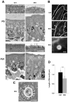
Fig. 4.. Microvillus atrophy and inclusions in Rab11a IKO mice.
(A) Electron micrographs of P5 and P21 intestinal cells. Apical microvilli (first and third panels from top) are shorter in knockout cells (IKO) than in control (Ctrl) cells. (B) Sections of intestinal cells at P21 stained with phalloidin. F-actin localisation is prominent in the apical plasma membranes in villi of control intestines (top panel). Phalloidin-positive intracellular spheroids are evident in mutant villi (arrowheads in the middle panel). An enlarged view is shown in the bottom panel. (C) A microvillus inclusion body from a P21 IKO intestinal epithelial cell visualised using electron microscopy. (D) A graph showing microvillus length in epithelial cells at P5. Values represent mean ± s.d. (n>40 cells randomly selected from three mice per group; *** P<0.001; Student's t-test). Scale bars: 2 µm (A,C), 20 µm (B, middle), 2 µm (B, bottom).
Fig. 5.
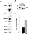
Fig. 5.. Quantification of a starvation marker and protein levels.
(A) Western blot analysis of protein levels in crude extracts from the small intestines of control (Ctrl) and IKO mice at P5. Small intestine extracts were analysed by Western blot analysis using antibodies against various apical proteins (DPPIV and AlP), basolateral proteins (LDL-R, Na+K+ATPase, and E-cadherin), Rabs (Rab8a, 10, 11a, and 11b), and a loading control (Lamin B). (B) Treatment of peptide N-glycosidase F (PNGase F) in lysates from the small intestine of control (Ctrl) and IKO mice at P5. The samples were analysed by Western blotting using anti-DPPIV antibody. (C) Amounts of total ketone bodies in control (Ctrl) and IKO mice at P5. Values represent means ± s.d. from 4–10 mice. ***P<0.001; Student's t-test.
Fig. 6.
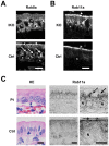
Fig. 6.. Spatial interdependence between Rab11a, Rab8a, and myosin Vb.
(A) Localisation of Rab8a in epithelial cells of the small intestine in control (Ctrl) and Rab11a IKO mice at P5. Rab8a is mainly localised on perinuclear punctate structures (arrowheads) below the nucleus (N) in IKO cells, whereas it is localised on large vacuolar structures (arrows) above the nucleus in control cells. (B) Localisation of Rab11a in epithelial cells of the small intestine in Ctrl and Rab8a KO mice at P7. Rab11a is exclusively localised below the apical plasma membrane (an arrowhead) in Ctrl epithelial cells, but it is also localised punctate structures (an arrow) above the nucleus in Rab8a KO epithelial cells. (C) Localisation of Rab11a in epithelial cells of the small intestine from a control (Ctrl) and a microvillus atrophy patient (Pt). Rab11a is localised below the apical plasma membrane in control intestinal cells (arrowheads), but it is localised perinuclear structures in patient's intestinal cells (arrows). HE Scale bars: 10 µm. N, nucleus.
Fig. 7.
Fig. 7.. Localisation of Rab8a and Rab11a and quantification of Rab11a, Rab8a, and Rab10 in the small intestine during postnatal development.
(A) Localisation of Rab8a (top), Rab11a (middle) and non-immune rabbit IgG (bottom), as determined by immunofluorescence, during postnatal (P) days in wild-type epithelial cells of the small intestine. (B) Levels of Rab11a, Rab8a, and Rab10 in the wild-type small intestine during postnatal development. Scale bar: 10 µm.

Similar articles

Cited by

References

    1. Ang A. L., Taguchi T., Francis S., Fölsch H., Murrells L. J., Pypaert M., Warren G., Mellman I. (2004). Recycling endosomes can serve as intermediates during transport from the Golgi to the plasma membrane of MDCK cells. J. Cell Biol. 167, 531–543 10.1083/jcb.200408165 - DOI - PMC - PubMed
    1. Benli M., Döring F., Robinson D. G., Yang X., Gallwitz D. (1996). Two GTPase isoforms, Ypt31p and Ypt32p, are essential for Golgi function in yeast. EMBO J. 15, 6460–6475. - PMC - PubMed
    1. Casanova J. E., Wang X., Kumar R., Bhartur S. G., Navarre J., Woodrum J. E., Altschuler Y., Ray G. S., Goldenring J. R. (1999). Association of Rab25 and Rab11a with the apical recycling system of polarized Madin-Darby canine kidney cells. Mol. Biol. Cell 10, 47–61 10.1091/mbc.10.1.47 - DOI - PMC - PubMed
    1. Cresawn K. O., Potter B. A., Oztan A., Guerriero C. J., Ihrke G., Goldenring J. R., Apodaca G., Weisz O. A. (2007). Differential involvement of endocytic compartments in the biosynthetic traffic of apical proteins. EMBO J. 26, 3737–3748 10.1038/sj.emboj.7601813 - DOI - PMC - PubMed
    1. Duman J. G., Tyagarajan K., Kolsi M. S., Moore H. P., Forte J. G. (1999). Expression of rab11a N124I in gastric parietal cells inhibits stimulatory recruitment of the H+-K+-ATPase. Am. J. Physiol. 277, C361–C372. - PubMed

LinkOut - more resources