. 2014 Dec 20:14:46.
doi: 10.1186/s12861-014-0046-5.

Isoform-specific functions of Mud/NuMA mediate binucleation of Drosophila male accessory gland cells

Affiliations

Isoform-specific functions of Mud/NuMA mediate binucleation of Drosophila male accessory gland cells

Kiichiro Taniguchi et al. BMC Dev Biol. .

Abstract

Background: In standard cell division, the cells undergo karyokinesis and then cytokinesis. Some cells, however, such as cardiomyocytes and hepatocytes, can produce binucleate cells by going through mitosis without cytokinesis. This cytokinesis skipping is thought to be due to the inhibition of cytokinesis machinery such as the central spindle or the contractile ring, but the mechanisms regulating it are unclear. We investigated them by characterizing the binucleation event during development of the Drosophila male accessory gland, in which all cells are binucleate.

Results: The accessory gland cells arrested the cell cycle at 50 hours after puparium formation (APF) and in the middle of the pupal stage stopped proliferating for 5 hours. They then restarted the cell cycle and at 55 hours APF entered the M-phase synchronously. At this stage, accessory gland cells binucleated by mitosis without cytokinesis. Binucleating cells displayed the standard karyokinesis progression but also showed unusual features such as a non-round shape, spindle orientation along the apico-basal axis, and poor assembly of the central spindle. Mud, a Drosophila homolog of NuMA, regulated the processes responsible for these three features, the classical isoform Mud(PBD) and the two newly characterized isoforms Mud(L) and Mud(S) regulated them differently: Mud(L) repressed cell rounding, Mud(PBD) and Mud(S) oriented the spindle along the apico-basal axis, and Mud(S) and Mud(L) repressed central spindle assembly. Importantly, overexpression of Mud(S) induced binucleation even in standard proliferating cells such as those in imaginal discs.

Conclusions: We characterized the binucleation in the Drosophila male accessory gland and examined mechanisms that regulated unusual morphologies of binucleating cells. We demonstrated that Mud, a microtubule binding protein regulating spindle orientation, was involved in this binucleation. We suggest that atypical functions exerted by three structurally different isoforms of Mud regulate cell rounding, spindle orientation and central spindle assembly in binucleation. We also propose that Mud(S) is a key regulator triggering cytokinesis skipping in binucleation processes.


Figures
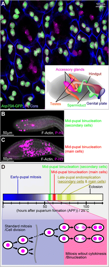
Figure 1
Figure 1
Synchronous binucleation of Drosophila male accessory gland cells occurs in the pupal stage. (A) Adult accessory gland epithelium labeled as indicated at the bottom left. Main cells (nuclei stained both green and magenta) and secondary cells (nuclei stained only magenta) are shown. The inset at the bottom right depicts an adult male abdomen (gray) and the reproductive systems around the hindgut. Posterior is to the right. Scale bar, 10 μm. (B and C) Synchronous entry into M phase in secondary (B) and main (C) cells in the accessory glands during mid-pupal binucleation stages. Labels as indicated at the bottom right. Scale bars, 50 μm. (D) Schematic diagram showing cell cycle transition of epithelial cells in accessory gland during the pupal stage.
Figure 2
Figure 2
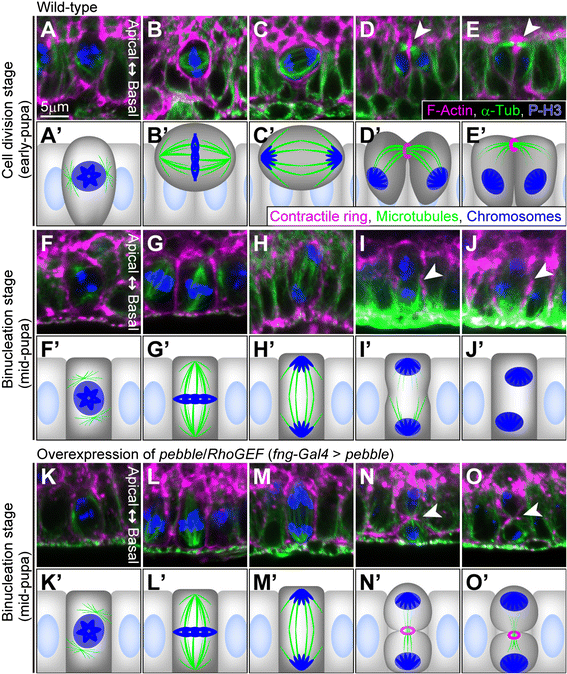
Central spindle and contractile ring are not formed during binucleation. Photomicrographs showing cross-sectional views of cells (A–O) and their schematic diagrams (A’–O’) are arrayed from left to right according to the M phase progression. (A–E) Main cells during cell division stage in early pupa (30–35 hours APF). (F–J) Main cells during binucleation stage in mid-pupa (60–65 hours APF). (K–O) Binucleation-stage main cells in which pebble was overexpressed just before binucleation. Arrowheads in (D, E, I, J, N and O) indicate equatorial planes in late anaphase and telophase during cell division and binucleation. Cells are labeled as indicated at the bottom of (E). Scale bar in (A), 5 μm, is applicable to (A–O).
Figure 3
Figure 3
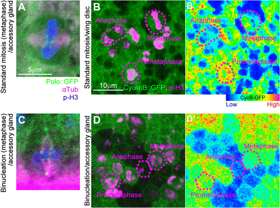
Metaphase spindle formation and metaphase-anaphase transition are normal during binucleation. (A and C) Localization of polo to kinetochores in main cells in metaphase during cell division in early pupa (A) and during binucleation in mid-pupa (C). Cells are labeled as indicated at the bottom of (A). En face views (A) and cross-sectional views (C). Scale bar, 5 μm, is applicable to (A and C). (B, B’, D and D’) Levels of cyclin B. En face images of the wing disc epithelium in third-instar larva as an example of proliferating tissue (B and B’) and the accessory gland epithelium in mid-pupa as an example of binucleating tissue (D and D’). Cells in (B and D) are labeled as indicated at the bottom right of (B). Intensities of cyclin B::GFP in (B) and (D) are represented by a rainbow-color scale, with red meaning high intensity and blue meaning low intensity. Magenta dashed lines indicate outlines of mitotic cells in various M-phase subphases. Scale bar in (B), 10 μm, is applicable to (B, B’, D and D’).
Figure 4
Figure 4
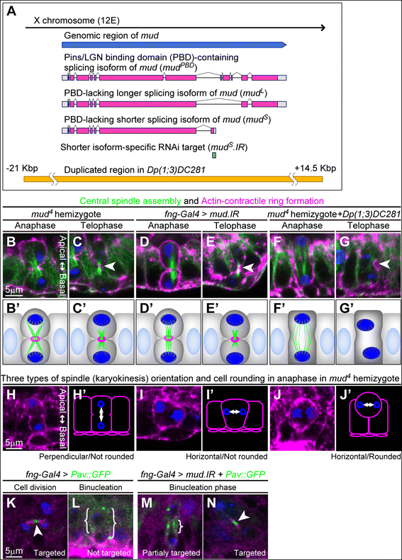
Loss-of-function for mud erases various characteristics of binucleation. (A) Schematic diagram of the mud transcriptional unit and three representative splicing variants of mud (coding regions are in magenta). Regions corresponding to mud S .IR (green) and chromosomal duplication in Dp(1;3)DC281 (yellow) are also shown. (B–G) Cross-sectional views of main cells in late anaphase (B, D and F) and telophase (C, E and G) during the binucleation stage in mutants hemizygous for mud 4 (B and C), in knockdown for mud (D and E) and in mutants hemizygous for mud 4 rescued by one copy of Dp(1;3)DC281 (F and G). Cells are labeled with phalloidin (magenta), anti-α-Tub antibody (green), and anti-P-H3 antibody (blue). Arrowheads in (C, E and G) indicate equatorial planes. Scale bar in (B), 5 μm, is applicable to (B–G). (B’–G’) Schematic diagrams of (B and G). (H–J) Three representative types of spindle orientation and cell shapes (bottom). Cross-sectional views of main cells in late anaphase during the binucleation stage in mutants hemizygous for mud 4 are shown. Cells are labeled with phalloidin (magenta) and anti-P-H3 antibody (blue). Scale bar in (H), 5 μm, is applicable to (H–J). (H’–J’) Schematic diagrams of (H–J). (K–N) Main cells expressing Pav::GFP plus ends marker in telophase in wild-type (K,L) and mud-knockdown (M,N) cells. Cell division stage in early pupa (K, en face view) and binucleation stage in mid-pupa (L–N, cross sectional views) are shown. Cells are labeled with anti-α-Tub antibody (magenta), Pav::GFP fluorescence with anti-GFP antibody (green) and anti-P-H3 antibody (blue). Arrowheads in (K and N) and curly brackets in (L and M) indicate the localization of Pav::GFP on microtubules. Scale bar in (K), 5 μm, is applicable to (K–N).
Figure 5
Figure 5
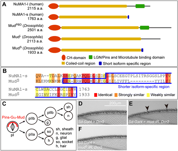
mud S is not required for the spindle orientation during asymmetric cell division. (A) Schematic diagram of molecular structures of human NuMA1 and Drosophila Mud. Shared domains are as indicated at the bottom. (B) Amino acid sequence alignment of the C-terminal regions of NuMA1-s and MudS. The blue box indicates short isoform- specific regions in NuMA1-s (amino acids 1701–1763) and MudS (amino acids 1880–1933). Red, orange and yellow overlays indicate similarities as indicated at the bottom. (C) Schematic diagram of SOP lineage in Drosophila wing margin. Cells indicated at the bottom right are produced in this lineage. Mud forms a complex with Gα and Pins that is localized asymmetrically (red crescent). (D–F) Bristles on adult anterior wing margin in wild-type (D), in knockdown of all mud isoforms (E), and in knockdown specific to mud S (F). Arrowheads in (E) indicate multi-bristle phenotypes.
Figure 6
Figure 6
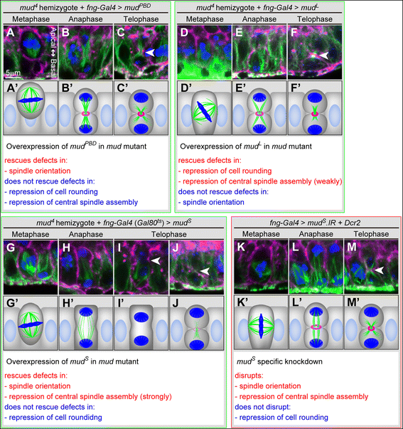
Three types of mud splicing isoforms differently regulate morphologies of binucleating cells. (A–J) Rescue of mud mutant phenotypes by each mud isoform. Cross-sectional views of main cells in metaphase (A, D and G), late anaphase (B, E and H) and telophase (C, F, I and J) during the binucleation stage in mutants hemizygous for mud 4 with overexpression of FLAG::mud PBD (A–C), FLAG::mud L (D–F) or FLAG::mud S (G–J). Cells are labeled with phalloidin (magenta), anti-α-Tub antibody (green) and anti-P-H3 antibody (blue). The cell in (I) shows neither a central spindle assembly nor furrow progression. The cell in (J) shows furrow progression but no central spindle assembly. Arrowheads in (C, F, I and J) indicate equatorial planes. Scale bar in (A), 5 μm, is applicable to (A–J). (A’–J’) Schematic diagrams of (A–J). Cross-sectional views of mud S- knockdown main cells in metaphase (K), late anaphase (L) and telophase (M) during the binucleation stage. Cells are labeled with phalloidin (magenta), anti-α-Tub antibody (green) and anti-P-H3 antibody (blue). Arrowhead in (M) indicates an equatorial plane. Scale bar in (A), 5 μm, is applicable to (K–M). (K’–M’) Schematic diagrams of (K–M). Effects on cell morphologies in each of the three rescued genotypes ((A–C): mud 4 hemizygotes rescued by mud PBD, (D–F): mud 4 hemizygotes rescued by mud L, (G–J): mud 4 hemizygote rescued by mud S) and in mud S-knockdown cells (K–M) are listed under each set of diagrams.
Figure 7
Figure 7
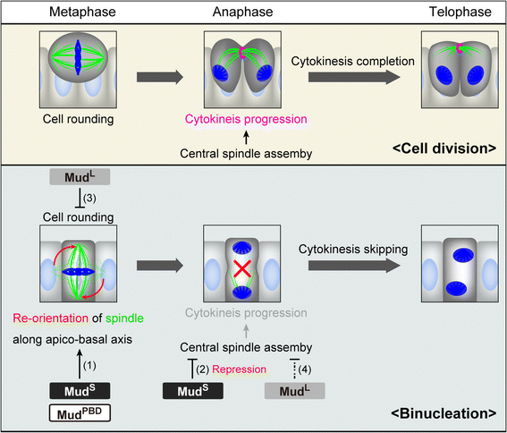
Model for binucleation and isoform-specific functions of Mud. Schematic diagrams of cell division and binucleation are shown. In binucleation, MudS changes mitosis from cell division to binucleation by (1) reorienting the mitotic spindle from horizontal to vertical along the apico-basal axis and (2) repressing assembly of the central spindle. MudPBD is also required at this time for orienting the spindle along the apico-basal axis. MudL, in contrast, (3) represses mitotic cell rounding and may assist in the process of cytokinesis skipping by (4) partially repressing the assembly of the central spindle.
Figure 8
Figure 8
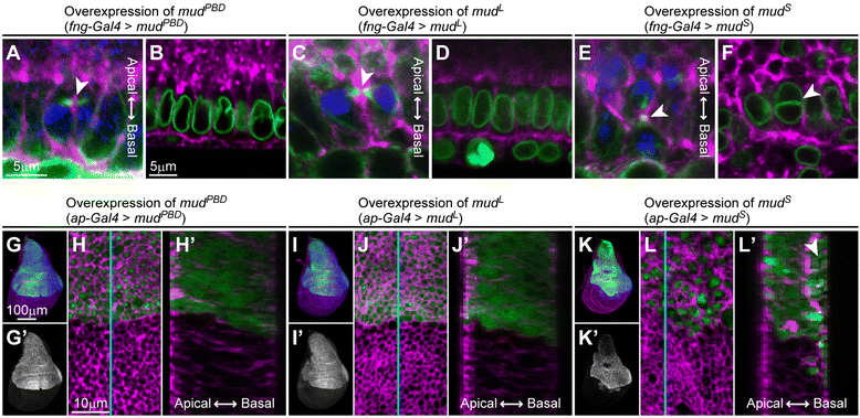
Overexpression of mud S is sufficient for converting dividing cells into binucleating ones. (A–F) Cross-sectional views of main cells in early pupal proliferating stage in which mud PBD (A and B), mud L (C and D) or mud S (E and F) are overexpressed. Cells in telophase (A, C and E) or interphase (B, D and F) are shown. Cells are labeled with phalloidin (magenta), anti-α-Tub antibody (green in A, C and E), anti-LamDm0 antibody (green in B, D and F) and anti-P-H3 antibody (blue). Arrowheads in (A, C and E) indicate equatorial planes. The arrowhead in (F) indicates a binucleate cell. Scale bar in (A), 5 μm, is applicable to (A, C and E). Scale bar in (B), 5 μm, is applicable to (B, D and F). (G, G’, I, I’, K and K’) Wing imaginal discs in which FLAG::mud PBD (G) , FLAG::mud L (I) or FLAG::mud S (K) is induced in their dorsal compartment (labeled with GFP). Cells are labeled with phalloidin (magenta), GFP (green) and anti-FLAG antibody (blue). Gray-scale images in (G’, I’ and K’) are of the blue channels in (G, I and K). Scale bar in (G), 100 μm, is applicable to (G, G’, I, I’, K and K’). (H, J and L) Magnified views of (G, I and K) around the dorso-ventral boundaries. Magenta and green channels are shown. Cells with expression of mud S were enlarged in volume (L), possibly as a result of cytokinesis defects. (H’, J’ and L’) Cross-sectional images reconstructed by using a stack of confocal sections at cyan lines in (H, I and L). The epithelium with expression of mud S has an abnormally folded or layered structure (arrowhead in L’). Scale bar in (H), 10 μm, is applicable to (H, H’, J, J’, L and L’).

Similar articles

Cited by

References

    1. Pasumarthi KB, Field LJ. Cardiomyocyte cell cycle regulation. Circ Res. 2002;90:1044–54. doi: 10.1161/01.RES.0000020201.44772.67. - DOI - PubMed
    1. Celton-Morizur S, Merlen G, Couton D, Margall-Ducos G, Desdouets C. The insulin/Akt pathway controls a specific cell division program that leads to generation of binucleated tetraploid liver cells in rodents. J Clin Invest. 2009;119:1880–7. - PMC - PubMed
    1. Bersell K, Arab S, Haring B, Kuhn B. Neuregulin1/ErbB4 signaling induces cardiomyocyte proliferation and repair of heart injury. Cell. 2009;138:257–70. doi: 10.1016/j.cell.2009.04.060. - DOI - PubMed
    1. Kwon M, Scholey JM. Spindle mechanics and dynamics during mitosis in Drosophila. Trends Cell Biol. 2004;14:194–205. doi: 10.1016/j.tcb.2004.03.003. - DOI - PubMed
    1. D'Avino PP, Savoian MS, Glover DM. Cleavage furrow formation and ingression during animal cytokinesis: a microtubule legacy. J Cell Sci. 2005;118:1549–58. doi: 10.1242/jcs.02335. - DOI - PubMed