. 2014 Dec 18;9(12):e115423.
doi: 10.1371/journal.pone.0115423. eCollection 2014.

Live cell imaging reveals novel functions of Salmonella enterica SPI2-T3SS effector proteins in remodeling of the host cell endosomal system

Affiliations

Live cell imaging reveals novel functions of Salmonella enterica SPI2-T3SS effector proteins in remodeling of the host cell endosomal system

Roopa Rajashekar et al. PLoS One. .

Abstract

Intracellular Salmonella enterica induce a massive remodeling of the endosomal system in infected host cells. One dramatic consequence of this interference is the induction of various extensive tubular aggregations of membrane vesicles, and tubules positive for late endosomal/lysosomal markers are referred to as Salmonella-induced filaments or SIF. SIF are highly dynamic in nature with extension and collapse velocities of 0.4-0.5 µm x sec-1. The induction of SIF depends on the function of the Salmonella Pathogenicity Island 2 (SPI2) encoded type III secretion system (T3SS) and a subset of effector proteins. In this study, we applied live cell imaging and electron microscopy to analyze the role of individual effector proteins in SIF morphology and dynamic properties of SIF. SIF in cells infected with sifB, sseJ, sseK1, sseK2, sseI, sseL, sspH1, sspH2, slrP, steC, gogB or pipB mutant strains showed a morphology and dynamics comparable to SIF induced by WT Salmonella. SIF were absent in cells infected with the sifA-deficient strain and live cell analyses allowed tracking of the loss of the SCV membrane of intracellular sifA Salmonella. In contrast to analyses in fixed cells, in living host cells SIF induced by sseF- or sseG-deficient strains were not discontinuous, but rather continuous and thinner in diameter. A very dramatic phenotype was observed for the pipB2-deficient strain that induced very bulky, non-dynamic aggregations of membrane vesicles. Our study underlines the requirement of the study of Salmonella-host interaction in living systems and reveals new phenotypes due to the intracellular activities of Salmonella.


Conflict of interest statement

Competing Interests: The authors have declared that no competing interests exist.

Figures

Figure 1
Figure 1. Analysis of the integrity of the Salmonella-containing vacuole in living or fixed host cells.
For analyses of fixed cells, HeLa cells were infected with Salmonella wild-type or various mutant strains. Following incubation for 16 h under standard cell culture conditions, infected cells were fixed with 3% PFA in PBS for 15 min. Immuno-staining was performed for LAMP1 (green) and Salmonella O-antigen (red). For analyses of living cells, HeLa cells were transfected with a plasmid for the expression of LAMP1-GFP (green) and infected with Salmonella strains expressing mCherry (red). Representative fixed (A) or living cells (B) infected with WT or sifA strains were imaged using a Zeiss LSM 700 confocal microscope. Micrographs of live cells infected with further strains are shown in S1 Figure. Arrowheads indicate the positions of individual bacteria and SCV membranes. Scale bars, 5 µm and 1 µm in overview and detail micrographs, respectively. Z sections of live cell infected with WT or sifA strains are shown in S2 Figure and animated 3D projections for live cells infected with WT or sifA strains are shown in S1 Movie and S2 Movie, respectively. C) For quantitative analyses, at least 20 infected cells per indicated strain were randomly selected for image acquisition and micrographs were used for the analysis of presence or absence of a continuous LAMP1-positive membrane around the bacteria. Black and gray bars represent the number of continuous SCV in fixed and live cells, respectively. Means and standard deviations shown are representative for three independent experiments. Statistical significance between WT and various mutant strains was determined by one-way ANOVA and is indicated as: ns, not significant; *, P <0.05; **, P <0.01; ***, P<0.001.
Figure 2
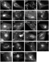
Figure 2. Systematic analyses of role of effector proteins of the SPI2-T3SS for the formation and morphology of tubular endosomes in living host cells.
HeLa cells were transiently transfected with a vector for the expression of LAMP1-GFP (white). Salmonella WT and a set of isogenic mutant strains with deletions of specific SPI1-T3SS or SPI2-T3SS effector proteins as indicated were used to infect transfected cells. The bacteria harbored plasmids for the constitutive expression of GFP or mCherry. Time lapse series for transfected and infected cells are recorded for the early and late phases of intracellular life, i.e. 4-5 h and 7–9 h post infection (p.i.), respectively. Infection and live cell imaging has been performed independently at least three times per strain, and still images from representative time lapse series are shown. Scale bar, 10 µm. Corresponding movies for each of the experiments shown are available as S3 Movie to S25 Movie.
Figure 3
Figure 3. Effect of fixation on morphology of tubular endosomal aggregates.
HeLa cells were transfected with LAMP1-GFP (green) and subsequently infected with Salmonella WT, sseF or pipB2 strains harboring a plasmid for the expression of mCherry (red). Imaging of infected cells in chamber slides was performed prior fixation and positions were stored for repositioning of a motorized stage. Chamber slides were removed from the stage, cell were washed and fixed by addition of 3% PFA in PBS. Subsequently, the chamber slide was repositioned on the microscopy stage and the positions were relocated in order to image the same cell after fixation. Representative infected cells were imaged at 6-8 h p.i. prior and after fixation. The appearance of tubular structures in an sseF-infected cell is shown in detail micrographs and indicated by arrowheads. Scale bars, 10 µm.
Figure 4
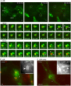
Figure 4. Intracellular fate of the sifA strain in living host cells.
HeLa cells were transfected with LAMP1-GFP (green) and infected with the sifA strain (A, B) or completed sifA strain (C) constitutively expressing mCherry (red). A) Live cell imaging of infected cells was performed over 10 h and time lapse series with intervals of 5 min were obtained. The stills correspond to a movie shown as S26 Movie. Detailed micrographs of two clusters of bacteria are shown that lost the integrity of the SCV about 7 h 35 min (SCV1) or 8 h 15 min (SCV2) p.i. B) A sifA-infected HeLa cell at 7 h p.i. showing intracellular microcolonies enclosed by a continuous LAMP1-GFP-positive SCV but lacking tubular compartments. C) Restoration of tubular LAMP1-GFP-positive compartments by sifA complemented with psifA. Inserts show the GFP channel. The time point of imaging is indicated as hh:mn:ss. Scale bars, 10 µm and 1 µm in overview and detail micrographs in A), respectively; 10 µm in B) and C).
Figure 5
Figure 5. Intracellular fates of sseF- or sseG-deficient strains in living host cells.
Infection was performed as described for Fig. 4 with strains deficient in sseF (A), sseG (B), or the complemented sseF strain (C). Time lapse series were generated at 6 to 7 h p.i. as indicated. Note the appearance of thin, highly dynamic LAMP1-GFP-positive tubules in sseF- or sseG-infected cells. Scale bars, 10 µm and 2 µm in overview and detail micrographs, respectively. The stills correspond to a movie shown as S27 Movie.
Figure 6
Figure 6. Intracellular fate of the pipB2 strain in living host cells.
The experiment was performed as described for Fig. 4 with a strain deficient in pipB2 (A), or the complemented pipB2 strain (B). Time lapse series were generated at 7 or 8 h p.i. as indicated. Note the appearance of bulky, non-dynamic LAMP1-GFP-positive tubules in pipB2-infected cells. Scale bars, 10 µm and 2 µm in overview and detail micrographs, respectively. The stills correspond to a time lapse series shown as S28 Movie.
Figure 7
Figure 7. Access of endocytic cargo to SIF.
HeLa cells were transfected with a vector for expression of LAMP1-GFP (green) and infected with WT, sseF or pipB2 strains expressing mCherry (red) as indicated. At 4 h p.i., the cells were pulse-chased with Dextran-Alexa568 (red) for 3 h. Live cell imaging was performed at 7–8 h p.i. Note the appearance of dynamic extended tubular structures double positive for LAMP1-GFP and Dextran-Alexa568 in cells infected with WT or sseF strains, as well as double positive bulky structures in pipB2-infected cells. Scale bars, 2 µm.
Figure 8
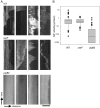
Figure 8. Dynamics of SIF in HeLa cells infected with Salmonella WT and various mutant strains.
HeLa cells were transfected with the LAMP1-GFP construct and infected with Salmonella WT, sseF-deficient or pipB2-deficient strains. At 4–6 h p.i., time lapse series of infected cells were recorded. SIF were identified and the length of individual SIF was determined in 100 images of the time lapse series, representing 500 msec time delay between each frame. Kymographs SIF were generated using the EMBL Image J software. A) Four representative Kymographs are shown each for cells infected with Salmonella WT, sseF, or pipB2 strains as indicated. Scale bar, 5 µm. B) The velocities of SIF extension or collapse were calculated from kymographs generated for cells 5 h p.i., combining 100 events of extension and contraction per strain. The data are displayed as box and whisker plot.
Figure 9
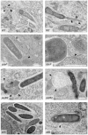
Figure 9. Ultrastructural features of tubular membrane compartments induced by Salmonella WT and various mutant strains.
HeLa cells were infected with WT Salmonella or mutant strains lacking SPI2 effectors as indicated. The cells were processed for TEM analyses at 10 h p.i. Representative cells for each infecting strain are shown. Arrowheads indicate the membranes of tubular compartments. Scale bars, 500 nm.

Similar articles

Cited by

References

    1. Haraga A, Ohlson MB, Miller SI (2008) Salmonellae interplay with host cells. Nat Rev Microbiol 6:53–66. - PubMed
    1. Kuhle V, Hensel M (2004) Cellular microbiology of intracellular Salmonella enterica: functions of the type III secretion system encoded by Salmonella pathogenicity island 2. Cell Mol Life Sci 61:2812–2826. - PubMed
    1. Figueira R, Holden DW (2012) Functions of the Salmonella pathogenicity island 2 (SPI-2) type III secretion system effectors. Microbiology 158:1147–1161. - PubMed
    1. Schroeder N, Mota LJ, Meresse S (2011) Salmonella-induced tubular networks. Trends Microbiol 19:268–277. - PubMed
    1. Garcia-del Portillo F, Zwick MB, Leung KY, Finlay BB (1993) Salmonella induces the formation of filamentous structures containing lysosomal membrane glycoproteins in epithelial cells. Proc Natl Acad Sci U S A 90:10544–10548. - PMC - PubMed

Publication types

Grants and funding

This work was supported by the Deutsche Forschungsgemeinschaft as project grant HE1964/17-1 and continued by grant HE1964/18-1. The funders had no role in study design, data collection and analysis, decision to publish, or preparation of the manuscript.

LinkOut - more resources