. 2014 Dec 15;28(24):2726-38.
doi: 10.1101/gad.246546.114.

The scaffold protein WRAP53β orchestrates the ubiquitin response critical for DNA double-strand break repair

Affiliations

The scaffold protein WRAP53β orchestrates the ubiquitin response critical for DNA double-strand break repair

Sofia Henriksson et al. Genes Dev. .

Abstract

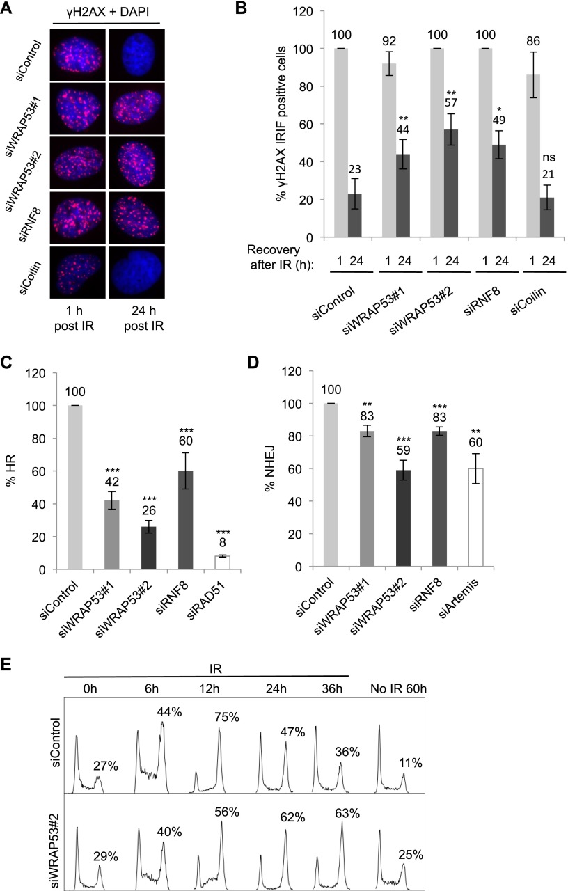
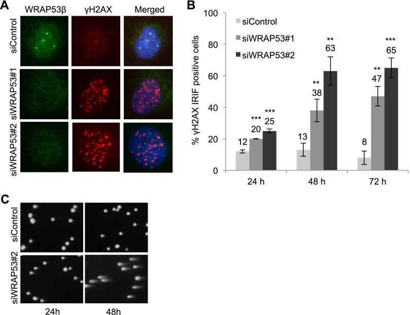
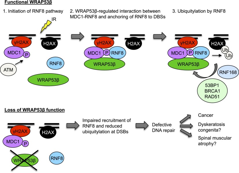
The WD40 domain-containing protein WRAP53β (WD40 encoding RNA antisense to p53; also referred to as WDR79/TCAB1) controls trafficking of splicing factors and the telomerase enzyme to Cajal bodies, and its functional loss has been linked to carcinogenesis, premature aging, and neurodegeneration. Here, we identify WRAP53β as an essential regulator of DNA double-strand break (DSB) repair. WRAP53β rapidly localizes to DSBs in an ATM-, H2AX-, and MDC1-dependent manner. We show that WRAP53β targets the E3 ligase RNF8 to DNA lesions by facilitating the interaction between RNF8 and its upstream partner, MDC1, in response to DNA damage. Simultaneous binding of MDC1 and RNF8 to the highly conserved WD40 scaffold domain of WRAP53β facilitates their interaction and accumulation of RNF8 at DSBs. In this manner, WRAP53β controls proper ubiquitylation at DNA damage sites and the downstream assembly of 53BP1, BRCA1, and RAD51. Furthermore, we reveal that knockdown of WRAP53β impairs DSB repair by both homologous recombination (HR) and nonhomologous end-joining (NHEJ), causes accumulation of spontaneous DNA breaks, and delays recovery from radiation-induced cell cycle arrest. Our findings establish WRAP53β as a novel regulator of DSB repair by providing a scaffold for DNA repair factors.

Keywords: DNA repair; MDC1; RNF8 E3 ligase; WD40 scaffold; WRAP53β; ubiquitin.


Figures

Figure 1.
Figure 1.
WRAP53β accumulates at sites of DNA damage in an ATM/H2AX/MDC1-dependent manner. (A) U2OS cells were microirradiated, fixed 5 min later, and immunostained for WRAP53β and γH2AX, a marker for DNA DSBs. Nuclei were stained with DAPI in all immunofluorescence experiments. (B) U2OS cells were treated with siControl or two different WRAP53β targeting oligonucleotides (siWRAP53#1 and siWRAP53#2) for 48 h, irradiated (6 Gy, 1-h recovery) or left untreated, fixed after pre-extraction with cytoskeleton (CSK) buffer, and immunostained for WRAP53β and γH2AX. (C) U2OS cells were irradiated (6 Gy), fixed, and immunostained for WRAP53β at the indicated time points. Quantification is given as the percentage of the 100 cells counted in each experiment whose nuclei contained WRAP53β IRIF. (D) U2OS cells were treated with the inhibitors or siRNAs, as indicated, for 6 h or 48 h, respectively; irradiated (6 Gy, 1-h recovery); fixed after pre-extraction with CSK buffer; and immunostained for WRAP53β and γH2AX. (E) Quantification of the results in D, as the percentage of the 100 cells counted in each experiment whose nuclei contained WRAP53β IRIF. The error bars depict the SEM; n = 3; (***) P < 0.001 as determined by Student’s t-test. (F) ChIP assay showing the recruitment of WRAP53β to the I-PpoI-induced DSB at chromosome 1 in MCF7 cells stably expressing ddI-PpoI. The time indicated is hours after the addition of 4-OHT. The I-PpoI cleavage site on chromosome 1 is located at distance 0. Cells were cultivated in medium containing 0.1% FBS for 24 h before DSB induction. Data are shown as the mean of two independent experiments. The Y-axis displays the fold change in relative occupancy normalized to the control.
Figure 2.
Figure 2.
WRAP53β promotes recruitment of repair proteins to DSBs. (A) U2OS cells were transfected with siControl or siWRAP53#2 oligonucleotides for 24 h, exposed to IR (6 Gy) or left untreated, and, 1 h later, immunostained for γH2AX, MDC1, BRCA1, 53BP1, and RAD51. (B) U2OS cells treated as in A and then immunostained for RNF168 and conjugated ubiquitin (with the FK2 antibody). In the case of GFP-RNF8 staining, following treatment with oligonucleotides for 24 h, the cells were transiently transfected with the GFP-RNF8 plasmid for 8 h, exposed to IR (6 Gy), allowed to recover for 1 h, and then fixed and analyzed. (C) Quantification of the results in A and B as the percentage of 200 cells counted in each experiment whose nuclei contained IRIF. In the case of GFP-RNF8, only successfully transfected cells were counted. (D) U2OS cells were treated with the siRNAs indicated for 24 h, irradiated (6 Gy), allowed to recover for 1 h, and then subjected to Western blotting for WRAP53β, H2AX, and β-actin. The error bars depict the SEM. n = 3; (**) P < 0.01; (***) P < 0.001, as determined by Student’s t-test.
Figure 3.
Figure 3.
WRAP53β binds MDC1 and RNF8 via their FHA domains. (A) U2OS cells were either left untreated or irradiated with 6 Gy of IR, and, 30 min, later immunoprecipitation of WRAP53β was performed, followed by immunoblotting of WRAP53β, MDC1, GFP-RNF8, and β-actin. (B) U2OS cells were transfected with the indicated HA-MDC1 constructs for 16 h and irradiated with 2 Gy, and, 30 min later, immunoprecipitation of WRAP53β was performed, followed by immunoblotting of WRAP53β and HA-MDC1. (C) Schematic illustration of RNF8 deletion constructs. (D) U2OS cells were transiently transfected with EGFP-RNF8 plasmids, HA-MDC1, and Flag-WRAP53β for 16 h; irradiated; and subjected to immunoprecipitation of GFP followed by immunoblotting for GFP-RNF8, Flag-WRAP53β, and HA-MDC1. (HC) Heavy chain of the antibody. U2OS (E) and H1299 (F) cells were transiently transfected with Flag-RNF8 plasmids, HA-MDC1, and EGFP-WRAP53β for 16 h; irradiated; and subjected to Flag immunoprecipitation followed by immunoblotting for the indicated proteins. (G) Schematic illustration of the domain architecture of MDC1 and RNF8, where black lines mark WRAP53β- and MDC1-binding sites. Numbers indicate amino acids.
Figure 4.
Figure 4.
WRAP53β facilitates MDC1–RNF8 interaction through its WD40 domain. (A) U2OS cells were treated with the siRNAs indicated for 48 h and with GFP-RNF8 for 24 h (all samples), irradiated with 6 Gy, and, 30-min later, subjected to immunoprecipitation of WRAP53β followed by immunoblotting of WRAP53β, MDC1, RNF8, γH2AX, and β-actin. (B) Immunoprecipitation of MDC1 in irradiated (6 Gy, 15-min recovery) U2OS cells treated with the siRNA indicated for 48 h or ATM inhibitor (ATMi) for 24 h. All samples were transfected with GFP-RNF8 for 16 h. (C) U2OS cells were treated with the siRNAs indicated for 48 h or ATM inhibitor for 16 h, irradiated with 6 Gy, allowed to recover for 15 min, and then subjected to Western blotting of MDC1, WRAP53β, γH2AX, and β-actin. (D) Schematic illustration of EGFP-tagged deletion constructs of WRAP53β. (E) U2OS cells were transiently transfected with the indicated EGFP-WRAP53β plasmids and Flag-RNF8 for 16 h, irradiated, and subjected to GFP immunoprecipitation followed by immunoblotting for MDC1, Flag-RNF8, and GFP-WRAP53β. (HC) Heavy chain of the antibody. (F) U2OS cells were transfected with siControl or siWRAP53#2 oligonucleotides for 8 h followed by transfection of EGFP-Empty or EGFP-WRAP53β WD40 (1–7) for 16 h, exposed to IR (6 Gy), and, after 1 h, immunostained for 53BP1 followed by quantification of the results. The graph in A shows the percentage of 100 GFP transfected cells in each experiment whose nuclei were 53BP1-positive. The error bars depict the SEM. n = 3; (*) P < 0.05, as determined by Student’s t-test. (G) U2OS cells were transiently transfected with the indicated EGFP-WRAP53β plasmids, HA-MDC1, and Flag-RNF8 for 16 h; irradiated; and subjected to immunoprecipitation of GFP followed by immunoblotting for HA-MDC1, Flag-RNF8, and GFP-WRAP53β. (H) Schematic illustration of how WRAP53β scaffolds the MDC1–RNF8 complex. Upon DNA damage, WRAP53β binds MDC1 and RNF8 simultaneously via its WD40 domain and facilitates their interaction.
Figure 5.
Figure 5.
WRAP53β promotes HR and NHEJ. (A) U2OS cells were treated with the siRNAs indicated for 24 h, exposed to 6 Gy of IR, fixed 1 h or 24 h later, and immunostained for γH2AX. (B) Quantification of the results in A showing the percentage of nuclei containing >10 γH2AX foci (n = 200). (C,D) HR (C) and NHEJ (D) efficiency following treatment of the cells with the siRNA indicated for 48 h. DR-GFP (HR) and EJ5-GFP (NHEJ) reporter systems were used in the FACS analysis, with siRAD51 and siArtemis as positive controls. (E) Cells were transfected with siRNA for 24 h, exposed to IR (3 Gy), harvested at the time points indicated, and subjected to flow cytometry. Nonirradiated cells were treated with siRNA alone for 60 h. The error bars depict the SEM. n = 3; (*) P < 0.05; (**) P < 0.01; (***) P < 0.001, as determined by Student’s t-test.
Figure 6.
Figure 6.
WRAP53β protects cells against accumulation of spontaneous DNA damage. (A) U2OS cells were treated with siControl or two different siWRAP53 oligonucleotides (siWRAP53#1 and siWRAP53#2) for 24 h, fixed, and immunostained for WRAP53β and γH2AX. (B) The percentage of nuclei in A containing >10 γH2AX foci was quantified in the 200 cells counted for each experiment. (C) After treating U2OS with siWRAP53#2 or siControl for 24 h or 48 h, DNA damage was assessed by the alkaline comet assay. The error bars depict the SEM. n = 3; (**) P < 0.01; (***) P < 0.001, as determined by Student’s t-test.
Figure 7.
Figure 7.
Schematic model of WRAP53β function in the DDR pathway. (Step 1) In response to IR, γH2AX and MDC1 accumulate at DSBs independently of WRAP53β. ATM-mediated phosphorylation of MDC1 makes MDC1 competent to bind RNF8. However, RNF8 is not yet localized at DSBs. (Step 2) WRAP53β is recruited to sites of DNA damage in an ATM-, H2AX-, and MDC1-dependent manner. Simultaneous binding of MDC1 and RNF8 to WRAP53β facilitates their direct interaction and retention of RNF8 at DSBs. (Step 3) Once assembled at DSBs, RNF8 catalyzes ubiquitylation of H2AX. Ubiquitylation at DSBs enables recruitment and accumulation of 53BP1, BRCA1, and RAD51 and subsequent DNA repair.

Similar articles

Cited by

References

    1. Adamson B, Smogorzewska A, Sigoillot FD, King RW, Elledge SJ. 2012. A genome-wide homologous recombination screen identifies the RNA-binding protein RBMX as a component of the DNA-damage response. Nat Cell Biol 14: 318–328. - PMC - PubMed
    1. Coster G, Goldberg M. 2010. The cellular response to DNA damage: a focus on MDC1 and its interacting proteins. Nucleus 1: 166–178. - PMC - PubMed
    1. d’Adda di Fagagna F, Teo SH, Jackson SP. 2004. Functional links between telomeres and proteins of the DNA-damage response. Genes Dev 18: 1781–1799. - PubMed
    1. Doil C, Mailand N, Bekker-Jensen S, Menard P, Larsen DH, Pepperkok R, Ellenberg J, Panier S, Durocher D, Bartek J, et al. . 2009. RNF168 binds and amplifies ubiquitin conjugates on damaged chromosomes to allow accumulation of repair proteins. Cell 136: 435–446. - PubMed
    1. Falck J, Coates J, Jackson SP. 2005. Conserved modes of recruitment of ATM, ATR and DNA-PKcs to sites of DNA damage. Nature 434: 605–611. - PubMed

Publication types

MeSH terms