Enhanced skeletal muscle expression of extracellular superoxide dismutase mitigates streptozotocin-induced diabetic cardiomyopathy by reducing oxidative stress and aberrant cell signaling
- PMID: 25504759
- PMCID: PMC4445759
- DOI: 10.1161/CIRCHEARTFAILURE.114.001540
Enhanced skeletal muscle expression of extracellular superoxide dismutase mitigates streptozotocin-induced diabetic cardiomyopathy by reducing oxidative stress and aberrant cell signaling
Abstract
Background: Exercise training enhances extracellular superoxide dismutase (EcSOD) expression in skeletal muscle and elicits positive health outcomes in individuals with diabetes mellitus. The goal of this study was to determine if enhanced skeletal muscle expression of EcSOD is sufficient to mitigate streptozotocin-induced diabetic cardiomyopathy.
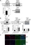
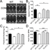
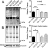
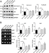
Methods and results: Exercise training promotes EcSOD expression in skeletal muscle and provides protection against diabetic cardiomyopathy; however, it is not known if enhanced expression of EcSOD in skeletal muscle plays a functional role in this protection. Here, we show that skeletal muscle-specific EcSOD transgenic mice are protected from cardiac hypertrophy, fibrosis, and dysfunction under the condition of type 1 diabetes mellitus induced by streptozotocin injection. We also show that both exercise training and muscle-specific transgenic expression of EcSOD result in elevated EcSOD protein in the blood and heart without increased transcription in the heart, suggesting that enhanced expression of EcSOD from skeletal muscle redistributes to the heart. Importantly, cardiac tissue in transgenic mice displayed significantly reduced oxidative stress, aberrant cell signaling, and inflammatory cytokine expression compared with wild-type mice under the same diabetic condition.
Conclusions: Enhanced expression of EcSOD in skeletal muscle is sufficient to mitigate streptozotocin-induced diabetic cardiomyopathy through attenuation of oxidative stress, aberrant cell signaling, and inflammation, suggesting a cross-organ mechanism by which exercise training improves cardiac function in diabetes mellitus.
Keywords: antioxidants; cardiomyocyte; diabetic cardiomyopathies; exercise; hypertrophy; oxidative stress.
© 2014 American Heart Association, Inc.
Figures


Similar articles
-
Extracellular superoxide dismutase ameliorates skeletal muscle abnormalities, cachexia, and exercise intolerance in mice with congestive heart failure.Circ Heart Fail. 2014 May;7(3):519-30. doi: 10.1161/CIRCHEARTFAILURE.113.000841. Epub 2014 Feb 12. Circ Heart Fail. 2014. PMID: 24523418 Free PMC article.
-
Enhanced phosphoinositide 3-kinase(p110α) activity prevents diabetes-induced cardiomyopathy and superoxide generation in a mouse model of diabetes.Diabetologia. 2012 Dec;55(12):3369-81. doi: 10.1007/s00125-012-2720-0. Epub 2012 Sep 22. Diabetologia. 2012. PMID: 23001375
-
Muscle-derived IL-1β regulates EcSOD expression via the NBR1-p62-Nrf2 pathway in muscle during cancer cachexia.J Physiol. 2024 Sep;602(17):4215-4235. doi: 10.1113/JP286460. Epub 2024 Aug 21. J Physiol. 2024. PMID: 39167700
-
Therapeutic targeting of oxidative stress with coenzyme Q10 counteracts exaggerated diabetic cardiomyopathy in a mouse model of diabetes with diminished PI3K(p110α) signaling.Free Radic Biol Med. 2015 Oct;87:137-47. doi: 10.1016/j.freeradbiomed.2015.04.028. Epub 2015 Apr 30. Free Radic Biol Med. 2015. PMID: 25937176
-
Extracellular superoxide dismutase, a molecular transducer of health benefits of exercise.Redox Biol. 2020 May;32:101508. doi: 10.1016/j.redox.2020.101508. Epub 2020 Mar 19. Redox Biol. 2020. PMID: 32220789 Free PMC article. Review.
Cited by
-
Tumor necrosis factor-α decreases EC-SOD expression through DNA methylation.J Clin Biochem Nutr. 2017 May;60(3):169-175. doi: 10.3164/jcbn.16-111. Epub 2017 Apr 7. J Clin Biochem Nutr. 2017. PMID: 28584398 Free PMC article.
-
Muscle-derived extracellular superoxide dismutase inhibits endothelial activation and protects against multiple organ dysfunction syndrome in mice.Free Radic Biol Med. 2017 Dec;113:212-223. doi: 10.1016/j.freeradbiomed.2017.09.029. Epub 2017 Oct 2. Free Radic Biol Med. 2017. PMID: 28982599 Free PMC article.
-
Antioxidant Properties of Whole Body Periodic Acceleration (pGz).PLoS One. 2015 Jul 2;10(7):e0131392. doi: 10.1371/journal.pone.0131392. eCollection 2015. PLoS One. 2015. PMID: 26133377 Free PMC article.
-
YiXin-Shu, a ShengMai-San-based traditional Chinese medicine formula, attenuates myocardial ischemia/reperfusion injury by suppressing mitochondrial mediated apoptosis and upregulating liver-X-receptor α.Sci Rep. 2016 Mar 11;6:23025. doi: 10.1038/srep23025. Sci Rep. 2016. PMID: 26964694 Free PMC article.
-
Exendin-4 promotes extracellular-superoxide dismutase expression in A549 cells through DNA demethylation.J Clin Biochem Nutr. 2016 Jan;58(1):34-9. doi: 10.3164/jcbn.15-16. Epub 2015 Nov 20. J Clin Biochem Nutr. 2016. PMID: 26798195 Free PMC article.
References
-
- Wild S, Roglic G, Green A, Sicree R, King H. Global Prevalence of Diabetes: Estimates for the year 2000 and projections for 2030. Diabetes Care. 2004;27:1047–1053. - PubMed
-
- Hayat Sa, Patel B, Khattar RS, Malik Ra. Diabetic cardiomyopathy: mechanisms, diagnosis and treatment. Clin Sci (Lond) 2004;107:539–557. - PubMed
-
- Role of cardiovascular risk factors in prevention and treatment of macrovascular disease in diabetes. American Diabetes Association. Diabetes Care. 1989;12:573–579. - PubMed
-
- Rubler S, Dlugash J, Yuceoglu YZ, Kumral T, Branwood AW, Grishman A. New type of cardiomyopathy associated with diabetic glomerulosclerosis. Am J Cardiol. 1972;30:595–602. - PubMed
-
- Choi KM, Zhong Y, Hoit BD, Grupp IL, Hahn H, Dilly KW, Guatimosim S, Lederer WJ, Matlib MA. Defective intracellular Ca(2+) signaling contributes to cardiomyopathy in Type 1 diabetic rats. Am J Physiol Heart Circ Physiol. 2002;283:H1398–H1408. - PubMed
Publication types
MeSH terms
Substances
Grants and funding
LinkOut - more resources
Full Text Sources
Medical
Molecular Biology Databases

