. 2015 Jan;125(1):337-49.
doi: 10.1172/JCI77172. Epub 2014 Dec 8.

Glucokinase activity in the arcuate nucleus regulates glucose intake

Glucokinase activity in the arcuate nucleus regulates glucose intake

Syed Hussain et al. J Clin Invest. 2015 Jan.

Abstract

The brain relies on a constant supply of glucose, its primary fuel, for optimal function. A taste-independent mechanism within the CNS that promotes glucose delivery to the brain has been postulated to maintain glucose homeostasis; however, evidence for such a mechanism is lacking. Here, we determined that glucokinase activity within the hypothalamic arcuate nucleus is involved in regulation of dietary glucose intake. In fasted rats, glucokinase activity was specifically increased in the arcuate nucleus but not other regions of the hypothalamus. Moreover, pharmacologic and genetic activation of glucokinase in the arcuate nucleus of rodent models increased glucose ingestion, while decreased arcuate nucleus glucokinase activity reduced glucose intake. Pharmacologic targeting of potential downstream glucokinase effectors revealed that ATP-sensitive potassium channel and P/Q calcium channel activity are required for glucokinase-mediated glucose intake. Additionally, altered glucokinase activity affected release of the orexigenic neurotransmitter neuropeptide Y in response to glucose. Together, our results suggest that glucokinase activity in the arcuate nucleus specifically regulates glucose intake and that appetite for glucose is an important driver of overall food intake. Arcuate nucleus glucokinase activation may represent a CNS mechanism that underlies the oft-described phenomena of the "sweet tooth" and carbohydrate craving.


Figures
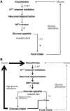
Figure 6
Figure 6. Schematic representation of the proposed mechanism by which arcuate nucleus glucokinase regulates glucose intake.
(A) Representation when only one diet is available. When mixed food is ingested, glucose is released and enters the arcuate nucleus. Metabolism of the glucose by glucokinase results in closure of the KATP channel and increased NPY release. The increase in NPY stimulates further food intake. The normal satiety process would terminate the feeding response. (B) Representation when a rich source of glucose is available in addition to normal diet. If pure glucose or a food high in glucose is available, the positive feedback loop will be more active. This will result in a preferential intake of the food with a high glucose concentration and reduced intake of the mixed diet.
Figure 5
Figure 5. Effect of increased arcuate nucleus glucokinase on NPY and α-MSH and effect of NPY antagonist and calcium channel blockade on glucokinase activator–stimulated glucose intake.
(A) Hypothalamic Ampk, Acc, and Fas expression in iARC-GFP and iARC-GK rats (n = 9). (B) Hypothalamic AMPK activity in hypothalami from iARC-GFP and iARC-GK rats (n = 10). (C) 2% w/v glucose solution intake 1 hour after intra-arcuate injection of vehicle ω-agatoxin-IVA (Agatox) or nifedipine (Nifed) and subsequent injection of CpdA or control in rats (n = 15). (D) Glucose-induced NPY release from hypothalamic slices of iARC-GFP or iARC-GK rats (n = 7 iARC-GFP; n = 8 iARC-GK). (E) Glucose-induced α-MSH release from hypothalamic slices of iARC-GFP or iARC-GK rats (n = 7 iARC-GFP; n = 8 iARC-GK). (F) NPY release from rat hypothalamic slices, following treatment with CpdA, glibenclamide, or diazoxide (n = 10–12). (G) α-MSH release from rat hypothalamic slices, following treatment with CpdA, glibenclamide, or diazoxide (n = 6–9). (H) NPY release from rat hypothalamic slices, following treatment with CpdA or diazoxide alone and in combination (CpdA+Dia) (n = 10–12). (I) 2% w/v glucose solution intake 1 hour after intra-peritoneal injection of vehicle CGP-71683 (CGP) or BMS-193885 (BMS) and subsequent intra-arcuate injection of CpdA or control in rats (n = 14). Data are presented as mean ± SEM. *P < 0.05 versus corresponding control values; #P < 0.05 versus control values from 8 mM and 15 mM glucose; **P < 0.00001 versus corresponding CpdA-injected group; †††P < 0.0001 versus vehicle CpdA-injected group; ***P < 0.0001.
Figure 4
Figure 4. Effect of activating and inhibiting arcuate nucleus KATP channels on food intake and glucose appetite.
(A) Chow intake relative to that of control-treated rats 4 hours after intra-arcuate injection of 2 nmol glibenclamide (Glib) or vehicle (control) in rats (n = 12). (B) 2% w/v glucose solution intake relative to that of control-treated rats 4 hours after intra-arcuate injection of 2 nmol glibenclamide or vehicle in rats (n = 11). (C) Chow intake and (D) 2% w/v glucose intake relative to that of control-treated rats 4 hours after intra-arcuate injection of 2 nmol glibenclamide or vehicle in rats with ad libitum access to 2% w/v glucose and normal chow (n = 10). (E) Chow intake relative to that of control-treated rats 1 hour after intra-arcuate injection of 1 nmol diazoxide (Dia) or vehicle in rats (n = 9). (F) 2% w/v glucose intake relative to that of control-treated rats 1 hour after intra-arcuate injection of 1 nmol diazoxide or vehicle in rats (n = 9). (G) Chow intake and (H) glucose intake relative to that of control-treated rats 1 hour after intra-arcuate injection of 1 nmol diazoxide or vehicle in rats with ad libitum access to 2% w/v glucose and normal chow (n = 9). (I) 2% w/v glucose solution intake 1 hour after intra-arcuate injection of vehicle CpdA or 1 nmol diazoxide administered alone (control) or followed by injection of 0.5 nmol CpdA in rats (n = 10). Data are presented as mean ± SEM. *P < 0.05; ***P < 0.001 versus control; †††P < 0.001 versus CpdA vehicle injected.
Figure 3
Figure 3. Effect of decreased arcuate nucleus glucokinase activity on food and glucose intake.
(A) Glucokinase (GK) activity in Hep G2 cell lysates after transfection with either rAAV-GFP or rAAV-ASGK (n = 6). (B) Glucokinase activity in arcuate nucleus, VMN, and PVN of male Wistar rats following intra-arcuate injection of either rAAV-GFP (iARC-GFP) or rAAV-ASGK (iARC-ASGK). (C) Cumulative food intake and (D) weight changes in iARC-GFP (black diamonds) and iARC-ASGK (white diamonds) rats after recovery from surgery (n = 11). (E) 2% w/v glucose solution intake, (F) food intake, and (G) energy intake in iARC-GFP (black diamonds) and iARC-ASGK (white diamonds) rats during a 24-hour feeding study, with ad libitum access to 2% w/v glucose solution and normal chow intake (n = 11). (H) 10% w/v glucose, (I) food intake with normal chow, (J) total energy intake, and (K) weight changes in iARC-GFP (black diamonds) and iARC-ASGK (white diamonds) rats, with ad libitum access to normal chow diet and 10% w/v glucose given 4 weeks after rAAV microinjection (n = 11). Data presented as mean ± SEM *P < 0.05, ***P < 0.001 versus corresponding control values.
Figure 2
Figure 2. Effect of increased arcuate nucleus glucokinase activity on glucose intake.
(A) 2% w/v glucose solution intake 1 hour after intra-arcuate injection of CpdA or control in rats (n = 7). Twenty-four hour intake of (B) 2% w/v and (C) 10% w/v glucose and fructose solutions in iARC-GFP (white bars) and iARC-GK rats (black bars) (n = 8). (D) 2% w/v glucose solution intake and (E) food intake 1 hour after intra-arcuate injection of CpdA or vehicle in rats with ad libitum access to 2% w/v glucose solution and normal chow (n = 7). (F) 2% w/v glucose solution intake, (G) food intake, and (H) energy intake in iARC-GFP (black circles) and iARC-GK (white squares) rats during a 24-hour feeding study, with ad libitum access to 2% w/v glucose solution and normal chow intake (n = 7). (I) Food intake in iARC-GFP (black circles) and iARC-GK (white squares) rats (n = 7) during a 24-hour feeding study, with ad libitum access to normal chow diet only. (J) 10% w/v glucose, (K) food intake with normal chow, (L) total energy intake, and (M) weight changes in iARC-GFP (black circles) and iARC-GK (white squares) rats with ad libitum access to normal chow diet and 10% w/v glucose given 4 weeks after rAAV microinjection (n = 8). Data are presented as mean ± SEM *P < 0.05, **P < 0.01, ***P < 0.001.
Figure 1
Figure 1. Effect increased arcuate nucleus glucokinase activity on food intake.
(A) Arcuate nucleus glucokinase activity in ad libitum chow–fed and 24-hour–fasted male Wistar rats (n = 8). (B) Normal chow intake 1 hour after intra-arcuate injection of 0.5 nmol CpdA or vehicle (control) in male Wistar rats (n = 10). (C) Glucokinase activity in HEK293 cell lysates after transfection with either rAAV-GFP or rAAV-GK (n = 6). (D) Glucokinase activity in arcuate nucleus (ARC), VMN, and PVN of male Wistar rats following intra-arcuate injection of either rAAV-GFP (iARC-GFP, white bars) or rAAV-GK (iARC-GK, black bars) (n = 8–11). (E) Cumulative food intake and (F) weight changes in iARC-GFP (black squares) and iARC-GK (white squares) rats after recovery from surgery (n = 12 iARC-GFP; n = 15 iARC-GK). Data are presented as mean ± SEM. *P < 0.05, **P < 0.01, ***P < 0.001 versus corresponding control values; #P < 0.0001 versus all other groups.

Similar articles

Cited by

References

    1. Mayer J. Glucostatic mechanism of regulation of food intake. N Engl J Med. 1953;249(1):13–16. doi: 10.1056/NEJM195307022490104. - DOI - PubMed
    1. Thompson DA, Campbell RG. Hunger in humans induced by 2-deoxy-D-glucose: glucoprivic control of taste preference and food intake. Science. 1977;198(4321):1065–1068. doi: 10.1126/science.929188. - DOI - PubMed
    1. Sclafani A. Oral and postoral determinants of food reward. Physiol Behav. 2004;81(5):773–779. doi: 10.1016/j.physbeh.2004.04.031. - DOI - PubMed
    1. Jacobs HL. Some physical, metabolic, and sensory components in the appetite for glucose. Am J Physiol. 1962;203:1043–1054. - PubMed
    1. Sclafani A, Ackroff K. Role of gut nutrient sensing in stimulating appetite and conditioning food preferences. Am J Physiol Regul Integr Comp Physiol. 2012;302(10):R1119–R1133. doi: 10.1152/ajpregu.00038.2012. - DOI - PMC - PubMed

Publication types