. 2015 Jun;22(6):1035-46.
doi: 10.1038/cdd.2014.194. Epub 2014 Dec 5.

CHCHD2 inhibits apoptosis by interacting with Bcl-x L to regulate Bax activation

Affiliations

CHCHD2 inhibits apoptosis by interacting with Bcl-x L to regulate Bax activation

Y Liu et al. Cell Death Differ. 2015 Jun.

Abstract

Mitochondrial outer membrane permeabilization (MOMP) is a critical control point during apoptosis that results in the release of pro-apoptotic mitochondrial contents such as cytochrome c. MOMP is largely controlled by Bcl-2 family proteins such as Bax, which under various apoptotic stresses becomes activated and oligomerizes on the outer mitochondrial membrane. Bax oligomerization helps promote the diffusion of the mitochondrial contents into the cytoplasm activating the caspase cascade. In turn, Bax is regulated primarily by anti-apoptotic Bcl-2 proteins including Bcl-xL, which was recently shown to prevent Bax from accumulating at the mitochondria. However, the exact mechanisms by which Bcl-xL regulates Bax and thereby MOMP remain partially understood. In this study, we show that the small CHCH-domain-containing protein CHCHD2 binds to Bcl-xL and inhibits the mitochondrial accumulation and oligomerization of Bax. Our data show that in response to apoptotic stimuli, mitochondrial CHCHD2 decreases prior to MOMP. Furthermore, when CHCHD2 is absent from the mitochondria, the ability of Bcl-xL to inhibit Bax activation and to prevent apoptosis is attenuated, which results in increases in Bax oligomerization, MOMP and apoptosis. Collectively, our findings establish CHCHD2, a previously uncharacterized small mitochondrial protein with no known homology to the Bcl-2 family, as one of the negative regulators of mitochondria-mediated apoptosis.


Figures
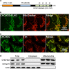
Figure 1
Figure 1
CHCHD2 is a CHCH domain-containing protein that localizes to the mitochondria. (a) Schematic representation of the CHCHD2 protein with putative mitochondrial-targeting signal (MTS), predicted transmembrane domain (TM) and CHCH (coiled-coil-helix coiled-coil helix) domain shown. The CHCH domain is comprised of four evenly spaced cysteines with the positions indicated. (b) C-terminally tagged CHCHD2–FLAG localizes to the mitochondria in U2OS cells as indicated by co-staining with MitoTracker dye. CHCHD2–FLAG was detected by immunofluorescence with anti-FLAG antibody. (c) Mitochondrial localization of endogenous CHCHD2 in U2OS cells detected by immunofluorescence staining using anti-CHCHD2 antibody as demonstrated by co-staining with the mitochondrial inner membrane protein Tim23 (Tim). (d) Subcellular fractionation of untreated U2OS cells using a differential detergent method shows CHCHD2 present in the mitochondria-enriched heavy membrane fraction (three replicates are shown). Mitochondrial hsp70 (mtHsp70) is included as a marker for the mitochondrial fraction, and actin is included as a marker for the cytoplasmic fraction
Figure 2
Figure 2
CHCHD2 is important for cell survival. (ae) CHCHD2 knockdown sensitizes U2OS cells to apoptosis induced by a variety of agents. Cells were treated with the indicated siRNA targeting CHCHD2 for 48 h, after which the cells were treated with the following apoptotic stimuli: (b) UV (6 mJ/cm2 for 24 h), (c) cisplatin (10 μg/ml for 18 h), (d) doxorubicin (7.5 μM for 18 h) and (e) staurosporine (200 nM for 18 h). The number of cells that survived each treatment was counted in at least 4 random fields of view under each condition. The relative survival is shown as a percentage of the siNS control cells from three independent experimental repeats. (si-#1 and si-#2 target two distinct regions of CHCHD2 mRNA as described in the Experimental Procedures section). Error bars denote the S.E. (f) Overexpression of CHCHD2 protects U2OS cells from apoptosis. Cells were infected for 24 h with control adenovirus expressing GFP (Ad-Ctl) or adenovirus expressing untagged CHCHD2 (Ad-CHCHD2), after which the cells were treated with UV to induce apoptosis (6 mJ/cm2 for 24 h). The number of cells that survived the treatment is shown as a percentage of the Ad-GFP-infected control cells
Figure 3
Figure 3
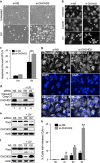
CHCHD2 inhibits mitochondrial apoptosis and MOMP. (a) CHCHD2 knockdown sensitizes U2OS cells to cell death induced by UV (6 mJ/cm2). Phase-contrast images are shown for cells treated for 48 h with nonspecific siRNA (siNS) or CHCHD2 siRNA (si-CHCHD2) with or without UV treatment. (b) CHCHD2 knockdown enhances UV-induced nuclear fragmentation in U2OS cells. Representative DAPI staining is shown for U2OS cells treated for 48 h with siNS or si-CHCHD2 followed by UV (6 mJ/cm2 for 24 h) or cisplatin (10 μg/ml for 24 h) treatment. (c) CHCHD2 knockdown enhances UV-induced phosphatidylserine exposure. U2OS cells were treated with UV (6 mJ/ cm2) for 24 h, followed by Annexin-V-FITC and PI-double staining. The cells were then analyzed by flow cytometry. FITC-positive or FITC–PI-double positive cells were counted as apoptotic cells, and the percentages of apoptotic cells from three independent experiments are shown in the graph. The error bars denote the S.E. (d) Caspase-3 cleavage is enhanced in UV-treated cells upon knockdown of CHCHD2. HeLa cells were transfected with CHCHD2 siRNA (D2) or control siRNA (NS) followed by UV treatment (2.5 mJ/cm2 for 18 h). Cells were then lysed, resolved by SDS-PAGE and probed by western blot for cleaved caspase-3 and CHCHD2. Actin is shown as a loading control. (e) CHCHD2 knockdown increases PARP cleavage in response to apoptotic stimuli. HeLa cells were treated with the indicated siRNA for 48 h and then treated with UV (2.5 mJ/cm2 for 18 h) to induce apoptosis. Actin is shown as a loading control. (f) CHCHD2 overexpression inhibits PARP cleavage in response to apoptotic stimuli. HeLa cells were infected with adenovirus expressing empty vector (Ctl) or CHCHD2 (D2) for 48 h and then treated with UV (2.5 mJ/cm2 for 18 h) to induce apoptosis. Actin is shown as a loading control. (g, h) CHCHD2 knockdown increases the rate of MOMP as indicated by cytochrome c release. U2OS cells were pretreated with the caspase inhibitor QVD to prevent post-MOMP cell death and then treated with the indicated siRNA for 48 h followed by UV irradiation (50 mJ/cm2) for the indicated amount of time. (g) Representative immunofluorescence images are shown for endogenous cytochrome c. (h) Quantification of cells with diffuse cytochrome c staining as a result of UV irradiation at various time points. Cells were counted in at least four random fields of view, and more than 400 cells were counted in total. The graph depicts the percentage of diffusely staining cells of the total cells from three independent experiments. The error bars denote the S.E.
Figure 4
Figure 4
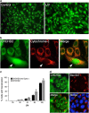
Loss of mitochondrial CHCHD2 correlates with Bax activation. (a) Mitochondrial CHCHD2 is depleted in response to apoptotic stimuli. U2OS cells were pretreated with QVD for 1 h and then treated with UV (6 mJ/cm2) for 24 h where indicated. Endogenous CHCHD2 was detected by immunofluorescence staining. (b) Loss of mitochondrial CHCHD2 occurs prior to cytochrome c release in response to apoptotic stimuli. Immunofluorescence staining of endogenous CHCHD2 (green) and cytochrome c (red) in UV-treated (6 mJ/cm2 for 12 h) U2OS cells shows that in some cells cytochrome c remains in the mitochondria after mitochondrial CHCHD2 is depleted (indicated by the arrow). (c) Loss of mitochondrial CHCHD2 occurs more rapidly than cytochrome c release. U2OS cells were pretreated with the caspase inhibitor QVD (10 μM for 1 h), after which the cells were treated with UV (6 mJ/cm2) to induce apoptosis and then fixed at the indicated time points. Cells were subjected to immunofluorescence staining for endogenous CHCHD2 and cytochrome c. The percentage of cells with diffuse CHCHD2 staining and cytochrome c staining was quantified at each time point and are represented on the graph as the averages of three independent experiments. For each experiment, at least four random fields of view were counted comprising at least 400 cells. Error bars denote the S.E. (d) Bax activation correlates with the loss of mitochondrial CHCHD2 in U2OS cells. Cells were treated with UV (6 mJ/cm2 for 24 h) to induce apoptosis. Immunofluorescence staining was performed to detect endogenous CHCHD2 (green) and activated Bax (6A7 antibody, red). Activated Bax is observed only in cells in which mitochondrial CHCHD2 is depleted. Nuclei were stained with DAPI
Figure 5
Figure 5
Bcl-xL is required for the anti-apoptotic function of CHCHD2. (a, b) CHCHD2 interacts with Bcl-xL. U2OS cells stably expressing nonspecific (sh-NS) or CHCHD2-specific (sh-CHCHD2) shRNA were transfected with FLAG–Bcl-xL for 24 h and then lysed with 1% CHAPS buffer. Lysates containing 1 mg of total protein were immunoprecipitated with antibodies against CHCHD2 (a) or FLAG (b). Samples were analyzed by western blotting with the indicated antibodies. Input loading represents 5% of the total cell lysate used for IP. (c) CHCHD2 enhances, but is not required for, the apoptosis-inhibiting function of Bcl-xL. U2OS cells were treated with siNS or si-CHCHD2 for 48 h, followed by transient transfection with FLAG–Bcl-xL or empty vector for another 24 h and then were treated by UV irradiation (6 mJ/cm2) for 24 h. The cells were then stained with Annexin-V–FITC/PI and analyzed by flow cytometry. The means of the percentages of apoptotic cells from three independent experiments are shown, and the error bars denote the S.E. (d, e) The inhibitory function of CHCHD2 on apoptosis is compromised by knockdown of Bcl-xL. U2OS cells stably expressing transfected CHCHD2 were treated with the indicated siRNA constructs for 48 h followed by UV irradiation treatment (25 mJ/cm2) for 18 h as indicated. Cells were then stained with Annexin-V–FITC/PI and analyzed by flow cytometry. (d) The mean of the percentages of apoptotic cells from three independent experiments are shown, and the error bars denote the S.E. (e) The apoptosis inhibition ratio was calculated based on the apoptosis percentages shown in d. The difference between the percentage of apoptotic cells with CHCHD2 overexpression and the percentage of apoptotic cells with empty vector was divided by the percentage of apoptosis in cells transfected with empty vector for each UV-treated sample. The error bars denote the S.E.
Figure 6
Figure 6
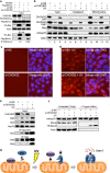
CHCHD2 cooperates with Bcl-xL to regulate Bax translocation, oligomerization and activation. (a) CHCHD2 knockdown reduces Bcl-xL–Bax interaction. U2OS cells stably expressing nonspecific (sh-NS) or CHCHD2-specific (sh-CHCHD2) shRNA were transfected with HA-Bax and FLAG-Bcl-xL for 24 h and then were lysed with 1% CHAPS buffer. Cell lysates containing 1 mg of total protein were immunoprecipitated with HA-specific antibody. Samples were analyzed by western blotting with the indicated antibodies. (b) CHCHD2 knockdown increases mitochondrial translocation of Bax. HeLa cells were transfected with the indicated siRNA for 48 h and then were treated with UV irradiation (25 mJ/cm2 for 3 h) to induce apoptosis. Subcellular fractionation was performed, and the total cell lysates (Total), cytosolic fractions (Cyto) and mitochondrial fractions (Mito) were analyzed by western blotting with the indicated antibodies. (c, d) CHCHD2 knockdown augments Bax activation. U2OS cells were treated with the indicated siRNA for 48 h followed by UV irradiation to induce apoptosis (6 mJ/cm2). Cells were fixed 24 h later and stained for activated Bax (6A7). Red staining indicates the presence of activated Bax accumulated on the mitochondrial surface. (e) CHCHD2 knockdown augments mitochondrial Bak accumulation and Bax–Bak hetero-oligomerization. Mitochondria were enriched by fractionation and then were lysed with 1% CHAPS lysis buffer. Following lysis, 500 μg of mitochondrial lysate was immunoprecipitated with Bax (6A7) or Bak antibody. Samples were analyzed by western blotting with the indicated antibodies. (f) CHCHD2 knockdown increases trypsin-resistant, oligomerized Bax (TR-Bax). A trypsin resistance assay was conducted on isolated mitochondria after treating HeLa cells with the indicated siRNA for 48 h followed by UV irradiation. Mitochondria were treated with trypsin to digest monomeric Bax, lysed, resolved by SDS-PAGE and then probed by immunoblotting. (g) Schematic depiction of CHCHD2 function. Apoptotic stimuli such as UV induce loss of mitochondria CHCHD2, thereby facilitating release of Bax from Bcl-xL-dependent inhibition. Bax then accumulates on the mitochondria and forms oligomers to induce MOMP and apoptosis

Similar articles

Cited by

References

    1. Kerr JF, Wyllie AH, Currie AR. Apoptosis: a basic biological phenomenon with wide-ranging implications in tissue kinetics. Br J Cancer. 1972;26:239–257. - PMC - PubMed
    1. Chipuk JE, Moldoveanu T, Llambi F, Parsons MJ, Green DR. The BCL-2 family reunion. Mol Cell. 2010;37:299–310. - PMC - PubMed
    1. Wei MC, Zong WX, Cheng EH, Lindsten T, Panoutsakopoulou V, Ross AJ, et al. Proapoptotic BAX and BAK: a requisite gateway to mitochondrial dysfunction and death. Science. 2001;292:727–730. - PMC - PubMed
    1. Kim H, Tu HC, Ren D, Takeuchi O, Jeffers JR, Zambetti GP, et al. Stepwise activation of BAX and BAK by tBID, BIM, and PUMA initiates mitochondrial apoptosis. Mol Cell. 2009;36:487–499. - PMC - PubMed
    1. Gavathiotis E, Reyna DE, Davis ML, Bird GH, Walensky LD. BH3-triggered structural reorganization drives the activation of proapoptotic BAX. Mol Cell. 2010;40:481–492. - PMC - PubMed

Publication types

MeSH terms