Review
. 2015 Mar;244(3):342-66.
doi: 10.1002/dvdy.24234. Epub 2014 Dec 29.

Wnt and FGF mediated epithelial-mesenchymal crosstalk during lung development

Affiliations
Review

Wnt and FGF mediated epithelial-mesenchymal crosstalk during lung development

Thomas Volckaert et al. Dev Dyn. 2015 Mar.

Abstract

Background: The adaptation to terrestrial life required the development of an organ capable of efficient air-blood gas exchange. To meet the metabolic load of cellular respiration, the mammalian respiratory system has evolved from a relatively simple structure, similar to the two-tube amphibian lung, to a highly complex tree-like system of branched epithelial airways connected to a vast network of gas exchanging units called alveoli. The development of such an elaborate organ in a relatively short time window is therefore an extraordinary feat and involves an intimate crosstalk between mesodermal and endodermal cell lineages.

Results: This review describes the molecular processes governing lung development with an emphasis on the current knowledge on the role of Wnt and FGF signaling in lung epithelial differentiation.

Conclusions: The Wnt and FGF signaling pathways are crucial for the dynamic and reciprocal communication between epithelium and mesenchyme during lung development. In addition, some of this developmental crosstalk is reemployed in the adult lung after injury to drive regeneration, and may, when aberrantly or chronically activated, result in chronic lung diseases. Novel insights into how the Wnt and FGF pathways interact and are integrated into a complex gene regulatory network will not only provide us with essential information about how the lung regenerates itself, but also enhance our understanding of the pathogenesis of chronic lung diseases, as well as improve the controlled differentiation of lung epithelium from pluripotent stem cells.

Keywords: Fgf10; Hippo; Wnt; differentiation; epithelium; progenitor.


Conflict of interest statement

The authors declare no conflict of interest

Figures

Fig. 1
Fig. 1. An overview of lung development
The respiratory system (trachea and lungs) originates from a population of endoderm progenitor cells located in the ventral region of the anterior foregut, which becomes specified towards respiratory fate (marked by Nkx2.1 expression, blue domain) at approximately embryonic day (E) 9 in mouse. Lung development initiates at E9.5 in mouse (~4 weeks in humans) with the outpocketing of two primary lung buds into the surrounding mesenchyme, followed by branching morphogenesis to generate the arborised respiratory tree. Historically and mostly based on histology, lung development was subdivided into four main stages: pseudoglandular, canalicular, saccular and alveolar stage. A more modern view of lung development involves two stages: the branching stage which corresponds to the pseudoglandular stage, and the alveolar differentiation stage. The conducting airway epithelium starts to differentiate during the branching stage, whereas alveolar epithelial differentiation initiates at ~E16.5 when the distal epithelium gives rise to bipotential alveolar progenitors (dashed lines). Goblet cells arise only after birth and are rare during normal homeostasis. Airway smooth muscle cell (ASMC) differentiation initiates early during the branching stage and is closely coordinated with the outgrowth of the epithelium. Note that the lung contains many more mesenchymal cell types, which are omitted from this figure and review. ATI, alveolar type I; NE, neuroendocrine.
Fig. 2
Fig. 2. The FGF signaling pathway
(A) The structure of the FGF receptor (FGFR). (B) FGFs signal through receptor tyrosine kinases, which have intrinsic protein tyrosine kinase activity in their cytosolic domains. Binding of dimeric FGFs causes receptor dimerization, which leads to a conformational shift in the receptor structure that activates the intracellular kinase domains and causes intermolecular transphosphorylation. These phosphorylated tyrosine residues then function as docking sites for adaptor proteins which themselves may be directly phosphorylated by FGFR, leading to the activation of multiple signal transduction pathways, including the PI3K-AKT pathway (blue), the Ras-Raf-Mek-Erk (MAPK) pathway (orange) and the phospholipase C/Ca2+ pathway (green). (C) Proposed model for the different epithelial responses to FGF10 (migration) versus FGF7 (proliferation) via FGFR2b. FGF10 specifically induces phosphorylation of Y734 of FGFR2b, which leads to the recruitment of PI3K and SH3bp4 resulting in receptor recycling and sustained signaling. Y734 does not get phosphorylated in response to FGF7 leading to receptor degradation and transient signaling. See main text for details.
Fig. 3
Fig. 3. The canonical Wnt signaling pathway
In the absence of Wnt ligands (Wnt OFF), cytosolic β-catenin that does not participate in adherens junction (AJ) stabilization is captured by the destruction complex, where it is first phosphorylated, and then ubiquitinated by recruiting β-TrCP followed by its proteasome-mediated degradation. According to the latest model, binding of Wnt ligands to their Lrp5/6-Frizzled receptor (Wnt ON) recruits the intact destruction complex to the activated receptor where it is still able to capture and phosphorylate β-catenin. Binding of Axin to activated LRP dislodges the Hippo co-transcriptonal effectors YAP/TAZ from the destruction complex, rendering β-catenin invisible to β-TrCP. As a result, β-catenin can no longer be ubiquitinated and newly synthesized β-catenin accumulates in the cytoplasm and translocates to the nucleus where it replaces the repressor Groucho and binds to LEF/TCF trancription factors to induce Wnt target genes. Note that once dislodged from the destruction complex, YAP/TAZ can translocate to the nucleus as well where they interact with TEAD transcription factors to initiate gene expression. YAP/TAZ/TEAD-mediated gene expression is therefore an integral part of the Wnt response. DKK proteins inhibit Wnt signaling by preventing the interaction of Wnt ligands with LRP5/6.
Fig. 4
Fig. 4. Patterning of the developing endoderm and initiation of the respiratory system
(A) An overview of endoderm formation in the developing mouse embryo. (B) Formation of the lungs and trachea from a group of Nkx2.1+ progenitor cells (blue domain) located in the ventral region of the anterior foregut. The single foregut tube anterior to the outgrowing primary lung buds eventually seperates into a ventral trachea connecting to the lung buds, and a dorsal esophagus, which is connected to the digestive system. (C) Ventral-dorsal cross-section of the developing single tube foregut showing the molecular players involved in ventral-dorsal patterning events, which instructs the dorsal endoderm to become esophagus (Sox2 domain, yellow) and the ventral endoderm to acquire respiratory fate (Nkx2.1 domain, blue). (D) Simplified model of the gene regulatory network underlying epithelial-mesenchymal crosstalk during primary lung bud outgrowth, with a special emphasis on the central role of FGF10 and its regulation.
Fig. 5
Fig. 5. Patterning and differentiation of the developing lung epithelium
(A) (Upper part) Patterning of the developing lung epithelium during the branching stage (E9.5–E16.5). Shown is a schematic representiation of an E11.5 mouse lung. The distal tip of the developing lung buds contain a pool of multipotent Sox9+/Id2+ epithelial progenitor cells (green), which give rise to Sox2+ conducting airway epithelium (blue), but also have the potential to generate alveolar epithelium. (Lower part) Overview of the molecular players involved in the epithelial-mesenchymal crosstalk, which establishes and maintains a distal tip organizing center. FGF10 and Wnt signaling play a central role in this communication. Red arrows, negative regulation; green arrows, positive regulation; blue arrows, transcriptional activation. (B) Model showing the lineage relationships between epithelial progenitor cells and their differentiated progeny during early lung development. Note that basal cells can only be found in the cartilaginous (extrapulmonary) airways in the mouse. Basal cells most likely arise from Sox2+ progenitors but their origin has not been definitively established by lineage tracing experiments. The differentiated conducting airway epithelial cell types retain Sox2 expression throughout development and adult life (not shown). (C) Starting at around E16.5, the Sox9+ distal epithelial progenitor cells no longer give rise conducting airway epithelium, but instead become restricted to alveolar epithelial fate. This results in a stretch of Sox2/Sox9 bipotential epithelial cells, expressing both ATI (Pdpn) and ATII (Sftpc) markers, located between the Sox2-expressing conducting airways and the leading edge of Sox9+ progenitor cells. At this time, the bronchioalveolar duct junction (BADJ) is being established. (D) Model showing the lineage relationships between distal epithelial progenitors and their progeny during alveolar epithelial differentiation. ATI, alveolar type I; ATII, alveolar type II. See main text for details.
Fig. 6
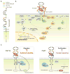
Fig. 6. Model for the coordinated differentiation of conducting airway epithelium and airway smooth muscle
A subset of Fgf10-expressing cells located in the distal submesothelial mesenchyme are progenitors for airway smooth muscle cells (ASMCs) and their amplification, as well as Fgf10 expression is dependent on mesenchymal Wnt/β-catenin signaling. FGF10 directly activates β-catenin in the distal epithelial progenitors, and induces Bmp4 and Sox9 expression, to maintain them and keep them from differentiating into Sox2+ conducting airway epithelium. As the epithelial tube grows towards the distal source of FGF10, progeny from the distal multipotent epithelial progenitors are left behind in the epithelial stalk and once they are out of FGF10’s reach they lose Sox9 expression, start expressing Sox2 and differentiate into conducting airway epithelium. At the same time, as ASMC progenitors passively acquire more proximal positions, they come in contact with epithelial-derived BMP4 and SHH causing them to stop expressing Fgf10 and ultimately differentiate into mature ASMCs that envelop the developing conducting airway epithelium.

Similar articles

Cited by

References

    1. Aberle H, Bauer A, Stappert J, Kispert A, Kemler R. beta-catenin is a target for the ubiquitin-proteasome pathway. EMBO J. 1997;16:3797–3804. - PMC - PubMed
    1. Abler LL, Mansour SL, Sun X. Conditional gene inactivation reveals roles for Fgf10 and Fgfr2 in establishing a normal pattern of epithelial branching in the mouse lung. Dev Dyn. 2009;238:1999–2013. - PMC - PubMed
    1. Alanis DM, Chang DR, Akiyama H, Krasnow MA, Chen J. Two nested developmental waves demarcate a compartment boundary in the mouse lung. Nat Commun. 2014;5:3923. - PMC - PubMed
    1. Alescio T, Cassini A. Induction in vitro of tracheal buds by pulmonary mesenchyme grafted on tracheal epithelium. J Exp Zool. 1962;150:83–94. - PubMed
    1. Ameri J, Stahlberg A, Pedersen J, Johansson JK, Johannesson MM, Artner I, Semb H. FGF2 specifies hESC-derived definitive endoderm into foregut/midgut cell lineages in a concentration-dependent manner. Stem Cells. 2010;28:45–56. - PubMed

Publication types

LinkOut - more resources