

Predictive value of 18F-fluorodeoxyglucose positron emission tomography - computed tomography compared to postoperative pathological findings for patients with non-small-cell lung cancer
- PMID: 25469279
- PMCID: PMC4251154
- DOI: 10.3892/mco.2014.408
Predictive value of 18F-fluorodeoxyglucose positron emission tomography - computed tomography compared to postoperative pathological findings for patients with non-small-cell lung cancer
Abstract
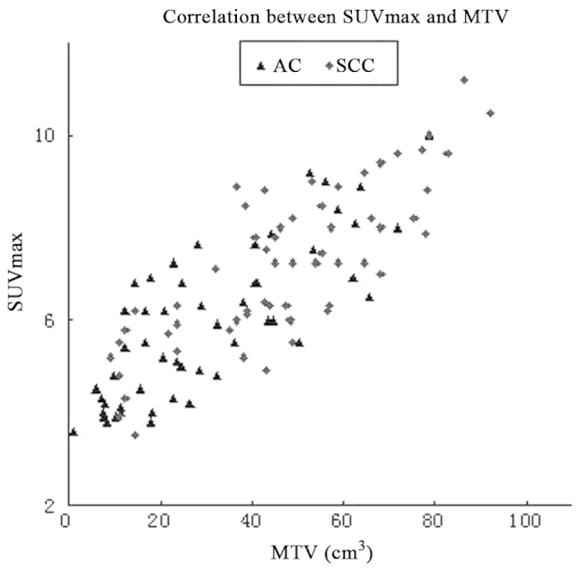
This study was conducted to investigate the predictive value of 18F-fluorodeoxyglucose positron emission tomography-computed tomography (18F-FDG PET-CT) in patients with non-small-cell lung cancer (NSCLC), compared to that of postoperative pathological findings, for T and N staging and the associations of the metabolic parameters of the primary tumor with histological type and differentiation. The preoperative contrast-enhanced CT and 18F-FDG PET-CT and postoperative pathological findings of 112 NSCLC patients treated with lobectomy or pneumonectomy combined with systematic mediastinal lymphadenectomy were retrospectively reviewed. Compared to the postoperative pathological findings, the effect of contrast-enhanced CT and 18F-FDG PET-CT on T and N staging were evaluated. The metabolic tumor volume (MTV) and maximum standardized uptake value (SUVmax) of the primary tumor were measured. The associations between these metabolic parameters and histological type and differentiation were also evaluated. The differences in the accuracy in overall staging and T staging between PET-CT and contrast-enhanced CT were significant (91.1 vs. 69.6%, P=0.000; and 92.9 vs. 76.8%, P=0.000, respectively). The sensitivity, specificity, positive predictive value, negative predictive value and accuracy of regional lymph node metastasis detection were 91.7, 93.0, 86.5, 95.8 and 92.6%, respectively, with PET-CT; and 71.3, 77.2, 60.6, 84.5 and 75.2%, respectively, with contrast-enhanced CT. The SUVmax (7.29±1.83 vs. 5.91±1.65, t=4.15, P=0.000) and MTV (48.20±22.47 vs. 30.21±19.72 cm3, t=4.48, P=0.000) were significantly higher for squamous cell carcinoma (SCC) compared to those for adenocarcinoma (AC). There was a positive correlation between the MTV and SUVmax of the primary tumor (Pearson's r=0.838, P=0.000). Significant differences were observed among differentiation subgroups in the SUVmax and MTV of the primary tumor for both SCC and AC. In conclusion, compared to the postoperative pathological findings, the predictive value of 18F-FDG PET-CT for T and N staging in NSCLC was higher compared to that of contrast-enhanced CT. The FDG uptake of the primary tumor was associated with histological type and differentiation and the difference was statistically significant. Therefore, the SUVmax and MTV of the primary tumor may be valuable indices to partly predict the histological type and grade of differentiation of NSCLC.
Keywords: computed tomography; metabolic tumor volume; non-small-cell lung cancer; positron emission tomography; staging; standardized uptake value.
Figures
Similar articles
-
The value on SUV-derived parameters assessed on 18F-FDG PET/CT for predicting mediastinal lymph node metastasis in non-small cell lung cancer.BMC Med Imaging. 2023 Apr 5;23(1):49. doi: 10.1186/s12880-023-01004-7. BMC Med Imaging. 2023. PMID: 37020286 Free PMC article.
-
Prognostic value of SUVmax and metabolic tumor volume on 18F-FDG PET/CT in early stage non-small cell lung cancer patients without LN metastasis.Biomed Mater Eng. 2014;24(6):3091-103. doi: 10.3233/BME-141131. Biomed Mater Eng. 2014. PMID: 25227018
-
The maximum standardized uptake value of fluorodeoxyglucose positron emission tomography of the primary tumour is a good predictor of pathological nodal involvement in clinical N0 non-small-cell lung cancer.Eur J Cardiothorac Surg. 2013 Jul;44(1):83-7. doi: 10.1093/ejcts/ezs604. Epub 2012 Dec 11. Eur J Cardiothorac Surg. 2013. PMID: 23233074
-
Potential predictors of the pathologic response after neoadjuvant chemoimmunotherapy in resectable non-small cell lung cancer: a narrative review.Transl Lung Cancer Res. 2024 May 31;13(5):1137-1149. doi: 10.21037/tlcr-24-142. Epub 2024 May 24. Transl Lung Cancer Res. 2024. PMID: 38854945 Free PMC article. Review.
-
Changes in Metabolism as a Diagnostic Tool for Lung Cancer: Systematic Review.Metabolites. 2022 Jun 14;12(6):545. doi: 10.3390/metabo12060545. Metabolites. 2022. PMID: 35736478 Free PMC article. Review.
Cited by
-
Correlation between 18-FDG standardized uptake value and tumor grade in patients with resectable non-small cell lung cancer.Transl Cancer Res. 2023 Dec 31;12(12):3530-3537. doi: 10.21037/tcr-23-798. Epub 2023 Nov 18. Transl Cancer Res. 2023. PMID: 38192987 Free PMC article.
-
N-staging in large cell neuroendocrine carcinoma of the lung: diagnostic value of [18F]FDG PET/CT compared to the histopathology reference standard.EJNMMI Res. 2021 Jul 22;11(1):68. doi: 10.1186/s13550-021-00811-9. EJNMMI Res. 2021. PMID: 34292419 Free PMC article.
-
Volumetric parameters on 18F-FDG PET/CT predict the survival of patients with gastric cancer associated with their expression status of c-MET.BMC Cancer. 2019 Aug 8;19(1):790. doi: 10.1186/s12885-019-5935-3. BMC Cancer. 2019. PMID: 31395059 Free PMC article.
-
The prognostic value of volume-based parameters using 18F-FDG PET/CT in gastric cancer according to HER2 status.Gastric Cancer. 2018 Mar;21(2):213-224. doi: 10.1007/s10120-017-0739-0. Epub 2017 Jun 22. Gastric Cancer. 2018. PMID: 28643145
References
-
- Antoch G, Saoudi N, Kuehl H, et al. Accuracy of whole-body dual-modality fluorine-18-2-fluoro-2-deoxy-D-glucose positron emission tomography and computed tomography (FDG-PET/CT) for tumor staging in solid tumors: comparison with CT and PET. J Clin Oncol. 2004;22:4357–4368. - PubMed
-
- MacDonald K, Searle J, Lyburn I. The role of dual time point FDG PET imaging in the evaluation of solitary pulmonary nodules with an initial standard uptake value less than 2.5. Clin Radiol. 2011;66:244–250. - PubMed
-
- Shum WY, Hsieh TC, Yeh JJ, et al. Clinical usefulness of dual-time FDG PET-CT in assessment of esophageal squamous cell carcinoma. Eur J Radiol. 2012;81:1024–1028. - PubMed
-
- Xiu Y, Bhutani C, Dhurairaj T, et al. Dual-time point FDG PET imaging in the evaluation of pulmonary nodules with minimally increased metabolic activity. Clin Nucl Med. 2007;32:101–105. - PubMed
-
- Hu Q, Wang W, Zhong X, et al. Dual-time-point FDG PET for the evaluation of locoregional lymph nodes in thoracic esophageal squamous cell cancer. Eur J Radiol. 2009;70:320–324. - PubMed
LinkOut - more resources
Full Text Sources
Other Literature Sources
Research Materials