. 2015 Jul;8(4):746-59.
doi: 10.1038/mi.2014.106. Epub 2014 Dec 3.

Control of pathogenic effector T-cell activities in situ by PD-L1 expression on respiratory inflammatory dendritic cells during respiratory syncytial virus infection

Affiliations

Control of pathogenic effector T-cell activities in situ by PD-L1 expression on respiratory inflammatory dendritic cells during respiratory syncytial virus infection

S Yao et al. Mucosal Immunol. 2015 Jul.

Abstract

Respiratory syncytial virus (RSV) infection is a leading cause of severe lower respiratory tract illness in young infants, the elderly and immunocompromised individuals. We demonstrate here that the co-inhibitory molecule programmed cell death 1 (PD-1) is selectively upregulated on T cells within the respiratory tract during both murine and human RSV infection. Importantly, the interaction of PD-1 with its ligand PD-L1 is vital to restrict the pro-inflammatory activities of lung effector T cells in situ, thereby inhibiting the development of excessive pulmonary inflammation and injury during RSV infection. We further identify that PD-L1 expression on lung inflammatory dendritic cells is critical to suppress inflammatory T-cell activities, and an interferon-STAT1-IRF1 axis is responsible for increased PD-L1 expression on lung inflammatory dendritic cells. Our findings suggest a potentially critical role of PD-L1 and PD-1 interactions in the lung for controlling host inflammatory responses and disease progression in clinical RSV infection.


Conflict of interest statement

DISCLOSURE

The authors declared not conflict of interest.

Figures

Figure 1
Figure 1
Murine and human T cells express programmed cell death 1 (PD-1) within the respiratory tract during respiratory syncytial virus (RSV) infection. (ac) Wild-type Balb/c mice were infected with RSV. PD-1 expression (a) and % of PD-1 + cells in CD4 + and CD8+ T cells (b) at different days post infection are depicted. (c) PD-1 expression on activated CD4 and CD8+ T cells (CD44hi) of lung and mediastinal lymph nodes at day 5 post RSV infection. (d) IL-10-eGFP reporter (Vert-X) mice were infected with RSV. Mean fluorescence intensity (MFI) of PD-1 expression on naïve, IL-10 , or IL-10+ (CD44hi) lung T cells is depicted (day 5 post infection). (e, f) Peripheral blood mononuclear cells and nasal washes were collected on healthy children or RSV-infected children. (e) PD-1 expression on CD8+ T cells is depicted. (f) % of PD-1+ cells in CD8+ T cells is depicted. (ad) Data are representative of two to three independent experiments. (e, f) Data are from four healthy and six RSV-infected donors. *, P<0.05 as determined by nonparametric Mann–Whitney test.
Figure 2
Figure 2
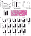
Programmed cell death 1 (PD-1) blockade following respiratory syncytial virus (RSV) infection leads to enhanced host morbidity and pulmonary injury. Wild-type Balb/c mice were infected with RSV and treated with control antibody, phosphate-buffered saline (PBS) or α-PD-L1 as indicated. (a) Host morbidity was monitored through weight loss. (b) RSV replication in the lung was monitored through RSV-L gene expression at indicated days post infection. (c) Airway virus titers from indicated mice were determined by plaque assay. Day 3, airway RSV titers from day 3-infected mice; day 6 control, airway RSV titers from day 6-infected mice treated with Rat IgG; day 6 α-PD-L1, airway RSV titers from day 6-infected mice treated with α-PD-L1. (d) Lung vascular and airway leakage was monitored through Evans-Blue concentrations in the airway at day 7 post infection. (e) Lung histopathology was measured by hematoxylin and eosin staining at day 7 post infection. (f) Pro-inflammatory cytokine levels in BAL from control or α-PD-L1-treated mice (day 9 post infection). Data are pooled from two to three experiments (a, c), representative from two to three experiments, (b, d, e) or from one experiment with four to five mice each group (f). *, P<0.05 was determined by unpaired Student’s t-test (a, d, f), NS, non-significant.
Figure 3
Figure 3
PD-L1 blockade results in enhanced inflammatory activities of effector T cells. (ad) Wild-type Balb/c mice were infected with respiratory syncytial virus (RSV) and treated with phosphate-buffered saline (PBS) or α-PD-L1. At day 5 post infection, mice were injected with monensin to block the in vivo secretion of cytokines by T cells. (A) Direct interferon-γ (IFN-γ) and tumor-necrosis factor-α (TNF-α) staining of ex vivo lung CD4 + and CD8+ T cells without in vitro stimulation. (b) % of IFN-γ+ and TNF-α+ cells in ex vivo lung CD4+ and CD8+ T cells without in vitro stimulation. (c, d) Lung CD4+ and CD8+ T cells were stimulated with phorbol 12-myristate 13-acetate (PMA) and Ionomycin (c). IFN-γ and TNF-α production by T cells are depicted. (d) % of IFN-γ+ and TNF-α+ cells in lung CD4 + and CD8+ T cells following PMA and Ionomycin stimulation. (e) Effector CD8 + T cells were isolated from RSV-infected lungs (day 8) and stimulated in vitro with increasing amount of plate-bound α-CD3 (0.01 and 0.1 μg ml−1) in the absence or presence of recombinant PD-L1–Ig protein. IFN-γ concentrations in the supernatant following 24 h stimulation are depicted. Data are representative of three independent experiments. *, P<0.05 as determined by unpaired Student’s t-test.
Figure 4
Figure 4
Inflammatory dendritic cell (DC)-derived PD-L1 inhibits cytokine production by effector T cells. (ac) Wild-type Balb/c mice were infected with respiratory syncytial virus (RSV). (a) PD-L1 expression (mean fluorescence intensity (MFI)) in the lung cells. (b) PD-L1 expression on various cell types in the lung (day 5 post infection). AM, alveolar macrophages; CD45, CD45 lung resident cells; iDC, Inflammatory DCs; MC, Ly6GCD11b+CD11c monocytes; Neu, neutrophils. (c) Relative expression levels of PD-L1 on different cell types (PD-L1 relative expression was calculated by PD-L1 MFI × % of cells in the lungs). (d) iDC and CD45 resident cells were sorted from the infected lungs at day 5 post infection. Cells were then mixed with day 8 lung CD8+ effector T cells in the presence of control Ab or α-PD-L1. Interferon-γ concentrations in the supernatant following 24 h stimulation are depicted. Data are representative of two to three independent experiments.
Figure 5
Figure 5
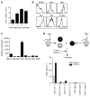
Type I and II interferon (IFNs) co-operatively induce PD-L1 expression on inflammatory dendritic cells (DCs) during respiratory syncytial virus (RSV) infection. (a, b) Wild-type (WT):Stat1 −/ − mixed bone marrow (BM) chimeric mice were infected with RSV. (a) Schematics of BM chimera construction and RSV infection. (b) PD-L1 expression (mean fluorescence intensity (MFI)) on WT and STAT1-deficient lung inflammatory DCs (iDC) in the same host at day 5 post infection. (c) WT and IFNAR1-deficient mice were infected with RSV. PD-L1 expression on WT and IFNAR1-deficient lung iDCs at day 5 post infection. (d) WT mice were infected with RSV and treated with control Ab or neutralizing α-IFN-γ. PD-L1 expression on lung iDCs at day 5 post infection. (e, f) WT and IFNAR1-deficient mice were infected with RSV and treated with α-IFN-γ as indicated. PD-L1 MFI (e) and messenger RNA message (f) on lung iDCs at day 5 post infection. (g) Bone marrow-derived DCs (BMDCs) were treated with phosphate-buffered saline, IFN-α, IFN-γ, or IFN-α plus IFN-γ. PD-L1 MFI on BMDCs is depicted. Data are representative of two to three independent experiments. *, P<0.05 as determined by paired Student’s t-test (b) or one way analysis of variance and Bonferroni post test (e).
Figure 6
Figure 6
IRF1 is required for PD-L1 expression on inflammatory DCs (iDC) during respiratory syncytial virus (RSV) infection. (a) IRF1 expression in wild-type (WT) or STAT1-deficient bone marrow-derived DCs (BMDCs) treated with phosphate-buffered saline (PBS), interferon-α (IFN-α) or IFN-γ. (b, c) PD-L1 expression (b) and mean fluorescence intensity (MFI) (c) on iDCs isolated from lungs of RSV-infected WT or IRF1-deficient mice (day 5 post infection). (d, e) WT: Irf1 −/ − mixed BM chimeric mice were infected with RSV. (d) Schematics of BM chimera construction and RSV infection. (e) PD-L1 expression (MFI) on WT and IRF1-deficient lung iDCs in the same host at day 5 post infection. (f, g) BMDCs were transduced with control or IRF1-expressing retroviruses. PD-L1 expression (f) and MFI (g) on control virus or IRF1-expressing-virus transduced DCs are depicted. (h) WT or IRF1-deficient lung iDCs was sorted from infected WT or Irf1 −/ − mice, respectively, at day 5 post infection. Cells were then mixed with day 8 lung CD8+ effector T cells in the presence of control Ab or α-PD-L1. IFN-γ concentrations in the supernatant following 24 h stimulation are depicted. Data are representative of three independent experiments. *, P<0.05 as determined by unpaired (c, g) or paired Student’s t-test (e).

Similar articles

Cited by

References

    1. Smyth RL, Openshaw PJ. Bronchiolitis. Lancet. 2006;368:312–322. - PubMed
    1. Falsey AR, Hennessey PA, Formica MA, Cox C, Walsh EE. Respiratory syncytial virus infection in elderly and high-risk adults. N Engl J Med. 2005;352:1749–1759. - PubMed
    1. Graham BS, Johnson TR, Peebles RS. Immune-mediated disease pathogenesis in respiratory syncytial virus infection. Immunopharmacology. 2000;48:237–247. - PubMed
    1. Peebles RS, Jr, Graham BS. Pathogenesis of respiratory syncytial virus infection in the murine model. Proc Am Thorac Soc. 2005;2:110–115. - PMC - PubMed
    1. Bennett BL, et al. Immunopathogenesis of respiratory syncytial virus bronchiolitis. J Infect Dis. 2007;195:1532–1540. - PubMed

Publication types

MeSH terms