. 2015 Jan 28;356(2 Pt B):656-68.
doi: 10.1016/j.canlet.2014.10.015. Epub 2014 Oct 18.

Synergistic antitumor interactions between MK-1775 and panobinostat in preclinical models of pancreatic cancer

Affiliations

Synergistic antitumor interactions between MK-1775 and panobinostat in preclinical models of pancreatic cancer

Guan Wang et al. Cancer Lett. .

Abstract

Pancreatic cancer remains a clinical challenge, thus new therapies are urgently needed. The selective Wee1 inhibitor MK-1775 has demonstrated promising results when combined with DNA damaging agents, and more recently with CHK1 inhibitors in various malignancies. We have previously demonstrated that treatment with the pan-histone deacetylase inhibitor panobinostat (LBH589) can cause down-regulation of CHK1. Accordingly, we investigated using panobinostat to down-regulate CHK1 in combination with MK-1775 to enhance cell death in preclinical pancreatic cancer models. We demonstrate that MK-1775 treatment results in increased H2AX phosphorylation, indicating increased DNA double-strand breaks, and activation of CHK1, which are both dependent on CDK activity. Combination of MK-1775 and panobinostat resulted in synergistic antitumor activity in six pancreatic cancer cell lines. Finally, our in vivo study using a pancreatic xenograft model reveals promising cooperative antitumor activity between MK-1775 and panobinostat. Our study provides compelling evidence that the combination of MK-1775 and panobinostat has antitumor activity in preclinical models of pancreatic cancer and supports the clinical development of panobinostat in combination with MK-1775 for the treatment of this deadly disease.

Keywords: CHK1; Drug combination; MK-1775; Pancreatic cancer; Panobinostat.


Conflict of interest statement

Conflict of interest

The authors declare no competing financial interests.

Figures
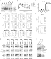
Fig. 1
Fig. 1
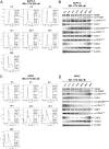
MK-1775 treatment induces DNA damage and activates the CHK1 pathway in pancreatic cancer cell lines. A, protein extracts from pancreatic cancer cell lines were subjected to Western blotting and probed with anti-Wee1, -PKMyt1, -p-CDK1, -CDK1, -p-CDK2, -CDK2, or -β-actin antibody. B, pancreatic cancer cell lines were cultured in 96-well plates at 37°C for 48 h in complete medium with variable concentrations of MK-1775 and viable cell numbers were determined using MTT reagent and a microplate reader. The data are presented as means ± standard errors from at least 3 independent experiments. C, pancreatic cancer cells were treated with vehicle control (Cont) or 500 nM MK-1775 (MK) for 48 h. The cells were fixed with ethanol, stained with PI, and subjected to flow cytometry analysis to determine cell cycle distribution. D, pancreatic cancer cells were treated with vehicle control or 500 nM MK-1775 for 48 h. Whole cell lysates were subjected to Western blotting and probed with anti-PARP, -p-H3, -γH2AX, -p-CHK1, -CHK1, -p-CDC25C, -p-CDK1, -CDK1, -p-CDK2, -CDK2, or -β-actin antibody. E, pancreatic cancer cells were treated with vehicle control or 500 nM MK-1775 for 48 h, then stained with PI and subjected to flow cytometry analysis. Apoptotic cells are expressed as the percentage of PI+ cells with sub-G1 DNA content. The data are presented as means of triplicates ± standard errors from one representative experiment. F, BxPC-3 cells were infected with Wee1 (designated Wee1-shRNA) or nontarget control (designated NTC-shRNA) shRNA lentivirus. The cells were fixed with ethanol, stained with PI, and subjected to flow cytometry analysis. G, BxPC-3 NTC- and Wee1-shRNA whole cell lysates were subjected to Western blotting and probed with anti-Wee1, -PARP, -p-CHK1, -CHK1, -p-CDC25C, -p-CDK1, -CDK1, -p-CDK2, -CDK2, -γH2AX, or -β-actin antibody. Representative Western blots and cell cycle histograms are shown.
Fig. 2
Fig. 2
MK-1775 induces p-CHK1 at early time points. A and C, BxPC-3 and HPAC cells were treated with 500 nM MK-1775 for up to 48 h. The cells were harvested, fixed with ethanol, stained with PI, and then subjected to flow cytometry analysis. B and D, the remaining cells were lysed and the protein extracts were subjected to Western blotting, and probed with anti-PARP, -p-CHK1, -CHK1, -p-CDC25C, -p-CDK1, -CDK1, -p-CDK2, -CDK2, -γH2AX, or -β-actin antibody. Experiments were performed at least three independent times and representative Western blots and cell cycle histograms are shown.
Fig. 3
Fig. 3
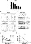
MK-1775-induced DNA damage, CHK1 phosphorylation, and cell death require active CDKs. A, HPAC cells were treated with 0–80 μM roscovitine (Rosc) for 48 h and viable cells were determined using MTT reagent and a microplate reader. The data are presented as means ± standard errors from at least 3 independent experiments. B, HPAC cells were treated with vehicle control, 500 nM MK-1775, 20μM roscovitine, or 500 nM MK-1775 plus 20 μM roscovitine for 8 h. Whole cell lysates were subjected to Western blotting and probed with anti-p-CHK1, -CHK1, -p-CDC25C, -p-CDK1, -CDK1, -p-CDK2, -CDK2, -γH2AX, or -β-actin antibody. C, HPAC cells were treated with MK-1775, roscovitine, or in combination for 48 h, stained with PI and subjected to flow cytometry analysis. Apoptotic cells were defined as the percentage of PI+ cells with sub-G1 DNA content. *** indicates p < 0.0005. D, HPAC cells were treated with variable concentrations of MK-1775 with or without 5 μM, 10μM or 20μM roscovitine. Viable cells were measured by MTT assays and the data are presented as means ± standard errors from at least 3 independent experiments. E, standard isobologram analyses of antitumor interactions between MK-1775 and roscovitine were performed in HPAC cells. The IC50 values of each drug are plotted on the axes; the solid line represents additive effect, while the points represent the concentrations of each drug resulting in 50% inhibition of growth. Points falling below the line indicate synergism, whereas those above the line indicate antagonism. F, BxPC-3 cells were treated with variable concentrations of roscovitine for 48 h and viable cells were determined by MTT assays. The data are presented as means ± standard errors from at least 3 independent experiments. G, BxPC-3 cells were treated with variable concentrations of MK-1775 with or without 5 μM, 10 μM or 20 μM roscovitine. Viable cells were measured by MTT assays and the data are presented as means ± standard errors from at least 3 independent experiments. H, standard isobologram analyses of antitumor interactions between MK-1775 and roscovitine were performed in BxPC-3 cells.
Fig. 4
Fig. 4
MK-1775 synergizes with LY2603618 in BxPC-3 cells. A, BxPC-3 cells were treated with variable concentrations of LY2603618 for 48 h and viable cells were determined by MTT assays. The data are presented as means ± standard errors from at least 3 independent experiments. B, BxPC-3 cells were treated with vehicle control, MK-1775 (MK), LY2603618 (LY) or MK-1775 plus LY2603618 for 48 h, stained with PI and subjected to flow cytometry analysis. C, protein extracts were subjected to Western blotting and probed with anti-PARP, -p-CHK1, -CHK1, -p-CDC25C, -p-CDK1, -CDK1, -p-CDK2, -CDK2, -γH2AX, or -β-actin antibody. D, BxPC-3 cells were treated with variable concentrations of MK-1775 with or without 0.25 μM, 0.5 μM or 1 μM LY2603618. Viable cells were measured by MTT assays and the data are presented as means ± standard errors from at least 3 independent experiments. E, standard isobologram analyses of antitumor interactions between MK-1775 and LY2603618 in BxPC-3 cells were performed.
Fig. 5
Fig. 5
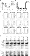
Panobinostat down-regulates CHK1 in pancreatic cancer cells. A, pancreatic cancer cell lines were cultured in 96-well plates at 37°C for 48 h in complete medium with variable concentrations of panobinostat and viable cell numbers were determined using MTT reagent and a microplate reader. The data are presented as means ± standard errors from at least 3 independent experiments. B–D, pancreatic cancer cells were treated with vehicle control or 40 nM panobinostat for 48 h, fixed with ethanol, stained with PI, and subjected to flow cytometry analysis. The sub-G1 data are presented as means of triplicates ± standard errors from one representative experiment (B). Representative cell cycle histograms are shown (C). Protein lysates were extracted from the remaining cells, subjected to Western blotting, and then probed with anti-PARP, -p-H3, -γH2AX, -ac-H4, -H4, -ac-tubulin, -p-CHK1, -CHK1, -p-CDC25C, -p-CDK1, -CDK1, -p-CDK2, -CDK2, or -β-actin antibody (D).
Fig. 6
Fig. 6
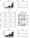
Panobinostat synergizes with MK-1775 in BxPC-3 cells. A, BxPC-3 cells were treated with panobinostat and MK-1775, alone or combined, for 48 h and subjected to PI staining and flow cytometry analysis. B, CI vs. Fa plot (combination index vs. fraction affected) for the apoptosis data presented in Panel A. The CI values were calculated using CompuSyn software. C, BxPC-3 cells were treated with panobinostat and/or MK-1775 for 48 h and subjected to PI staining and flow cytometry analysis. D, protein extracts were subjected to Western blotting and probed with anti-PARP, -p-H3, -p-CHK1, -CHK1, -p-CDC25C, -p-CDK1, -CDK1, -p-CDK2, -CDK2, -γH2AX, or -β-actin antibody. E, BxPC-3 cells were treated with panobinostat and MK-1775 for 48 h, then stained with annexin V/PI and subjected to flow cytometry analysis. The data are presented as means ± standard errors from one representative experiment. F, CI vs. Fa plot (combination index vs. fraction affected) for the apoptosis data presented in Panel E. The CI values were calculated using CompuSyn software.
Fig. 7
Fig. 7
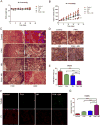
Panobinostat enhances the antitumor activity of MK-1775 in a BxPC-3 xenograft model. A, body weights were measured every 3–4 days. B, tumor volumes were calculated according to the following formula: m12 × m2× 0.5236 (m1: short diameter; m2: long diameter). C–E, tumor specimens were fixed in 10% formalin, embedded in paraffin, and cut into 4 μM-thick slides for H&E (C) and PCNA staining (D). The proliferation index was calculated as proliferation index = PCNA positive cells/observed cells × 100% and graphed as means ± standard errors (E). F, apoptosis was measured using the TUNEL assay in the tumor specimens. G, apoptosis index was calculated as TUNEL-positive cells/observed cells × 100% and graphed as means ± standard errors. *** indicates p < 0.0005.
Fig. 8
Fig. 8
Proposed mechanism for the antitumor interactions between MK-1775 and panobinostat in pancreatic cancer. Inhibition of Wee1 reduces phosphorylation of CDK1/CDK2, allowing CDK1/CDK2 to remain active, which leads to DNA damage. DNA damage triggers activation of ATM/ATR, which then activates CHK1. Active CHK1 inhibits CDC25s, leading to decreased removal of the inhibitory phosphorylation on CDK1/CDK2, resulting in increased p-CDK1/p-CDK2. This limits the amount of active CDK1/CDK2 and the resulting DNA damage following MK-1775 treatment. Panobinostat inhibits activation of CHK1, thus maintaining the active CDK1/CDK2 pools, enhancing DNA damage, and eventually leading to apoptosis. Others have suggested that CHK1 can activate Wee1, as indicated by the dashed line.

Similar articles

Cited by

References

    1. Siegel R, Ma J, Zou Z, Jemal A. Cancer statistics, 2014. CA Cancer J Clin. 2014;64:9–29. - PubMed
    1. Burris HA, 3rd, Moore MJ, Andersen J, Green MR, Rothenberg ML, Modiano MR, et al. Improvements in survival and clinical benefit with gemcitabine as first-line therapy for patients with advanced pancreas cancer: a randomized trial. J Clin Oncol. 1997;15:2403–2413. - PubMed
    1. Conroy T, Desseigne F, Ychou M, Bouche O, Guimbaud R, Becouarn Y, et al. FOLFIRINOX versus gemcitabine for metastatic pancreatic cancer. N Engl J Med. 2011;364:1817–1825. - PubMed
    1. Parker LL, Piwnica-Worms H. Inactivation of the p34cdc2-cyclin B complex by the human WEE1 tyrosine kinase. Science. 1992;257:1955–1957. - PubMed
    1. Watanabe N, Broome M, Hunter T. Regulation of the human WEE1Hu CDK tyrosine 15-kinase during the cell cycle. EMBO J. 1995;14:1878–1891. - PMC - PubMed

Publication types

MeSH terms