. 2015 Jan:61:62-75.
doi: 10.1016/j.exger.2014.11.018. Epub 2014 Nov 28.

Plasminogen activator inhibitor 1, fibroblast apoptosis resistance, and aging-related susceptibility to lung fibrosis

Affiliations

Plasminogen activator inhibitor 1, fibroblast apoptosis resistance, and aging-related susceptibility to lung fibrosis

Wen-Tan Huang et al. Exp Gerontol. 2015 Jan.

Abstract

Idiopathic pulmonary fibrosis (IPF) is a fatal lung disorder with unknown cause and no effective treatment. The incidence of and mortality from IPF increase with age, suggesting that advanced age is a major risk factor for IPF. The mechanism underlying the increased susceptibility of the elderly to IPF, however, is unknown. In this study, we show for the first time that the protein level of plasminogen activator inhibitor 1 (PAI-1), a protease inhibitor which plays an essential role in the control of fibrinolysis, was significantly increased with age in mouse lung homogenate and lung fibroblasts. Upon bleomycin challenge, old mice experienced augmented PAI-1 induction and lung fibrosis as compared to young mice. Most interestingly, we show that fewer (myo)fibroblasts underwent apoptosis and more (myo)fibroblasts with increased level of PAI-1 accumulated in the lung of old than in young mice after bleomycin challenge. In vitro studies further demonstrate that fibroblasts isolated from lungs of old mice were resistant to H2O2 and tumor necrosis factor alpha-induced apoptosis and had augmented fibrotic responses to TGF-β1, compared to fibroblasts isolated from young mice. Inhibition of PAI-1 activity with a PAI-1 inhibitor, on the other hand, eliminated the aging-related apoptosis resistance and TGF-β1 sensitivity in isolated fibroblasts. Moreover, we show that knocking down PAI-1 in human lung fibroblasts with PAI-1 siRNA significantly increased their sensitivity to apoptosis and inhibited their responses to TGF-β1. Together, the results suggest that increased PAI-1 expression may underlie the aging-related sensitivity to lung fibrosis in part by protecting fibroblasts from apoptosis.

Keywords: Aging; Fibroblast apoptosis; Idiopathic pulmonary fibrosis; Plasminogen activator inhibitor 1.


Figures

Fig 1
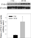
Fig 1. Age-dependent increase in PAI-1 protein level in mouse lung
Top panel, representative Western blotting pictures; Bottom panel, semi-quantified Western blotting data. *Significantly different from 3-month old mice (p<0.05, n = 4–5).
Fig 2
Fig 2. Age-dependent effect of bleomycin on mouse body weight
Mice were instilled with bleomycin or saline as described in the Material and Method section. Body weights before instillation and 7 or 14 days after instillation were recorded. The average body weight before instillation was 29.8g for 3-month old and 32.7 for 18-month old mice. The percentages of changes in body weights after treatment were presented (the body weights of the saline groups were from 14 days after saline instillation). α, Significantly different from the same age saline control; β, significantly different from 3-month old mice treated with bleomycin for the same length of time (p < 0.05, n = 5).
Fig 3
Fig 3. Age-dependent effects of bleomycin on BALF cell counts
Total and differential cell counts in BALF were determined as described in Material and Method section. A) Total cell numbers; B) Percentages of neutrophils; C) Percentages of lymphocytes; D) Percentages of macrophages in BALF. The data for the saline groups were from 14 days after saline instillation. α, Significantly different from the same age saline control; β, significantly different from 3-month old mice treated with bleomycin for the same length of time; χ, significantly different from the same aged mice treated with bleomycin for 7 days (p < 0.05, n = 5).
Fig 4
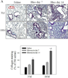
Fig 4. Age-dependent lung fibrotic responses in mice
A) Trichrome staining of collagens: Top panels, representative trichrome staining pictures; Bottom panel, semi-quantified collagen staining data. B) Hydroxyproline content in mouse lung. C) Western analyses of proteins: Top panels, Western blotting pictures (β-actin was used as protein loading control); Bottom panels, semi-quantified Western blotting data expressed as percentage of the same age saline controls. α, Significantly different from the same age saline control; β, significantly different from 3-month old mice treated with bleomycin for the same length of time; χ, significantly different from the same aged mice treated with bleomycin for 7 days (p<0.05, n=4–6).
Fig 5
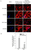
Fig 5. (Myo)fibroblast apoptosis in the lung of young and old mice treated with bleomycin
A) Left panels: representative pictures of immunostaining of mouse lung tissue with anti-fibroblast specific protein 1 (FSP-1) antibody (blue); Middle panels, representative overlay pictures of TUNEL (green) and nuclear (red) staining (yellow, TUNEL positive cells); Right panels: Overlay of FSP-1 (blue), TUNEL (green), and nuclear (red) staining (light blue color, TUNEL positive fibroblasts, arrows pointed). B) Quantitative data of apoptotic (myo)fibroblasts, expressed as the percentage of total (myo)fibroblasts as described in Material and Method section. α, Significantly different from the same age saline control; β, significantly different from 3-month old, bleomycin-treated mice (p<0.05, n=4–5). C) Co-localization of α-SMA (red), cleaved caspase-3 (green), and nuclear (blue) immunostaining. Yellow color stained cells (arrows pointed) are apoptotic myofibroblasts.
Fig 6
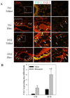
Fig 6. Age-dependent accumulation of PAI-1 expressing (myo)fibroblasts in mouse lung upon bleomycin challenge
A) Double immunofluorescence staining of mouse lung slides with antibodies to PAI-1 (red) and FSP-1 (green). Yellow color stained cells are PAI-1 expressing fibroblasts (overlay of green FSP-1 and red PAI-1). Central column: enlarged view of the white boxes in the left column. Right column: lung parenchyma. B) Quantification of PAI-1 expressing (myo)fibroblasts, expressed as the percentage of total (myo)fibroblasts. α, Significantly different from the same age saline control; β, significantly different from 3-month old, bleomycin-treated mice (p<0.05, n=4–5).
Fig 7
Fig 7. Responses of lung fibroblasts from young and old mice to apoptotic stimuli in vitro
Fibroblasts isolated from lungs of 3 month and 22 month old mice were challenged with 200 µM of H2O2 or 25 µM of TNF-α for 24 hours. Apoptosis was analyzed by flow cytometry. A) Representative flow cytometry graphic pictures. Q2: late apoptotic cell death labeled with both annexin V and propidium iodide; Q4: early apoptotic cell death labeled with annexin V only. B&C) Quantitative data of the early and late apoptotic cell death induced by H2O2 (B) or TNF-α (C). α, Significantly different from the corresponding 3M control fibroblasts (p<0.05, n=3).
Fig 8
Fig 8. Responses of lung fibroblasts from young and old mice to TGF-β1 in vitro
Isolated lung fibroblasts were treated with 1 ng/ml TGF-β1 for 24 hours and then Western analyses were conducted with cell lysates. Top panel: representative Western blotting pictures; bottom panels, semi-quantified Western blotting data, expressed as the percentage of 3M control fibroblasts. α, Significantly different from 3M control group; β, significantly different from TGF-β1 treated 3M fibroblasts (p<0.05, n=3).
Fig 9
Fig 9. Inhibition of PAI-1 activity eliminated the aging-related increase in apoptosis resistance and TGF-β1 sensitivity in isolated fibroblasts
Fibroblasts isolated from 3- and 22-month old mouse lungs were pretreated with 75 µM of PAI-1 inhibitor TM5275 for 8 hours and then treated with 200 µM of H2O2 or 1ng/ml TGF-β1 in the presence of TM5275 for 24 hours. (A) Effects of TM5275 on apoptotic response. Top panels: representative flow cytometry graphic pictures. Bottom panels: quantitative data of apoptotic cell death (early plus late stages). (B) Effects of TM5275 on the sensitivity of fibroblasts to TGF-β. Top panels: representative Western blotting pictures; Bottom panels, semi-quantified Western blotting data, expressed as the percentages of untreated control fibroblasts from same aged mice. α, Significantly different from the same age untreated control; β, significantly different from TGF-β1 treated 3M fibroblasts; (p<0.05, n=3–5).
Fig 10
Fig 10. Knockdown of PAI-1 with PAI-1 siRNA increased apoptosis sensitivity and reduced fibrotic responses in human lung fibroblasts
Human lung fibroblasts (CCL-210 cells) were transfected with PAI-1 siRNA or non-target siRNA (NT-siRNA) and then treated with 1 ng/ml TGFβ-1 (A) or 200 µM H2O2 (B) for 24 hours. A) Western analyses of PAI-1, fibronectin, and α-SMA proteins. Top panel: representative Western blotting pictures; bottom panels, semi-quantified Western blotting data. B) Flow cytometry analysis of apoptosis. α, Significantly different from the corresponding untreated control fibroblasts; β, significantly different from NT-siRNA transfected TGF-β1 (A) or H2O2 (B) treated fibroblasts (p<0.05, n=6).

Similar articles

Cited by

References

    1. Abderrahmani R, Francois A, Buard V, Tarlet G, Blirando K, Hneino M, Vaurijoux A, Benderitter M, Sabourin JC, Milliat F. PAI-1-dependent endothelial cell death determines severity of radiation-induced intestinal injury. PloS one. 2012;7:e35740. - PMC - PubMed
    1. Aillaud MF, Pignol F, Alessi MC, Harle JR, Escande M, Mongin M, Juhan-Vague I. Increase in plasma concentration of plasminogen activator inhibitor, fibrinogen, von Willebrand factor, factor VIII:C and in erythrocyte sedimentation rate with age. Thrombosis and haemostasis. 1986;55:330–332. - PubMed
    1. Araki T, Katsura H, Sawabe M, Kida K. A clinical study of idiopathic pulmonary fibrosis based on autopsy studies in elderly patients. Intern Med. 2003;42:483–489. - PubMed
    1. Bajou K, Peng H, Laug WE, Maillard C, Noel A, Foidart JM, Martial JA, DeClerck YA. Plasminogen activator inhibitor-1 protects endothelial cells from FasL-mediated apoptosis. Cancer Cell. 2008;14:324–334. - PMC - PubMed
    1. Beier JI, Kaiser JP, Guo L, Martinez-Maldonado M, Arteel GE. Plasminogen activator inhibitor-1 deficient mice are protected from angiotensin II-induced fibrosis. Arch Biochem Biophys. 2011;510:19–26. - PMC - PubMed

Publication types

Substances