Review
. 2014 Nov 20;7(1):a015818.
doi: 10.1101/cshperspect.a015818.

The centromere: epigenetic control of chromosome segregation during mitosis

Affiliations
Review

The centromere: epigenetic control of chromosome segregation during mitosis

Frederick G Westhorpe et al. Cold Spring Harb Perspect Biol. .

Abstract

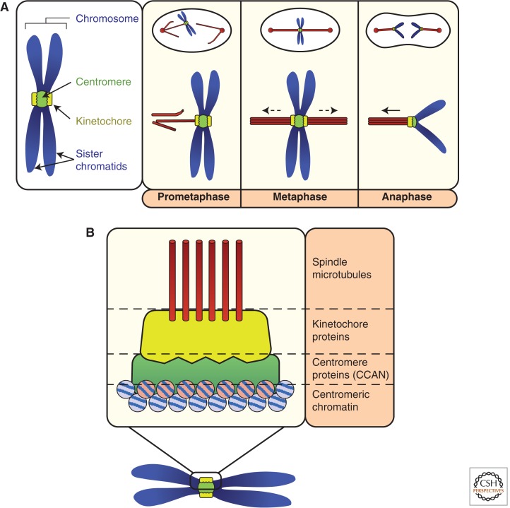
A fundamental challenge for the survival of all organisms is maintaining the integrity of the genome in all cells. Cells must therefore segregate their replicated genome equally during each cell division. Eukaryotic organisms package their genome into a number of physically distinct chromosomes, which replicate during S phase and condense during prophase of mitosis to form paired sister chromatids. During mitosis, cells form a physical connection between each sister chromatid and microtubules of the mitotic spindle, which segregate one copy of each chromatid to each new daughter cell. The centromere is the DNA locus on each chromosome that creates the site of this connection. In this review, we present a brief history of centromere research and discuss our current knowledge of centromere establishment, maintenance, composition, structure, and function in mitosis.


Figures

Figure 1.
Figure 1.
Introduction to centromere function and organization during mitosis. (A) Before and during the early stages of mitosis, centromeres (green circle) recruit kinetochore proteins (yellow discs). During prometaphase, the kinetochore forms the attachment sites for spindle microtubules (red rods). Once both kinetochores of all sister chromatid pairs are stably and correctly attached to microtubules, pulling forces exerted by microtubules (dashed arrows) cause migration of linked sister chromatids to the metaphase plate. At anaphase, sister chromatid cohesion is dissolved and the centromere and kinetochore harness microtubule-dependent forces that pull each sister chromatid to opposite ends of the dividing cell. (B) Basic architecture of the centromere in mitosis. Centromeric chromatin consists of specialized nucleosomes containing the histone H3 variant centromere protein (CENP)-A. CENP-A recruits a network of centromere proteins (green) that are collectively known as the constitutive centromere associated network (CCAN). Kinetochore proteins (yellow), specifically recruited by the CCAN for mitosis, attach to spindle microtubules.
Figure 2.
Figure 2.
CENP-A nucleosome structure and possible variants. A cartoon of a conventional octameric H3 nucleosome is shown, together with the most prominent models of CENP-A nucleosome composition, homotypic CENP-A octamers, heterotypic CENP-A/H3 octamers, and tetrameric CENP-A hemisomes. See main text for specific details.
Figure 3.
Figure 3.
Overview of centromere proteins and centromere architecture. Centromere proteins are grouped based on individual complexes, often based on the phenotypic consequence of protein depletion in cells and, more recently, biochemical characterization. CENP-A nucleosomes directly recruit CENP-C and possibly the CENP-N/L heterodimer. CENP-C recruits the CENP-H/I/K/M complex that in turn is required for CENP-T centromere localization. How this occurs remains unclear as specific CENP-C:CENP-H/I/K/M and CENP-H/I/K/M:CENP-T/W/S/X interactions have not been identified. The possibility that CENP-T/W/S/X proteins wrap centromeric DNA in nucleosome-like structures, and whether they associate with H3 nucleosomes at centromeres, is currently a topic of intense research. The functions and subcomplexes that comprise the remaining CENP proteins O/P/Q/R and U remain unclear. Note that both Saccharomyces cerevisiae and Saccharomyces pombe have centromere proteins not listed here (as they lack a known human homolog).
Figure 4.
Figure 4.
Functions of centromeric chromatin through the human cell cycle. Centromeric chromatin, defined by CENP-A-containing nucleosomes, is specifically assembled during mitotic exit and G1. Assembly causes the CENP-A copy number at centromeres to double (represented by transition from pink to red chromatin). During DNA replication/S phase, replication of centromere-DNA results in the distribution of CENP-A onto the two nascent DNA strands. This causes a twofold reduction in CENP-A at each centromere-DNA sequence (red to pink chromatin) (see Fig. 5 for more details). Note that it is this “diluted” centromeric chromatin (pink) that is responsible for building functional kinetochores before and during mitosis and, thus, for segregating chromosomes. Broadly speaking, centromere proteins retain localization at CENP-A chromatin through all stages of the cell cycle. How centromere proteins respond to changing CENP-A protein numbers within chromatin is not clear.
Figure 5.
Figure 5.
CENP-A assembly in humans. (1) “Parent” CENP-A nucleosomes (from the previous cell cycle), directly or indirectly specify the sites of new CENP-A assembly. (2) The centromere proteins shown in the figure; CENP-C, -N (which directly bind CENP-A nucleosomes), -H, -I, -K, and -M have been experimentally implicated in CENP-A assembly. (3) In addition to centromere proteins, the Mis18 complex, consisting of Mis18α, Mis18β, and M18BP1, is also required for CENP-A assembly, and most likely modifies (M) the chromatin and/or the recruitment of specialized loading factors. (4) Chromatin modifiers and chaperones, such as RbAp46 and 48, also have a role in modifying chromatin during CENP-A assembly. (5) Centromere proteins and the Mis18 complex somehow recruit HJURP, the CENP-A-specific chaperone. HJURP binding to CENP-A precludes CENP-A:H4 tetramer formation, and HJURP dimerization is required for CENP-A assembly, so one possible model is that nascent CENP-A is delivered as two dimers in a (HJURP:CENP-A:H4)2 complex, as shown. (6) The proteins or chromatin features that mark the site of new CENP-A assembly, the “placeholders,” remain unclear. (7) Finally, once CENP-A is assembled into chromatin, factors such as MgcRacGAP, which interacts with the Mis18 complex, stabilize incorporated CENP-A.
Figure 6.
Figure 6.
Models of CENP-A nucleosome distribution during DNA replication. (A) CENP-A (red) nucleosomes remain as homotypic octamers after passage of the replication fork. In the model shown, octameric histone H3.1 (cyan) nucleosomes occupy the gaps left by the twofold reduction in CENP-A nucleosomes on each DNA strand. Other possibilities include histone H3.3, naked DNA, or other species of specialized centromeric chromatin (such as CENP-T/W/S/X). (B) Each CENP-A nucleosome is split and segregated to both DNA strands. CENP-A:H4 dimers form heterotetramers with H3/H4 dimers, resulting in heterooctamer formation. (C) Octameric CENP-A nucleosomes are split and not replenished, resulting in the formation of tetrameric hemisomes. As hemisomes are incapable of wrapping as much DNA as octameric nucleosomes, this model results in an excess of free DNA, the fate of which is unclear (?). All these models assume octameric CENP-A nucleosomes are the prereplication conformation. Note that the composition of centromeric chromatin after DNA replication becomes the template for CENP-A assembly in the next cell cycle (see Figs. 3 and 4).

Similar articles

Cited by

References

    1. Amano M, Suzuki A, Hori T, Backer C, Okawa K, Cheeseman IM, Fukagawa T 2009. The CENP-S complex is essential for the stable assembly of outer kinetochore structure. J Cell Biol 186: 173–182. - PMC - PubMed
    1. Amaro AC, Samora CP, Holtackers R, Wang E, Kingston IJ, Alonso M, Lampson M, McAinsh AD, Meraldi P 2010. Molecular control of kinetochore-microtubule dynamics and chromosome oscillations. Nat Cell Biol 12: 319–329. - PMC - PubMed
    1. Bade D, Pauleau AL, Wendler A, Erhardt S 2014. The E3 ligase CUL3/RDX controls centromere maintenance by ubiquitylating and stabilizing CENP-A in a CAL1-dependent manner. Dev Cell 28: 508–519. - PubMed
    1. Bailey AO, Panchenko T, Sathyan KM, Petkowski JJ, Pai PJ, Bai DL, Russell DH, Macara IG, Shabanowitz J, Hunt DF, et al. 2013. Posttranslational modification of CENP-A influences the conformation of centromeric chromatin. Proc Natl Acad Sci 110: 11827–11832. - PMC - PubMed
    1. Baker AM, Fu Q, Hayward W, Lindsay SM, Fletcher TM 2009. The Myb/SANT domain of the telomere-binding protein TRF2 alters chromatin structure. Nucleic Acids Res 37: 5019–5031. - PMC - PubMed

Publication types

MeSH terms

LinkOut - more resources