. 2015 Jan 9;290(2):1129-40.
doi: 10.1074/jbc.M114.590943. Epub 2014 Nov 20.

p53 protein-mediated up-regulation of MAP kinase phosphatase 3 (MKP-3) contributes to the establishment of the cellular senescent phenotype through dephosphorylation of extracellular signal-regulated kinase 1/2 (ERK1/2)

Affiliations

p53 protein-mediated up-regulation of MAP kinase phosphatase 3 (MKP-3) contributes to the establishment of the cellular senescent phenotype through dephosphorylation of extracellular signal-regulated kinase 1/2 (ERK1/2)

Hui Zhang et al. J Biol Chem. .

Abstract

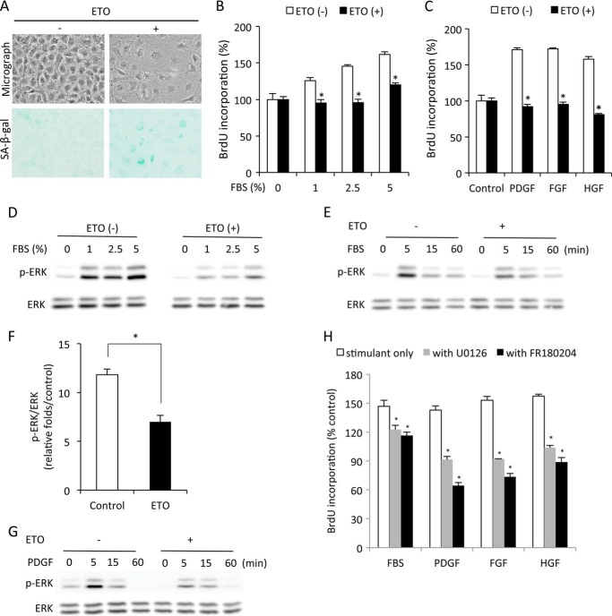
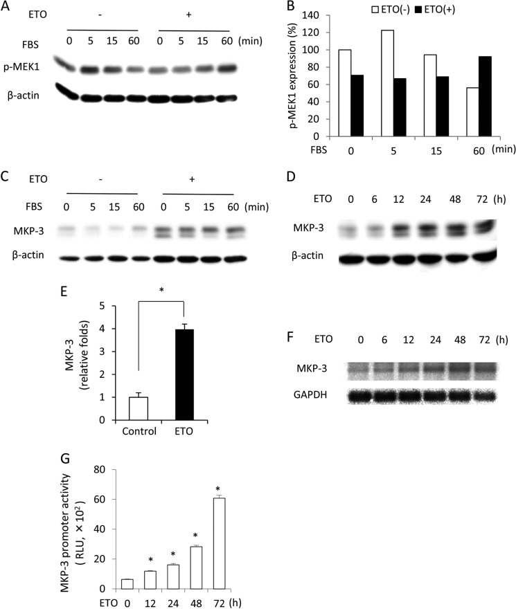
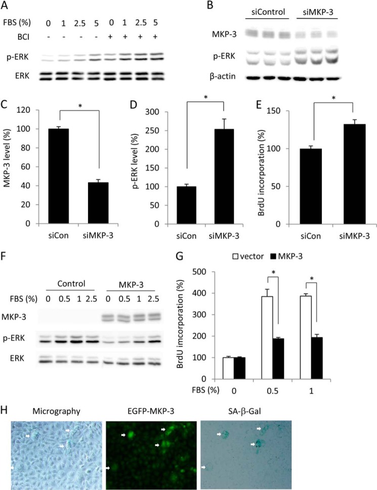
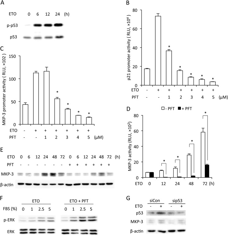
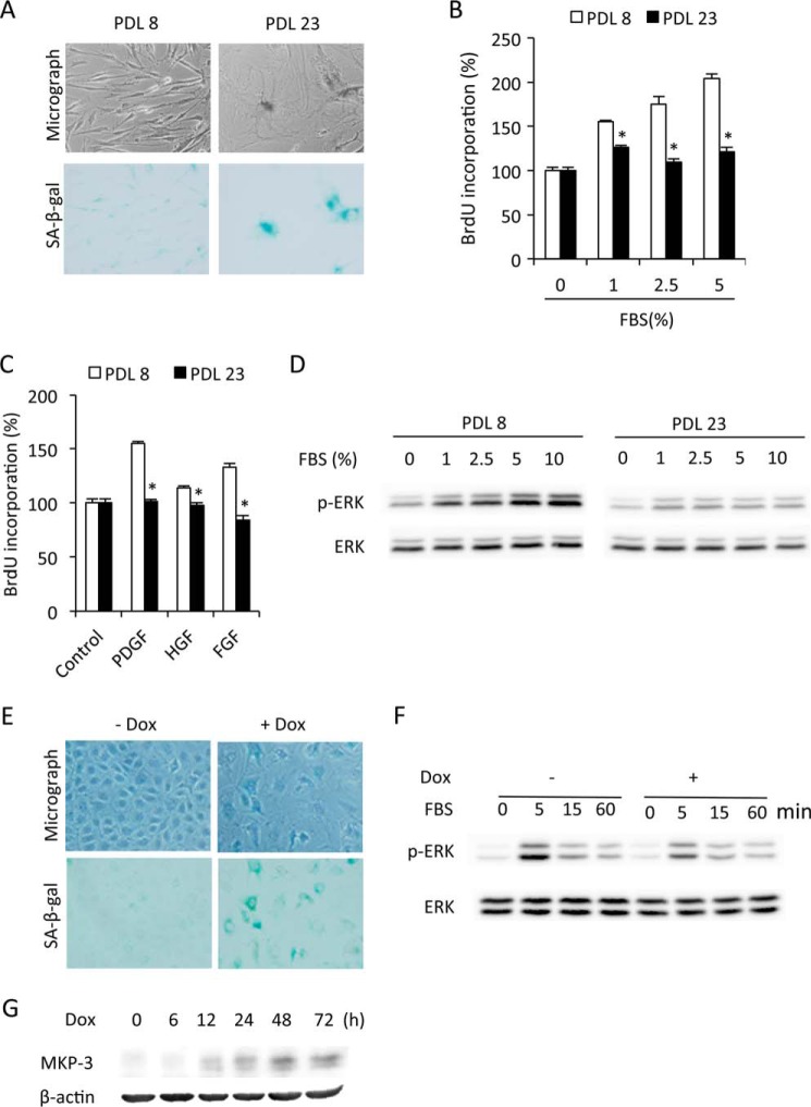
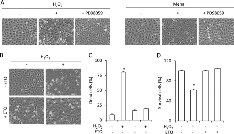
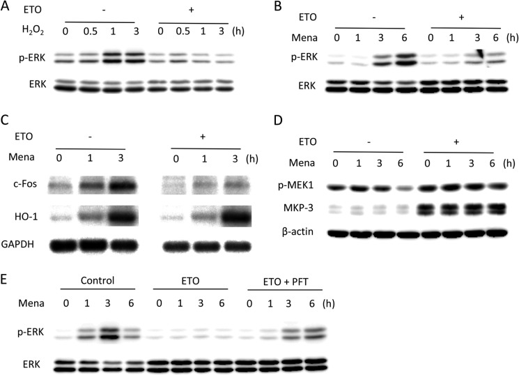
Growth arrest is one of the essential features of cellular senescence. At present, the precise mechanisms responsible for the establishment of the senescence-associated arrested phenotype are still incompletely understood. Given that ERK1/2 is one of the major kinases controlling cell growth and proliferation, we examined the possible implication of ERK1/2. Exposure of normal rat epithelial cells to etoposide caused cellular senescence, as manifested by enlarged cell size, a flattened cell body, reduced cell proliferation, enhanced β-galactosidase activity, and elevated p53 and p21. Senescent cells displayed a blunted response to growth factor-induced cell proliferation, which was preceded by impaired ERK1/2 activation. Further analysis revealed that senescent cells expressed a significantly higher level of mitogen-activated protein phosphatase 3 (MKP-3, a cytosolic ERK1/2-targeted phosphatase), which was suppressed by blocking the transcriptional activity of the tumor suppressor p53 with pifithrin-α. Inhibition of MKP-3 activity with a specific inhibitor or siRNA enhanced basal ERK1/2 phosphorylation and promoted cell proliferation. Apart from its role in growth arrest, impairment of ERK1/2 also contributed to the resistance of senescent cells to oxidant-elicited cell injury. These results therefore indicate that p53-mediated up-regulation of MKP-3 contributes to the establishment of the senescent cellular phenotype through dephosphorylating ERK1/2. Impairment of ERK1/2 activation could be an important mechanism by which p53 controls cellular senescence.

Keywords: Extracellular Signal-regulated Kinase (ERK); MAP Kinase Phosphatase 3 (MKP-3); Oxidative Stress; Proliferation; Senescence; p53.


Figures

FIGURE 1.
FIGURE 1.
ERK1/2 phosphorylation is impaired in senescent cells. A, induction of senescence by ETO. NRK cells were treated with 1 μg/ml ETO for 3 days and subjected to phase-contrast microscopy and SA-β-gal staining. B and C, reduced cell proliferation in senescent cells. NRK cells were treated with or without 1 μg/ml ETO for 3 days. Then cells were quiescent for 48 h and exposed to the indicated concentrations of FBS (B) or various growth factors (10 ng/ml, C) for 24 h. Cell proliferation was evaluated through BrdU incorporation. Data are mean ± S.E. (n = 4). *, p < 0.05 versus respective untreated control. HGF, hepatocyte-derived growth factor. D–G, impaired ERK1/2 phosphorylation in senescent cells. Cells were pretreated with or without ETP for 3 day and then exposed to the indicated concentration of FBS for 5 min or 5% FBS or 10 ng/ml PDGF for the indicated time intervals. Cellular lysates were extracted and subjected to Western blot analysis of p-ERK1/2 and ERK1/2 (D, E, and G). The densitometric data of p-ERK1/2 protein 5 min after exposure to 5% FBS was normalized to total ERK1/2 and control (F, data are mean ± S.E., n = 3). *, p < 0.05 versus untreated control. H, inhibition of ERK1/2 on cell proliferation. NRK cells were treated with 2.5% FBS, 10 ng/ml PDGF, FGF, or hepatocyte growth factor in the presence or absence of 20 μm U0126 or 100 μm FR180204 for 24 h and subjected to BrdU incorporation. Data are mean ± S.E. (n = 4). *, p < 0.05 versus respective untreated control.
FIGURE 2.
FIGURE 2.
Influence of the senescence-inducing agent ETO on MEK-1 activation and MKP-3 protein level. A–C, levels of phosphorylated MEK-1 and MKP-3 in control and senescent cells. NRK cells were treated with 1 μg/ml ETO for 3 days or left untreated. Then cells were exposed to 5% FBS for the indicated time. Cellular lysates were subjected to Western blot analysis of p-MEK1 (A) and MKP-3 (B). Densitometric quantitation of the level of the phosphorylated MEK-1 in A is shown in C. The result was normalized to zero point control. D–G, changes in MKP-3 during induction of senescence with ETO. Cells were treated with 1 μg/ml ETO for the indicated time and subjected to Western blot analysis of MKP-3. β-Actin was used as a loading control (D). Densitometric quantitation of the level of MKP-3 at the 48-h point following exposure to 1 μg/ml ETO is shown in E. The result was normalized to the control. Data are mean ± S.E. (n = 3). *, p < 0.05 versus control. F, cells were treated with 1 μg/ml ETO for the indicated time and subjected to Northern blot analysis of MKP-3. GAPDH was used as a loading control. G, cells transfected with pGL3B/DUSP6-luc were exposed to 1 μg/ml ETO for the indicated time. Cell lysates were subjected to a luciferase assay. Data are mean ± S.E. (n = 4). RLU, relative light unit. *, p < 0.05 versus zero point control.
FIGURE 3.
FIGURE 3.
Effect of MKP-3 on ERK1/2 phosphorylation, cell proliferation, cell size, and SA-β-gal activity. A–E, down-regulation of MKP-3 on ERK1/2 phosphorylation and cell proliferation. A, cells were pretreated with 1 μg/ml ETO for 3 days. After that, they were treated with 1 μm 2-benzylidene-3-(cyclohexylamino)-1-indanone hydrochloride (BCI) for 15 min or left untreated before being exposed to the indicated concentrations of FBS for an additional 5 min. Cell lysates were subjected to Western blot analysis of p-ERK1/2 and ERK1/2. B–E, cells were transfected with control or MKP-3 siRNA. Cell lysates were subjected to Western blot analysis of MKP-3 and p-ERK1/2. β-Actin was used as a loading control (B). Densitometric quantitations of the levels of MKP-3 and p-ERK1/2 in B are shown in C and D. The result was normalized to siRNA control (siCon). Data are mean ± S.E. (n = 3). *, p < 0.05 versus control. E, cells transfected with control or MKP-3 siRNA were subjected to BrdU incorporation. Data are mean ± S.E. (n = 4). *, p < 0.05 versus siRNA control. F and G, overexpression of MKP3 on ERK1/2 and cell proliferation. Cells that were transiently transfected with a control or MKP-3 plasmid were exposed to the indicated concentration of FBS for 5 min. Cellular protein was extracted and subjected to Western blot analysis of MKP-3, p-ERK, and ERK. G, cells transfected with control or MKP-3 plasmid were subjected to BrdU incorporation. Data are mean ± S.E.; n = 4; *, p < 0.05. H, transfection of NRK cells with a GFP-tagged MKP-3 gene on cell morphology and SA-β-gal activity. Cells were transfected with a GFP-tagged MKP-3 gene. After 4 days, cells were subjected to phase-contrast/immunofluorescence microscopy and SA-β-gal staining. Note the cell size and SA-β-gal activity in the GFP-positive cells (arrows).
FIGURE 4.
FIGURE 4.
Role of p53 in activation of MKP-3 transcription. A, change of p53 during senescence. NRK cells were treated with 1 μg/ml ETO for the indicated time. Cell lysate were extracted and subjected to Western blot analysis of p-p53 and p53. B–E, involvement of p53 in MKP-3 expression. Cells transfected with the pGL3B/DUSP6-luc (B) or pGL3B/p21WAF1/CIF1-Luc plasmids (C) were treated with 1 μg/ml ETO in the absence or presence of the indicated concentration of PFT for 24 h. Cell lysates were extracted and assayed for luciferase activity. Data are mean ± S.E. (n = 4). *, p < 0.05 versus ETO alone. RLU, relative light units. D, cells transfected with the pGL3B/DUSP6-luc plasmid were treated with 1 μg/ml ETO with or without 5 μm PFT for the indicated time. Cell lysates were extracted and assayed for luciferase activity. Data are mean ± S.E. (n = 4). *, p < 0.05 versus the respective time point control. E, cells were treated with 1 μg/ml ETO with or without 5 μm PFT for the indicated time. Cell lysates were detected for expression of MKP-3. β-Actin was used as a loading control. F, regulation of ERK1/2 by p53. Cells were treated with 1 μg/ml ETO with or without 5 μm PFT for 3 days. Then cells were stimulated with the indicated concentrations of FBS for 5 min. Cell lysates were analyzed for the level of p-ERK1/2 and total ERK1/2. G, down-regulation of p53 with siRNA on MKP-3 expression. Cells were transfected with control or p53 siRNA. Cell lysates were subjected to Western blot analysis of p53 and MKP-3. β-Actin was used as a loading control. siCon, control siRNA.
FIGURE 5.
FIGURE 5.
Impaired ERK1/2 activation in other models of cellular senescence. A, morphological changes in replicative senescent cells. WI-38 cells at passages 8 and 23 (PDL8 and PDL23) were subjected to SA-β-gal staining. B–D, impaired cell proliferation and ERK1/2 phosphorylation in replicative senescent cells. PDL8 and PDL23 cells were quiescent for 48 h. Then cells were exposed to the indicated concentrations of FBS (B) or 10 ng/ml growth factors (C) for 24 h. Cell proliferation was evaluated through BrdU incorporation. Data are mean ± S.E. (n = 4). *, p < 0.05 versus the respective control. HGF, hepatocyte-derived growth factor. D, PDL8 and PDL23 cells were stimulated with the indicated concentrations of FBS for 5 min and subjected to Western blot analysis of p-ERK1/2 and ERK1/2. E, induction of senescence by Dox. NRK cells were treated with 100 μg/ml Dox for 3 days and then subjected to phase-contrast microscopy and β-gal staining. F, impaired ERK1/2 activation in Dox-pretreated cells. NRK cells were treated with 100 μg/ml Dox for 3 days. Then cells were exposed to 5% FBS for the indicated time. The cellular lysates were subjected to Western blot analysis of p-ERK and ERK. G, effect of Dox on MKP-3 expression. NRK cells were treated with 100 μg/ml Dox for the indicated time and subjected to Western blot analysis of MKP-3. β-Actin was used as a loading control.
FIGURE 6.
FIGURE 6.
Resistance to oxidative cell injury in senescent cells. A, role of ERK1/2 in oxidative stress-induced cell injury. NRK cells were exposed to 100 μm H2O2 (left panel) or 50 μm menadione (Mena, right panel) in the presence or absence of 50 μm PD98059 for 12 h. The cell morphology under phase-contrast microscopy was photographed (magnification, ×200). B–D, resistance to oxidative injury in senescent cells. Cells were treated with or without 1 μg/ml ETO for 3 days, and then cells were exposed to 100 μm H2O2 for 12 h and subjected to phase-contrast microscopy (B) or formazan assay (D). The quantitative result of B is shown in C. Results are expressed as mean ± S.E. (n = 4). *, p < 0.05 versus untreated control.
FIGURE 7.
FIGURE 7.
Impaired ERK1/2 activation in oxidant-exposed senescent cells. A–C, ERK1/2 phosphorylation induced by oxidative stress in senescent cells. Control and senescent cells were exposed to 100 μm H2O2 (A) or 50 μm menadione (Mena, B) for the indicated times and subjected to Western blot analysis of p-ERK1/2 and ERK1/2. C, cells were exposed to 50 μm menadione for the indicated time and subjected to Northern blot analysis of c-Fos and Ho-1. GAPDH was used as a loading control. D and E, involvement of p53 in the regulation of ERK1/2 phosphorylation. D, control and senescent cells were treated with 50 μm menadione for the indicated time and subjected to Western blot analysis of p-MEK1 and MKP-3. β-Actin was used as a loading control. E, control cells were treated with 1 μg/ml ETO in the absence or presence of 5 μm PFT for 3 days, and then cells were exposed to 50 μm menadione for the indicated time. Cell lysates were subjected to Western blot analysis of p-ERK1/2 and ERK1/2.

Similar articles

Cited by

References

    1. Hayflick L. (1965) The limited in vitro lifetime of human diploid cell strains. Exp. Cell Res. 37, 614–636 - PubMed
    1. Shay J. W., Roninson I. B. (2004) Hallmarks of senescence in carcinogenesis and cancer therapy. Oncogene 23, 2919–2933 - PubMed
    1. Shay J. W., Wright W. E. (2004) Telomeres are double-strand DNA breaks hidden from DNA damage responses. Mol. Cell 14, 420–421 - PubMed
    1. d'Adda di Fagagna F., Reaper P. M., Clay-Farrace L., Fiegler H., Carr P., Von Zglinicki T., Saretzki G., Carter N. P., Jackson S. P. (2003) A DNA damage checkpoint response in telomere-initiated senescence. Nature 426, 194–198 - PubMed
    1. Herbig U., Jobling W. A., Chen B. P., Chen D. J., Sedivy J. M. (2004) Telomere shortening triggers senescence of human cells through a pathway involving ATM, p53, and p21(CIP1), but not p16(INK4a). Mol. Cell 14, 501–513 - PubMed

Publication types

MeSH terms

Substances