. 2014 Nov 21:5:5566.
doi: 10.1038/ncomms6566.

PKM2 phosphorylates MLC2 and regulates cytokinesis of tumour cells

Affiliations

PKM2 phosphorylates MLC2 and regulates cytokinesis of tumour cells

Yuhui Jiang et al. Nat Commun. .

Abstract

Pyruvate kinase M2 (PKM2) is expressed at high levels during embryonic development and tumour progression and is important for cell growth. However, it is not known whether it directly controls cell division. Here, we found that Aurora B phosphorylates PKM2, but not PKM1, at T45; this phosphorylation is required for PKM2's localization and interaction with myosin light chain 2 (MLC2) in the contractile ring region of mitotic cells during cytokinesis. PKM2 phosphorylates MLC2 at Y118, which primes the binding of ROCK2 to MLC2 and subsequent ROCK2-dependent MLC2 S15 phosphorylation. PKM2-regulated MLC2 phosphorylation, which is greatly enhanced by EGF stimulation or EGFRvIII, K-Ras G12V and B-Raf V600E mutant expression, plays a pivotal role in cytokinesis, cell proliferation and brain tumour development. These findings underscore the instrumental function of PKM2 in oncogenic EGFR-, K-Ras- and B-Raf-regulated cytokinesis and tumorigenesis.


Figures
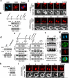
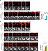
Figure 1
Figure 1. PKM2 Interacts with MLC2 and Is Critical for Cytokinesis
Immunoblotting (b, d-f, h) and immunofluorescence (a, c, g, i) analyses were performed with the indicated antibodies. Nuclei were stained with DAPI (blue). (a) U87 cells in cytokinesis were immunostained with the indicated antibodies. Scale bars, 10 μm. (b) U87/EGFRvIII cells, synchronized by thymidine double block (2 mM), were released for the indicated time periods. Doxycycline (500 ng/ml) was added at the indicated time point to induce PKM2 shRNA expression. MG132 (25 μM) was added at the indicated time point to sustain the cells in metaphase for 6 h. (c) U87/EGFRvIII cells expressing mCherry-histone H2B (for chromosome staining) were synchronized by thymidine double block. PKM2 shRNA was induced by doxycycline, as described in (b). MG132 was removed after 6 h incubation, followed by imaging analyses using a DeltaVision deconvolution microscope with a 20 × lens in a CO2 environment chamber. Selected time points are shown. Scale bars, 10 μm. (d) The indicated cells were treated with or without EGF (100 ng/ml) for 24 h. (e, f) U87 cells (e) or U87 cells expressing Flag-PKM2 (f), which had been synchronized by double thymidine block (2 mM) for 43 h, were unreleased or released for 9 h, followed by MG132 (25 μM) treatment for 1.5 h to arrest cells at metaphase. MG132 was removed for 30 min before cell harvesting. Immunoprecipitates with the indicated antibodies were incubated with or without CIP (10 units) for 30 min at 37°C and were washed with PBS three times. C, cytokinesis; I, interphase (no thymidine release). (g) Flag-MLC2-expressing U87 cells, synchronized by double thymidine block (2 mM) for 43 h, were released for 10 h (anaphase). Scale bars, 10 μm. (h) Control or MLC2 shRNA was expressed in U87/EGFRvIII cells. (i) U87/EGFRvIII cells expressing MLC2 shRNA and mCherry-histone H2B were synchronized. MG132 (25 μM) was added at the indicated time point as shown in (b) and incubated with the cells for 1.5 h to sustain the cells in metaphase. MG132 were then removed, followed by imaging analyses using a DeltaVision deconvolution microscope. Selected time points are shown. Scale bars, 10 μm.
Figure 2
Figure 2. Aurora B-phosphorylated PKM2 T45 interacts with MLC2
(a) PKM2, which was in the immunoprecipitate with an anti-MLC2 antibody from U87 cells in cytokinesis, was analyzed by mass spectrometry. The results of a fragment spectrometrum of a tryptic peptide at m/z 691.827 (mass error, −2.39 ppm) matched the doubly charged peptide 44-NTGIICTIGPASR-56, suggesting that T45 was phosphorylated. The Sequest score for this match was Xcorr = 3.19. The Mascot score was 46, and the expectation value was 5.1e-4. (b) U87 cells that had been synchronized by double thymidine block (2 mM) for 43 h were unreleased or released for 10 h. Immunoprecipitation analyses with the indicated antibodies were performed. C, cytokinesis; I, interphase (no thymidine release). (c) In vitro kinase assays were performed by mixing purified recombinant GST-Aurora B, His-PKM2 proteins, and 32P-ATP. (d) U87 cells that had been synchronized by double thymidine block (2 mM) for 43 h were unreleased or released for 9 h, followed by MG132 (25 μM) treatment for 1.5 h to arrest cells at metaphase. MG132 was removed, and the cells were treated with or without AZD1152 (100 nM), Y-27632 (10 μM), or ML-7 (10 μM) for 30 min before cell harvesting. (e) U87 cells that had been synchronized by double thymidine block (2 mM) for 43 h were released for 9 h, followed by MG132 (25 μM) treatment for 1.5 h. MG132 was removed, and the cells were treated with or without AZD1152 (100 nM) for 30 min before cell harvesting. Immunoprecipitation with the indicated antibodies was performed. (f) WT His-PKM2 or His-PKM2 T45A on agarose beads was incubated with or without purified Aurora B protein for a kinase assay; it was then washed in PBS three times and incubated with recombinant GST-MLC2.
Figure 3
Figure 3. PKM2 Phosphorylates MLC2 at Y118
a, c-g, Immunoblotting analyses were performed with the indicated antibodies. C, cytokinesis; I, interphase (no thymidine release). (a) WT His-PKM2, His-PKM2 K367M, or His-PKM1 on agarose beads was incubated with purified Aurora B protein for a kinase assay; it was then washed in PBS three times and incubated with recombinant GST-MLC2 to proceed the kinase assay in the presence of 32P-labeled PEP or ATP for autoradiography. (b) In vitro kinase assays were performed by mixing purified recombinant GST-MLC2 with His-PKM2. MLC2 was analyzed by mass spectrometry. The results of a mass spectrometric analysis of a tryptic fragment at m/z 550.779 (mass error, −0.16 ppm) matched those of the doubly charged peptide 111-GVLKADYVR-120, suggesting that Y118 was phosphorylated. The Sequest score for this match was Xcorr =2.83. The Mascot score was 51, and the expectation value was 1.1e-4. (c) In vitro kinase assays were performed by mixing the indicated GST-MLC2 proteins with WT His-PKM2 in the presence of 32P-PEP for autoradiography. (d) U87 cells that had been synchronized by double thymidine block (2 mM) for 43 h were released for the indicated time periods. (e) U87 cells, with and without MLC2 shRNA expression, were reconstituted with or without expression of WT rMLC2, rMLC2 Y118F, or rMLC2 Y152F (left panel). The cells were synchronized by double thymidine block (2 mM) for 43 h and then unreleased or released for 10 h (right panel). (f) U87 cells synchronized by double thymidine block (2 mM) for 43 h were unreleased or released for 9 h, followed by MG132 (25 μM) treatment for 1.5 h to arrest cells at metaphase. MG132 was replaced with AZD1152 (100 nM), Y-27632 (10 μM), or ML-7 (10 μM) for 30 min before cell harvesting. (g) U87 cells with PKM2 depletion were stably transfected with a vector expressing WT rPKM2 or rPKM2 T45A. These cells were synchronized by double thymidine block (2 mM) for 43 h and then unreleased or released for 10 h.
Figure 4
Figure 4. MLC2 pY118 Primes ROCK2-mediated MLC2 pS15
Immunoblotting analyses were performed with the indicated antibodies. C, cytokinesis; I, interphase (no thymidine release). (a, b) U87 cells with reconstituted expression of WT rMLC2, or rMLC2 Y118F (a), WT rPKM2, or rPKM2 T45A (b) were synchronized by double thymidine block (2 mM) for 43 h, with or without release for 10 h (left panel). Immunofluorescence analyses of cells in cytokinesis were performed with the indicated antibodies (middle panel). Scale bars, 10 μm. The relative fluorescence intensity of the indicated proteins in the contractile ring of 100 cells from each cell line was quantified. Data represent the mean ± SD of three independent experiments (right panel). *P<0.01: statistically significant value in relation to U87 cells with PKM2 depletion and reconstituted expression of WT rPKM2, Student's t-test. (c) U87 cells, synchronized by double thymidine block (2 mM) for 43 h, were released for 9 h, followed by MG132 (25 μM) treatment for 1.5 h to arrest cells at metaphase. MG132 was then replaced with Y-27632 (10 μM), compound C (20 μM), or ML-7 (10 μM) for 30 min before cell harvesting. (d) U87 cells were synchronized by double thymidine block (2 mM) for 43 h, followed by no release or release for 10 h. Immunoprecipitation analyses were performed. (e) In vitro kinase assays were performed by mixing purified recombinant active GST-ROCK2 with purified PKM2-phosphorylated MLC2 for autoradiography. (f) U87 cells with depleted PKM2 or MLC2 and reconstituted expression of WT rPKM2 or rPKM2 T45A (left panel), WT rMLC2 or rMLC2 Y118F (right panel) were synchronized and arrested at metaphase, as described in (c). MG132 was then replaced with AZD1152 (100 nM) for 30 min before cell harvesting (left panel). Immunoprecipitation analyses were performed. (g) WT GST-MLC2 on agarose beads was incubated with purified Aurora B-phosphorylated His-PKM2 protein for a kinase assay in the presence or absence of PEP, which was followed by incubation with the lysate of HeLa cells synchronized by double thymidine block (2 mM) for 43 h and release for 10 h. (h) U87 cells that expressed a control or two different ROCK2 shRNAs were synchronized by double thymidine block (2 mM) for 43 h and release for 10 h.
Figure 5
Figure 5. MLC2 Phosphorylation by PKM2 Is Critical for Cytokinesis
(a, b) U87/EGFRvIII cells with PKM2 depletion and reconstituted expression of WT rPKM2 or rPKM2 T45A (a) or with MLC2 depletion and reconstituted expression of WT rMLC2, rMLC2 Y118F, or rMLC2 S15A (b) were synchronized by double thymidine block (2 mM) for 43 h and released for 9 h. Imaging analyses were performed by using a DeltaVision deconvolution microscope with a 20×lens in a CO2 environment chamber. Pictures were taken at 4-min intervals. Selected time points are shown. Scale bars, 10 μm. Thirty cells from each cell line were analyzed. The data represent the mean ± SD of three independent experiments (right panel). *P<0.01: statistically significant value in relation to U87/EGFRvIII cells with reconstituted expression of WT rPKM2 (a) or WT rMLC2 (b), Student's t-test.
Figure 6
Figure 6. Oncogenic Signaling Promotes PKM2-regulated Cytokinesis
(a) U87 and U87/EGFRvIII cells, synchronized by thymidine double block (2 mM), were released for 10 h (anaphase). Immunoblotting analyses were performed with the indicated antibodies. (b, c) U87 and U87/EGFRvIII cells with reconstituted expression of WT rPKM2, rPKM2 T45A, WT rMLC2, rMLC2 Y118F, or rMLC2 S15A were synchronized by double thymidine block (2 mM) for 43 h and released for 36 h. Flow cytometric analyses were performed (b). The cells (2 × 10⁴) were plated and counted 7 days after being seeded in DMEM with 2% bovine calf serum (c). The data represent the mean ± SD of three independent experiments. *P<0.01: statistically significant value in relation to parental cells, Student's t-test. (d) MEFs and EGFRvIII-expressing MEFs with reconstituted expression of WT rPKM2, rPKM2 T45A, WT rMLC2, rMLC2 Y118F, or rMLC2 S15A were synchronized by double thymidine block (2 mM) for 43 h and released for 36 h. Flow cytometric analyses were performed. The data represent the mean ± SD of three independent experiments. (e) MEFs and EGFRvIII-expressing MEFs that had been synchronized by thymidine double block (2 mM) were unreleased or released for 10 h (anaphase). Immunoprecipitation and immunoblotting analyses were performed with the indicated antibodies. C, cytokinesis; I, interphase (no thymidine release). (f) The indicated cells (2 × 10⁴) were plated and counted 7 days after being seeded in DMEM with 2% bovine calf serum. The data represent the mean ± SD of three independent experiments. (g) BxPC3 and K-Ras G12V-expressing BxPC3 cells (top panel) or HS294T and B-Raf V600E-expressing HS294T cells (bottom panel) were treated with doxycycline (500 ng/ml) for the indicated time periods, as shown in Fig. 1b, to induce PKM2 shRNA expression. Flow cytometric analyses were performed. The data represent the mean ± SD of three independent experiments. Immunoblotting analyses were performed with the indicated antibodies.
Figure 7
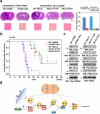
Figure 7. MLC2 Phosphorylation by PKM2 Promotes Tumorigenesis
(a, c) U87/EGFRvIII (5 × 10⁵) cells with PKM2 depletion and reconstituted expression of WT rPKM2 or rPKM2 T45A or with MLC2 depletion and reconstituted expression of WT rMLC2, rMLC2 Y118F, or rMLC2 S15A were intracranially injected into athymic nude mice. The mice were sacrificed 14 days after injection of cells and examined for tumor growth. Hematoxylin-and-eosin-stained coronal brain sections show representative tumor xenografts. Scale bars for original images, 0.2 cm; Scale bars for amplified images, 50 μm. Tumor volumes were measured using length (a) and width (b) and calculated using the equation: V = ab2/2. The data represent the means ± SD of seven mice (right panel). The tumor tissues normalized with expression levels of Flag-tagged rPKM2 or rMLC2 were immunoblotted with the indicated antibodies (c). (b) U87/EGFRvIII (5 × 10⁵) cells with PKM2 depletion and reconstituted expression of WT rPKM2 or rPKM2 T45A or with MLC2 depletion and reconstituted expression of WT rMLC2, rMLC2 Y118F, or rMLC2 S15A were intracranially injected into athymic nude mice. Data were presented by the Kaplan-Meier plots of 10 mice. (d) A Mechanism of PKM2-regulated cytokinesis. Aurora B-phosphorylated PKM2 at T45 results in PKM2's localization and its interaction with MLC2 at the equator/contractile ring region of mitotic cells, where PKM2 phosphorylates MLC2 at Y118. MLC2 Y118 phosphorylation primes ROCK2's binding to MLC2, ROCK2-dependent MLC2 S15 phosphorylation, and subsequent cytokinesis progression.

Similar articles

Cited by

References

    1. Pollard TD. Mechanics of cytokinesis in eukaryotes. Current opinion in cell biology. 2010;22:50–56. - PMC - PubMed
    1. Green RA, Paluch E, Oegema K. Cytokinesis in animal cells. Annual review of cell and developmental biology. 2012;28:29–58. - PubMed
    1. Sagona AP, Stenmark H. Cytokinesis and cancer. FEBS letters. 2010;584:2652–2661. - PubMed
    1. Glotzer M. The molecular requirements for cytokinesis. Science. 2005;307:1735–1739. - PubMed
    1. Matsumura F, Totsukawa G, Yamakita Y, Yamashiro S. Role of myosin light chain phosphorylation in the regulation of cytokinesis. Cell structure and function. 2001;26:639–644. - PubMed

Publication types

MeSH terms

Substances