. 2014 Nov 20;5(11):e1528.
doi: 10.1038/cddis.2014.479.

Activation of volume-sensitive outwardly rectifying chloride channel by ROS contributes to ER stress and cardiac contractile dysfunction: involvement of CHOP through Wnt

Affiliations

Activation of volume-sensitive outwardly rectifying chloride channel by ROS contributes to ER stress and cardiac contractile dysfunction: involvement of CHOP through Wnt

M Shen et al. Cell Death Dis. .

Abstract

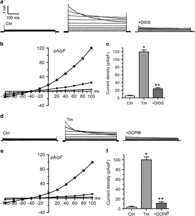
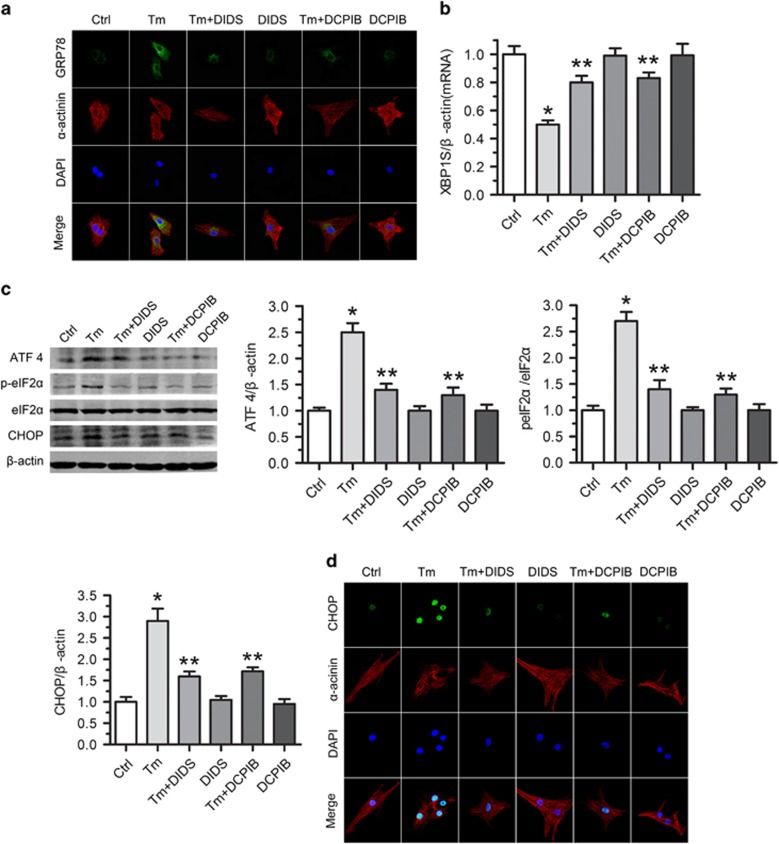
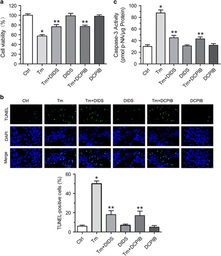
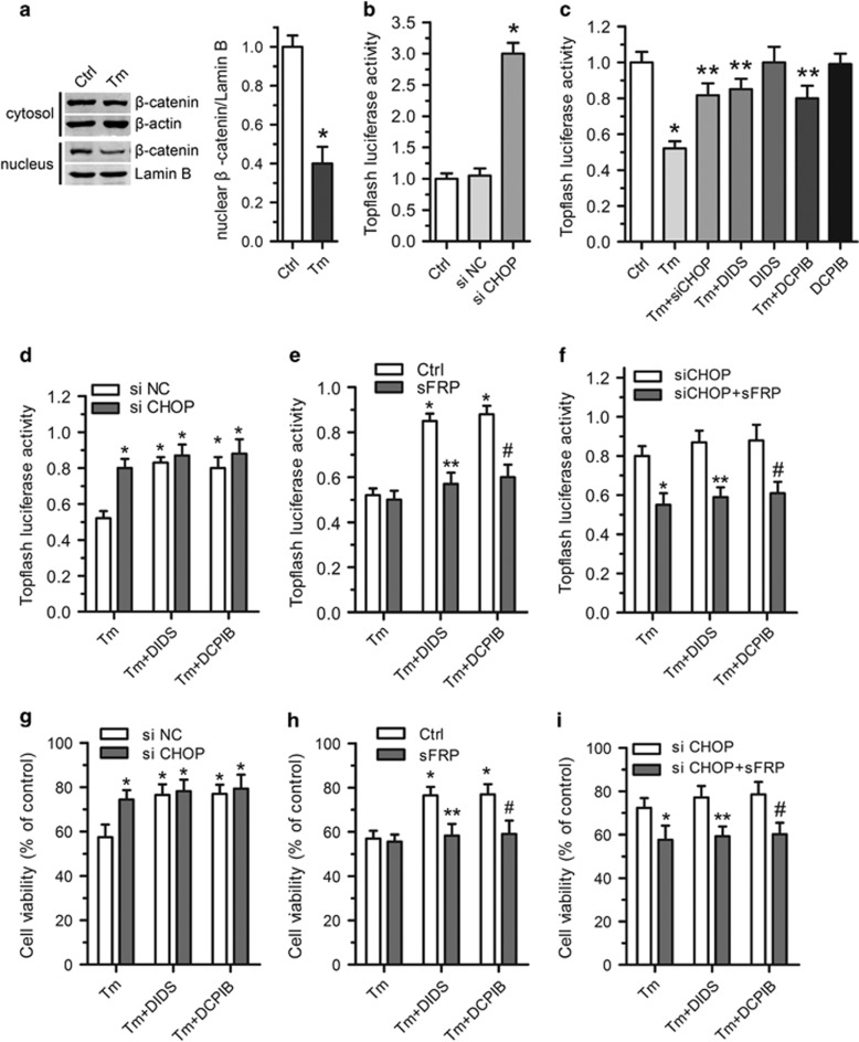
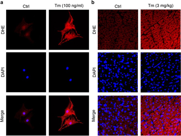
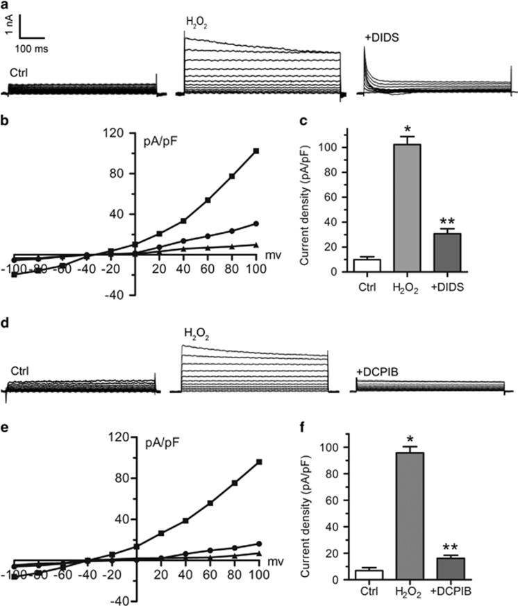
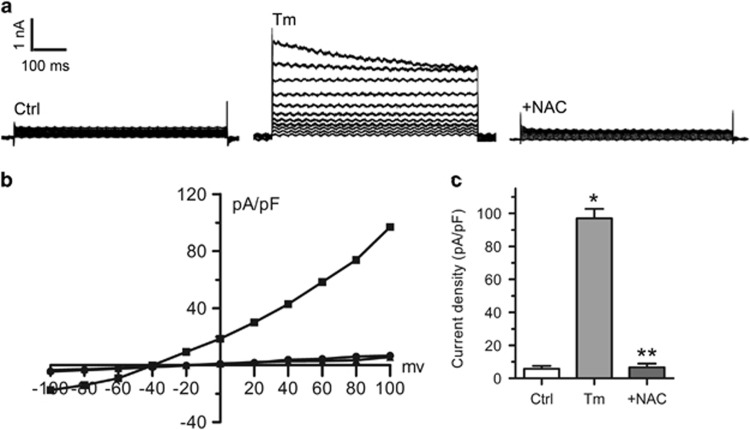
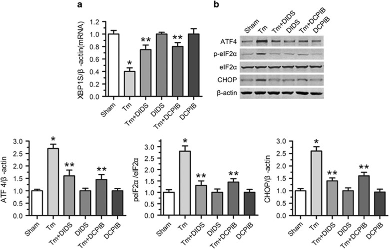
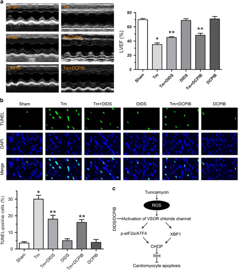
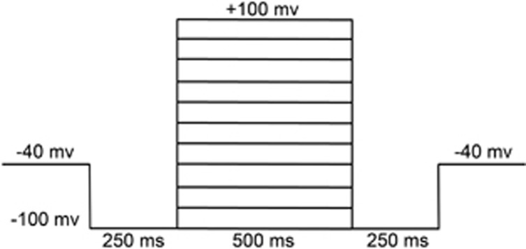
Endoplasmic reticulum (ER) stress occurring in stringent conditions is critically involved in cardiomyocytes apoptosis and cardiac contractile dysfunction (CCD). However, the molecular machinery that mediates cardiac ER stress and subsequent cell death remains to be fully deciphered, which will hopefully provide novel therapeutic targets for these disorders. Here, we establish tunicamycin-induced model of cardiomyocyte ER stress, which effectively mimicks pathological stimuli to trigger CCD. Tunicamycin activates volume-sensitive outward rectifying Cl(-) currents. Blockade of the volume-sensitive outwardly rectifying (VSOR) Cl(-) channel by 4,4'-diisothiocya-natostilbene-2,2'-disulfonic acid (DIDS), a non-selective Cl(-) channel blocker, and 4-(2-butyl-6,7-dichlor-2-cyclopentyl-indan-1-on-5-yl) oxybutyric acid (DCPIB), a selective VSOR Cl(-) channel blocker, improves cardiac contractility, which correlates with suppressed ER stress through inhibiting the canonical GRP78/eIF2α/ATF4 and XBP1 pathways, and promotes survival of cardiomyocytes by inverting tunicamycin-induced decrease of Wnt through the CHOP pathway. VSOR activation of tunicamycin-treated cardiomyocytes is attributed to increased intracellular levels of reactive oxygen species (ROS). Our study demonstrates a pivotal role of ROS/VSOR in mediating ER stress and functional impairment of cardiomyocytes via the CHOP-Wnt pathway, and suggests the therapeutic values of VSOR Cl(-) channel blockers against ER stress-associated cardiac anomalies.


Figures

Figure 1
Figure 1
Increased VSOR Cl currents in tunicamycin exposed cardiomyocytes. (a) Negligible background Cl currents recorded under isosmotic solution (Ctrl). Tm (3 μg/ml)-induced Cl currents exhibiting representative properties of VSOR Cl currents (Tm). Tm-induced VSOR Cl currents were inhibited by adding DIDS (500 μM); n=5 for each group. (b) Corresponding current-voltage (I-V) relationship for the mean current densities of Ctrl (▴), Tm (▪) and Tm with DIDS (●) conditions. (c) Current densities at +100 mV from b. *P<0.05 versus Ctrl; **P<0.05 versus Tm, n=5. (d) Negligible background Cl currents recorded under isosmotic solution (Ctrl). Tm (3 μg/ml)-induced Cl currents exhibiting representative properties of VSOR Cl currents (Tm). Tm-induced VSOR Cl currents were inhibited by adding DCPIB (10 μM); n=5 for each group. (e) Corresponding current-voltage (I-V) relationship for the mean current densities of Ctrl (▴), Tm (▪) and Tm with DCPIB (●) conditions. (f) Current densities at +100 mV from (e). *P<0.05 versus Ctrl; **P<0.05 versus Tm, n=5
Figure 2
Figure 2
VSOR Cl channel blockers rescue tunicamycin-induced ER stress in cardiomyocytes. Cardiomyocytes were treated with Tm (100 ng/ml) in the presence or absence of DIDS or DCPIB for 24 h. (a) Representative images of immunostaining for GRP78 (Green). Nuclei were labeled with DAPI; n=5 for each group. (b) qRT-PCR assay for XBP1S expression. β-Actin served as a loading control. *P<0.05 versus ctrl; **P<0.05 versus Tm, n=5. (c) Western blot analysis and quantitative assay for ATF4, p-eIF2α and CHOP protein expressions. β-Actin served as a loading control. *P<0.05 versus ctrl; **P<0.05 versus Tm, n=5. (d) Expression of CHOP by immunofluorescence (Green). Nuclei were counterstained with DAPI; n=5 for each group
Figure 3
Figure 3
Protective effect of VSOR Cl channel inhibitors on tunicamycin-induced cardiomyocyte death. Cardiomyocytes were treated with Tm (100 ng/ml) in the presence or absence of DIDS or DCPIB for 72 h. (a) Protective effect of DIDS and DCPIB on Tm-induced cardiomyocyte viability measured by MTT-assay, respecitvely. *P<0.05 versus ctrl; **P<0.05 versus Tm, n=5. (b) TUNEL staining of cardiomyocytes. Quantitative analysis of TUNEL-positive cardiomyocytes after Tm treatment with or without DIDS (75 μM) and DCPIB (2 μM) for 72 h. *P<0.05 versus ctrl; **P<0.05 versus Tm, n=5. (c) Activity analysis of caspase-3. *P<0.05 versus ctrl; **P<0.05 versus Tm, n=5
Figure 4
Figure 4
The CHOP-Wnt pathway in VSOR Cl channel blockers mediated protective role. (a) Western blot analysis and quantitative assay of both cytosol and nuclear expression of β-catenin in cardiomyocytes after Tm treatment with or without DIDS (75 μM) and DCPIB (2 μM) for 24 h; n=5 for each group. (b) Topflash and pTK-Rennila luciferase vector were transfected with siNC or siCHOP for 36 h in H9C2 cells. *P<0.05 versus ctrl, n=5. (c) H9C2 cells were transfected with Topflash and pTK-Rennila luciferase vector with or without siCHOP. After 12 h transfection, cells were additionally treated with tunicamycin with or without DIDS, DCPIB for 24 h, respectively. *P<0.05 versus ctrl; **P<0.05 versus Tm, n=5. (df) Topflash and pTK-Rennila luciferase vector with or without siCHOP were transfected for 12 h in H9C2 cells. After 12 h transfection, cells were additionally treated as indicated for 24 h, respectively. (d) *P<0.05 versus Tm+siNC, n=5; (e) *P<0.05 versus Tm; **P<0.05 versus Tm+DIDS; #P<0.05 versus Tm+DCPIB, n=5; (f) *P<0.05 versus Tm+siCHOP; **P<0.05 versus Tm+siCHOP+DIDS; #P<0.05 versus Tm+siCHOP+DCPIB, n=5. (gi) Cardiomyocytes were transfected with or without siCHOP for 24 h, and then cells were additionally treated as indicated for 72 h. Cell viability was measured by the MTT-assay in cardiomyocytes. (g) *P<0.05 versus Tm+siNC, n=5; (h) *P<0.05 versus Tm; **P<0.05 versus Tm+DIDS; #P<0.05 versus Tm+DCPIB, n=5; (i) *P<0.05 versus Tm+siCHOP; **P<0.05 versus Tm+siCHOP+DIDS; #P<0.05 versus Tm+siCHOP+DCPIB, n=5
Figure 5
Figure 5
Tunicamycin induced ROS production in vitro and in vivo. (a) Effect of Tm (100 ng/ml) for 48 h on the ROS level of cardiomyocytes monitored by dihydroethidine (DHE) staining (Red). Nuclei were counterstained with DAPI; n=5 for each group. (b) Increased ROS accumulation in tunicamycin (3 mg/kg, 48 h, i.p.)-exposed myocardium was revealed by DHE staining (Red). Nuclei were counterstained with DAPI; n=5 for each group
Figure 6
Figure 6
ROS induce VSOR Cl currents in cardiomyocytes. (a) Background Cl currents recorded under isosmotic solution (Ctrl). H2O2 (500 μM)-induced Cl currents exhibiting phenotypic properties of ICl,Vol (H2O2). H2O2-induced VSOR Cl currents were inhibited by adding DIDS (500 μM); n=5 for each group. (b) Corresponding current-voltage (I-V) relationship for the mean current densities of isosmotic (▴), H2O2 (▪) and H2O2 with DIDS (●) conditions. (c) Current densities at +100 mV from B. *P<0.05 versus Ctrl; **P<0.05 versus H2O2, n=5. (d) Negligible background Cl currents recorded under isosmotic solution (Ctrl). H2O2 (500 μM)-induced Cl currents exhibiting representative properties of VSOR Cl currents (H2O2). H2O2-induced VSOR Cl currents were inhibited by adding DCPIB (10 μM). n=5 for each group. (e) Corresponding current-voltage (I-V) relationship for the mean current densities of Ctrl (▴), Tm (▪) and Tm with DIDS (●) conditions. (f) Current densities at +100 mV from (e). *P<0.05 versus Ctrl; **P<0.05 versus H2O2, n=5
Figure 7
Figure 7
ROS production mediates tunicamycin induced VSOR Cl currents. (a) Background Cl currents recorded under isosmotic solution (Ctrl). Tm (3 μg/ml)-induced VSOR Cl currents exhibiting phenotypic properties of ICl,Vol (Tm). Tm-induced VSOR Cl currents were inhibited by the ROS scavenger NAC (10 mM). n=5 for each group. (b) Corresponding current-voltage (I-V) relationship for the mean current densities of isosmotic (▴), Tm (▪) and Tm with NAC (●) conditions. (c) Current densities at +100 mV from (b). *P<0.05 versus Ctrl; **P<0.05 versus Tm, n=5
Figure 8
Figure 8
VSOR Cl channel blockers rescue tunicamycin-induced ER stress in vivo. VSOR Cl channel was blocked with DIDS and DCPIB for 24 h before assessment of ER stress. (a) qRT-PCR assay for XBP1S expression. β-actin served as a loading control. *P<0.05 versus Sham; **P<0.05 versus Tm, n=5. (b) Western blot analysis and quantitative assay for ATF4, p-eIF2α and CHOP protein expressions. β-actin served as a loading control. *P<0.05 versus Sham; **P<0.05 versus Tm, n=5
Figure 9
Figure 9
VSOR Cl channel blockers improve cardiac function and attenuate tunicamycin-induced cardiomyocyte apoptosis. VSOR Cl channel was blocked with DIDS and DCPIB for 48 h before assessment of cardiac function and apoptosis, respectively. (a) Echocardiographic assessment of the left ventricular ejection fraction (LVEF). *P<0.05 versus Sham; **P<0.05 versus Tm, n=5. (b) TUNEL staining of apoptotic cells in myocardium, and quantified data displaying % of apoptosis. *P<0.05 versus Sham; **P<0.05 versus Tm, n=5. (c) Schematic representation of how VSOR Cl channel involved in tunicamycin-induced ER stress. Tunicamycin results in increased ROS production in the heart, which in turn activates VSOR Cl currents. VSOR Cl currents lead to increased ER stress, resulting in increased cell apoptosis and cardiac contractile dysfunction through CHOP-dependent regulation of Wnt expression.
Figure 10
Figure 10
Corresponding step protocol used to elicit current trace. To observe the current–voltage relationships, step pulses were generated from a holding potential of −40 mV to test potentials from −100 to +100 mV with 20 mV increments. To record the greater magnitude of pulse-induced currents, the −100 mv conditioning pulse was applied before and after test potentials.

Similar articles

Cited by

References

    1. Kaufman RJ. Stress signaling from the lumen of the endoplasmic reticulum: coordination of gene transcriptional and translational controls. Genes Dev. 1999;13:1211–1233. - PubMed
    1. Hetz C. The unfolded protein response: controlling cell fate decisions under ER stress and beyond. Nat Rev Mol Cell Biol. 2012;13:89–102. - PubMed
    1. Sciarretta S, Zhai P, Shao D, Zablocki D, Nagarajan N, Terada LS, et al. Activation of Nox4 in the Endoplasmic Reticulum Promotes Cardiomyocyte Autophagy and Survival During Energy Stress Through the PERK/eIF-2alpha/ATF4 Pathway. Circ Res. 2013;113:1253–1264. - PMC - PubMed
    1. He B. Viruses, endoplasmic reticulum stress, and interferon responses. Cell Death Differ. 2006;13:393–403. - PubMed
    1. Hotamisligil GS. Endoplasmic reticulum stress and atherosclerosis. Nat Med. 2010;16:396–399. - PMC - PubMed

Publication types

MeSH terms

Substances