Review
. 2014 Nov 4:8:348.
doi: 10.3389/fncel.2014.00348. eCollection 2014.

Connexons and pannexons: newcomers in neurophysiology

Affiliations
Review

Connexons and pannexons: newcomers in neurophysiology

Giselle Cheung et al. Front Cell Neurosci. .

Abstract

Connexin hemichannels are single membrane channels which have been traditionally thought to work in pairs to form gap junction channels across two opposing cells. In astrocytes, gap junction channels allow direct intercellular communication and greatly facilitate the transmission of signals. Recently, there has been growing evidence demonstrating that connexin hemichannels, as well as pannexin channels, on their own are open in various conditions. They allow bidirectional flow of ions and signaling molecules and act as release sites for transmitters like ATP and glutamate into the extracellular space. While much attention has focused on the function of connexin hemichannels and pannexons during pathological situations like epilepsy, inflammation, neurodegeneration or ischemia, their potential roles in physiology is often ignored. In order to fully understand the dynamic properties and roles of connexin hemichannels and pannexons in the brain, it is essential to decipher whether they also have some physiological functions and contribute to normal cerebral processes. Here, we present recent studies in the CNS suggesting emerging physiological functions of connexin hemichannels and pannexons in normal neuronal activity and behavior. We also discuss how these pioneer studies pave the way for future research to extend the physiological relevance of connexons and pannexons, and some fundamental issues yet to be addressed.

Keywords: astrocytes; connexins; hemichannels; learning and memory; neurons; pannexins; plasticity; synapses.


Figures

Figure 1
Figure 1
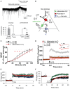
Astrocytic Cx43 HCs modulate synaptic transmission in hippocampal slices. (A–C) Basal astroglial Cx43 HC activity enhances excitatory synaptic transmission via ATP signaling. (A) Representative image showing ethidium bromide uptake (EtBr; red) in astrocytes (immunostained for GFAP; green) of the stratum radiatum in an acute hippocampal slice. Scale bar, 50 μm. (B) Bar graphs showing astrocytic EtBr uptake in brain slices obtained from wild-type (WT) and astroglial conditional Cx43 KO (Cx43−/−) mice normalized to control (untreated) conditions. Uptake was significantly deceased in WT slices treated with carbenoxolone (CBX, 200 μM) and Gap26 (100 μM), but not Gap26 scramble (100 μM) and 10panx (400 μM) peptides. In Cx43−/− slices, however, both CBX and Gap26 had no significant effect. (C) Bar graph on the left showing a decrease in amplitude of evoked EPSC recorded in CA1 pyramidal neurons during Gap26 application (red) as compared to before (Ct, black). Bar graph on the right showing that pretreatment with ATP P2 receptor antagonists (RB2 + PPADS, gray) occludes the effect of Gap26 (red). Sample traces of corresponding evoked EPSCs are shown above. Scale bar: 20 pA, 20 ms (left); 40 pA, 40 ms (right). (D–F) Cx43 HCs in astrocytes promote feedback inhibitory transmission by releasing ATP. (D) Representative recordings showing that photolysis of diazo-2, represented by lightning bolts, (i) evokes depolarization and bursting in interneurons, with the depolarization persisting with 1 μM TTX, and (ii) transiently increases the frequency of spontaneous inhibitory postsynaptic currents (IPSCs) in CA1 pyramidal neurons. (E) Bar graph indicating increased IPSC frequency in pyramidal neurons after diazo-2 photolysis compared to control condition. This effect was blocked by the P2Y1 receptor antagonist MRS2179 (50 μM) or in brain slices prepared from Cx43/Cx30KO mice. (F) Schematic diagram illustrating a proposed negative feedback mechanism during excitatory transmission. During glutamatergic signaling, Ca2+ influx into neurons results in a localized decrease in [Ca2+]e, which in turn opens Cx43 HCs on astrocytes through which ATP is released. ATP can either trigger slowly propagating astrocytic Ca2+ waves or, when degraded to ADP, depolarize and increase firing in interneurons via P2Y1 receptors, thereby enhancing inhibitory transmission. *p < 0.05; **p < 0.01; ***p < 0.001. Adapted, with permission, from Torres et al. (2012) (D–F).
Figure 2
Figure 2
Panx1 channels modulate neuronal excitability, synaptic transmission and plasticity in hippocampal slices. (A,B) Metabolic autocrine regulation of neuronal activity via Panx1 channels and adenosine. (A) Sample trace showing increased outward current upon reduced extracellular glucose (from 11 to 3 mM) and subsequent reversal to baseline with 10panx application (100 μM) in CA3 pyramidal neurons. Bar graphs showing the reversal of reduced glucose-induced outward current amplitude with 10panx (left), and that pretreatment with 10panx prevented reduced glucose-induced outward current. **p < 0.01. (B) Schematic showing a proposed model of purinergic autocrine regulation in CA3 pyramidal neurons. When [ATP]i is sufficient (1), low [Glucose]e (2) induces ATP release from Panx1 channels on neurons (3). ATP is then dephosphorylated to adenosine (4) which activates adenosice A1R rceptors (5). KATP channels are then opened leading to a decrease in neuronal excitability. (C–F) Panx1 regulates synaptic transmission, LTP and LTD. (C) Input-Output curves showing increased synaptic transmission in Panx1−/− (black line) compared to control Panx1+/+ (dashed line) mice. Such effect was abolished in Panx1−/− slices treated with 3 μM adenosine (red line). *p < 0.01; **p < 0.001. (D) LTP evoked by high frequency stimulation (four trains of 10 shocks at 100 Hz every 1 s; HFS) is enhanced in Panx1−/− (filled gray) compared to control Panx1+/+ (open gray) mice. Adenosine treatment in Panx1−/− slices (filled red) restores LTP levels to that of untreated control mice. Figure insets illustrate responses before and 30 min post HFS. Scale bar: 0.5 mV, 10 ms. (E) LTP induced by the delivery of theta burst stimulation protocol (TBS) is increased in adult Panx1−/− (green) compared to Panx1+/+ (black) mice, whereas no difference was observed between young mice (+/+, gray; −/−, blue). In the presence of 100 μM probenecid (Panx1 channel blocker; red), only transient LTP was enhanced. (F) Similarly, LTD induced by paired-pulse low frequency stimulation protocol (1Hz for 15 min; PP-LFS) are impaired in adult Panx1−/− (green) compared to Panx1+/+ (black) mice, whereas no difference was observed between young equivalent (+/+, gray; −/−, blue). In the presence of 100 μM probenecid, only a transient LTD was observed. Adapted, with permission, from Kawamura et al. (2010) (A,B), Prochnow et al. (2012) (C,D) and Ardiles et al. (2014) (E,F).
Figure 3
Figure 3
Cx HCs and Panx1 channels have significant roles in learning and memory. (A,B) Cx43 HC function is required in fear conditioning memory consolidation. (A) Diagram showing site of microinfusions of the TAT-Cx43L2, a selective Cx43 HC blocker, into the basolateral amydala (BLA). Asterisks indicate the tips of injection cannula (shaded regions). Enlargement is shown in insets. LA, lateral amygdala; CeA, central amygdala; SI, somatosensory primary; PRh, perirhinal; Ect, ectorhinal; Pir, piriform; and AI, auditory primary cortices; CPu, caudoputamen; ic, internal capsula. Scale bar: 1 mm. (B) Fear conditioning memory was tested by first training rats to associate a tone with a foot shock. Their memory of this association was then assessed by how long they remain immobile (freeze) in response to the same tone alone 24 h later. TAT-Cx43L2 (10 nM) microinfusion prior to training impaired fear conditioning memory (decreased in freezing time) compared to control. Such effect was rescued by co-microinfusion of a mixture of gliotransmitters including D-serine, glutamate, glutamine, glycine, ATP, and lactate (cocktail) rescued such effect. (C,D) Panx1 deletion leads to dysfunctions in learning and memory. (C) To assess object recognition, mice were allowed to explore two novel objects (A and B) for 5 min. 1 h later, they were allowed to explore the familiar object A together with a novel object C. Panx1−/− mice spent more time on object A than C compared to Panx1+/+ mice which did the opposite, indicating a deficit in object recognition. (D) Another memory test was carried out where mice were trained to remember locations of hidden cookies which were later removed. During the test, Panx1−/− mice explored locations further from the original locations of cookies compared to the Panx1+/+ mice but not as far as the untrained mice, indicating an impairment but not abolishment in memory. *p < 0.05; **p < 0.001. Adapted, with permission, from Stehberg et al. (2012) (A,B) and Prochnow et al. (2012) (C,D).
Figure 4
Figure 4
Cx HCs and Panx1 channels have significant roles in synaptic transmission essential for vision. (A,B) Cx55.5 HCs are important for contrast sensitivity in zebrafish retina. (A) To measure light-induced feedback responses, cones were first saturated with a 20 μm spot of light. A full-field light flash induced an inward current in cones due to negative feedback from horizontal cells. Cx55.5 mutant (red) cones showed a decreased feedback response compared to wild-type (black), as shown in sample traces. (B) Optokinetic gain, as a measure of contrast sensitivity, was determined by dividing the eye movement velocity by the velocity of the stimulus over a range of contrast in zebrafish larvae. This was significantly decreased in mutant compared to wild-type zebrafish. (C–E) Reciprocal regulation between resting microglia and neuronal activity via Panx1 channels. (C) Glutamate uncaging was performed in the intact zebrafish larvae to evoke Ca2+ activities of tectal neurons within 20 μm around the uncaging point of 1 μm in the soma layer of the optic tectum. From the side of microglia facing the uncaging point (“unc”), the proportion of the number of bulbous normalized to all process tips (“Bulbousunc/Tipunc”) is shown for larvae injected with splice morpholino oligonucleotides (MO) 6-min before (clear) and 24-min (gray) and 59-min (black) after uncaging. The increased in bulbous endings is shown in control MO, but abolished in Panx1 expression downregulation MO1 and MO2. (D) Normalized intensities of Ca2+ activities (light response amplitude) of tectal neurons in vivo evoked by moving bars at indicated frequencies are shown. Response is significantly reduced in neurons after microglial contact (red filled vs. clear bars) as compared to non-contact (black filled vs. clear bars). Numbers of neurons examined are shown on bars. (E) Schematic diagram showing a proposed model of microglial modulations of neuronal activity via Panx1 channels. During neuronal activity, neurons secrete “find me” signal locally (ATP being a candidate) via Panx1 channels, which steer microglial processes toward them (from “Surveying” to “I”). Bulbous endings are then formed on these processes promoting contact with neurons (“II”). Upon such contact, neuronal activity is downregulated (“III”). **p < 0.01; ***p < 0.001. Adapted, with permission, from Klaassen et al. (2011) (A,B), Li et al. (2012) (C,D).

Similar articles

Cited by

References

    1. Amzica F., Massimini M., Manfridi A. (2002). Spatial buffering during slow and paroxysmal sleep oscillations in cortical networks of glial cells in vivo. J. Neurosci. 22, 1042–1053. - PMC - PubMed
    1. Anselmi F., Hernandez V. H., Crispino G., Seydel A., Ortolano S., Roper S. D., et al. . (2008). ATP release through connexin hemichannels and gap junction transfer of second messengers propagate Ca2+ signals across the inner ear. Proc. Natl. Acad. Sci. U.S.A. 105, 18770–18775. 10.1073/pnas.0800793105 - DOI - PMC - PubMed
    1. Ardiles A. O., Flores-Muñoz C., Toro-Ayala G., Cárdenas A. M., Palacios A. G., Muñoz P., et al. (2014). Pannexin 1 regulates bidirectional hippocampal synaptic plasticity in adult mice. Front. Cell Neurosci. 8:326 10.3389/fncel.2014.00326 - DOI - PMC - PubMed
    1. Baranova A., Ivanov D., Petrash N., Pestova A., Skoblov M., Kelmanson I., et al. . (2004). The mammalian pannexin family is homologous to the invertebrate innexin gap junction proteins. Genomics 83, 706–716. 10.1016/j.ygeno.2003.09.025 - DOI - PubMed
    1. Bargiotas P., Krenz A., Hormuzdi S. G., Ridder D. A., Herb A., Barakat W., et al. . (2011). Pannexins in ischemia-induced neurodegeneration. Proc. Natl. Acad. Sci. U.S.A. 108, 20772–20777. 10.1073/pnas.1018262108 - DOI - PMC - PubMed

LinkOut - more resources