. 2014 Nov 18;9(1):24.
doi: 10.1186/1745-6150-9-24.

Minimization of extracellular space as a driving force in prokaryote association and the origin of eukaryotes

Affiliations

Minimization of extracellular space as a driving force in prokaryote association and the origin of eukaryotes

Scott L Hooper et al. Biol Direct. .

Erratum in

Abstract

Background: Internalization-based hypotheses of eukaryotic origin require close physical association of host and symbiont. Prior hypotheses of how these associations arose include chance, specific metabolic couplings between partners, and prey-predator/parasite interactions. Since these hypotheses were proposed, it has become apparent that mixed-species, close-association assemblages (biofilms) are widespread and predominant components of prokaryotic ecology. Which forces drove prokaryotes to evolve the ability to form these assemblages are uncertain. Bacteria and archaea have also been found to form membrane-lined interconnections (nanotubes) through which proteins and RNA pass. These observations, combined with the structure of the nuclear envelope and an energetic benefit of close association (see below), lead us to propose a novel hypothesis of the driving force underlying prokaryotic close association and the origin of eukaryotes.

Results: Respiratory proton transport does not alter external pH when external volume is effectively infinite. Close physical association decreases external volume. For small external volumes, proton transport decreases external pH, resulting in each transported proton increasing proton motor force to a greater extent. We calculate here that in biofilms this effect could substantially decrease how many protons need to be transported to achieve a given proton motor force. Based as it is solely on geometry, this energetic benefit would occur for all prokaryotes using proton-based respiration.

Conclusions: This benefit may be a driving force in biofilm formation. Under this hypothesis a very wide range of prokaryotic species combinations could serve as eukaryotic progenitors. We use this observation and the discovery of prokaryotic nanotubes to propose that eukaryotes arose from physically distinct, functionally specialized (energy factory, protein factory, DNA repository/RNA factory), obligatorily symbiotic prokaryotes in which the protein factory and DNA repository/RNA factory cells were coupled by nanotubes and the protein factory ultimately internalized the other two. This hypothesis naturally explains many aspects of eukaryotic physiology, including the nuclear envelope being a folded single membrane repeatedly pierced by membrane-bound tubules (the nuclear pores), suggests that species analogous or homologous to eukaryotic progenitors are likely unculturable as monocultures, and makes a large number of testable predictions.

Reviewers: This article was reviewed by Purificación López-García and Toni Gabaldón.


Figures
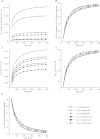
Figure 1
Figure 1
Effects of close association on respiration. A) The number of protons that need to pumped across the membrane to achieve a -200 mV Δp decrease as inter-cell spacing (gap) decreases. B) When normalized to the number of protons needed to achieve a -200 mV Δp with an infinite gap, the effects of cell shape essentially disappear and the effects of cell size are diminished (that is, the six curves now nearly overlap). This overlap shows that each cell type derives approximately the same relative energetic benefit from decreases in gap. These benefits are substantial, a 30% reduction in number of protons needed to be pumped (relative to the infinite case) at a 7 x 10⁻⁸ M gap and a 50% reduction at 3.5 x 10⁻⁸ M gap. C) Decreasing gap decreases the cytoplasmic alkalization necessary to achieve the Δp. D) Decreasing gap increases the acidification of the extracellular medium, but even with small gap this acidification is not extreme. E) Decreasing gap strongly increases the percentage of the Δp arising from the concentration term (and hence decreases the percentage arising from the electrical term) of the proton motor force equation. Key applies to all panels.
Figure 2
Figure 2
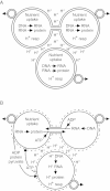
Two stages of proposed origin of eukaryotes. A) Pre-specialization close association. Three species closely associating because of respiratory energetic benefit, but maintaining independent nutrient uptake, respiration, DNA, RNA, and protein functions. B) Later stage in which cells have specialized to perform certain life functions (i.e., are obligate symbionts: left cell, protein factory; right cell, DNA repository/RNA factory; bottom cell, respirer), in which cell wall expression is regulated (and would be absent during symbiotic portions of existence, dashed outer large circles), but before internalization of any entity and while each still produces separate offspring. “H+ DNA” and “H+ RNA” represent the genetic material and abilities the respiratory symbiont maintains. In both panels small double-circled entities with outward pointing arrows represent offspring.
Figure 3
Figure 3
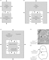
Internalization of the proto-mitochondrion cell. A1) Initial stage before proto-cytoplasmic cell has begun to surround proto-mitochondrion cell. A2) Intermediate stage at which proto-cytoplasmic cell has largely surrounded proto-mitochondrion cell. A3) Full internalization; proto-mitochondrion cell is now a mitochondrion; the two cells are now a single cell that possesses a respiratory organelle. The cell membrane of the proto-mitochondrion becomes the inner mitochondrial membrane and the outer mitochondrial membrane is derived from the cell membrane of the proto-cytoplasm cell. These are one-dimensional slices through the cells; in three dimensions the proto-cytoplasm cell surrounds the proto-mitochondrion cell on all sides. Filled arrows in A1-A3 represent non-respiratory (e.g., Na/K ATPase) pump activity. These pumps pump Na and Ca out of the cytoplasm into the external medium or lumen, and K out of the external medium or lumen into the cytoplasm. As the proto-cytoplasm cell surrounds the proto-mitochondrion cell, these pumps would thus automatically (i.e., without any change in their orientation in the membrane) work so as to maintain an external-like (high Na and Ca, low K) ionic environment in the lumen separating the cells. “L” shaped lines represent cell anchoring proteins. B1) Electron micrograph showing how the inner membrane enclosed entity of contemporary mitochondria can divide separately from the outer mitochondrial membrane, just as would a prokaryote enclosed by the membrane of another prokaryote. Figure taken with permission from [320]. B2) Tracing showing the membrane disposition in B1.
Figure 4
Figure 4
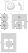
Internalization of the proto-nucleus. A) Schematic showing that the nuclear membrane is a single membrane due to the membrane-lined nuclear pores. B1) Initial stage before proto-cytoplasmic cell has begun to surround proto-nucleus cell. B2) Intermediate stage at which proto-cytoplasmic cell has largely surrounded proto-nucleus cell. B3) Full internalization; proto-nucleus is now a nucleus; the two cells are now a single cell that possesses a nucleus. These are one-dimensional slices through the cells; in three dimensions the proto-cytoplasm cell surrounds the proto-nucleus cell on all sides. Filled arrows in B1-B3 represent non-respiratory (e.g., Na/K ATPase) pump activity. These pumps pump Na and Ca out of the cytoplasm into the external medium or lumen, and K out of the external medium or lumen into the cytoplasm. As the proto-cytoplasm cell surrounds the proto-nucleus cell, these pumps would thus automatically (i.e., without any change in their orientation in the membrane) work so as to maintain an external-like (high Na and Ca, low K) ionic environment in the lumen separating the cells. “L” shaped lines represent cell anchoring proteins; lines are grey because it is possible the nanotubes alone could provide the physical support for internalization. Nanotubes are represented by the grey connections between the cells. Because these drawings are slices through the cell, it appears that the nanotubes separate the lumen into compartments. However, because these are tubes, the lumen is actually continuous (see Figure 5B2).
Figure 5
Figure 5
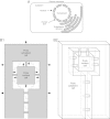
Origin of endoplasmic reticulum. A) Cross-section schematic showing that the endoplasmic reticulum is an outgrowth of the “outer” nuclear membrane and makes close appositions with the plasma membrane. Note that this outgrowth does not alter how the nuclear pores connect the cyto- and nucleoplasms. B1) Schematic showing how in the late stage of proto-nucleus cell internalization, an exterior-medium filled “tail” continuous with the lumen surrounding the proto-nucleus cell could form. Dashed lines show how the tail would eventually be separated from the external medium by fusion of proto-cytoplasm cell membrane. Filled arrows represent non-respiratory (e.g., Na/K ATPase) pump activity. These pumps pump Na and Ca out of the cytoplasm into the external medium or lumen, and K out of the external medium or lumen into the cytoplasm. As the proto-cytoplasm cell surrounds the proto-nucleus cell, these pumps would thus automatically (i.e., without any change in their orientation in the membrane) work so as to maintain an external-like (high Na and Ca, low K) ionic environment in the lumen separating the cells. “L” shaped lines represent cell anchoring proteins; lines are grey because the nanotubes alone possibly could provide the physical support for internalization. Nanotubes are represented by the grey connections between the cells. Because this drawing is a slice through the cells, it appears that the nanotubes separate the lumen into compartments. However, because the nanotubes are cylinders, the lumen is actually continuous (see B2). B2) Three dimensional drawing of a thick slice (a slab) through the cells at stage B1 showing how the lumen and tail form a single, contiguous compartment. B1 and B2 are slices through the cells; in full three dimensional drawings, the proto-cytoplasm cell would surround the proto-nucleus cell on all sides and the tail would be a pipe-like structure.
Figure 6
Figure 6
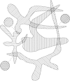
Theoretical implications for prokaryotic ecology and prokaryotic genetic diversity of hypotheses proposed here. The irregular shapes filled with different hatching represent different groups of related species; their extended shapes symbolize each group’s genetic diversity. The regions of overlap are subsets of each group genetically specialized to form symbiotic relationships with species in other groups. The circles connected to certain overlaps by dashed lines represent obligatorily symbiotic species, one from each group, that have lost the ability to fulfill complimentary life functions, and thus can reproduce (and be cultured) only as pairs.

Similar articles

Cited by

References

    1. Anderson SGE, Zomorodipour A, Anderson JO, Sicheritz-Ponten T, Alsmark UCM, Podowski RM, Naslun AK, Eriksson A, Winkler HH, Kurland CG. The genome sequence of Rickettsia prowazekii and the origin of mitochondria. Nature. 1998;396:133–140. doi: 10.1038/24094. - DOI - PubMed
    1. Atteia A, Adrait A, Brugière S, Tardif M, van Lis R, Deusch O, Degan T, Kuhn L, Gontero B, Martin W, Garin J, Joyard J, Rolland N. A proteomic survey of Chlamydomonas reinhardtii mitochondria sheds new light on the metabolic plasticity of the organelle and on the nature of the α-proteobacterial mitochondrial ancestor. Mol Biol Evol. 2009;26:1533–1548. doi: 10.1093/molbev/msp068. - DOI - PubMed
    1. Esser C, Ahmadinejad N, Wiegand C, Rotte C, Sebastiani F, Gelius-Dietrich G, Henze K, Kretschmann E, Richly E, Leister D, Bryant D, Steel MA, Lockhart PJ, Penny D, Martin W. A genome phylogeny for mitochondria among α-proteobacterial and a predominantly eubacterial ancestry of yeast nuclear genes. Mol Biol Evol. 2004;21:1643–1660. doi: 10.1093/molbev/msh160. - DOI - PubMed
    1. Lasek-Nesselquist E, Gogarten JP. The effects of model chioce and mitigating bias on the ribosomal tree of life. Mol Phylogenet Evol. 2013;69:17–38. doi: 10.1016/j.ympev.2013.05.006. - DOI - PubMed
    1. Martin W, Hoffmeister M, Rotte C, Henze K. An overview of endosymbiotic models for the origins of eukaryotes, their ATP-producing organelles (mitochondria and hydrogenosomes), and their heterotrophic lifestyle. Biol Chem. 2001;382:1521–1539. doi: 10.1515/BC.2001.187. - DOI - PubMed

LinkOut - more resources