

Diffusion tensor imaging of Parkinson's disease, multiple system atrophy and progressive supranuclear palsy: a tract-based spatial statistics study
- PMID: 25405990
- PMCID: PMC4236070
- DOI: 10.1371/journal.pone.0112638
Diffusion tensor imaging of Parkinson's disease, multiple system atrophy and progressive supranuclear palsy: a tract-based spatial statistics study
Abstract
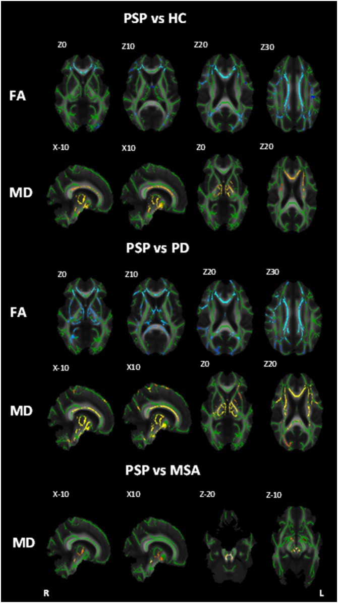
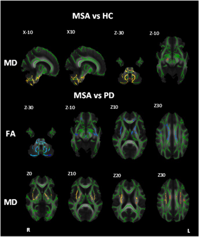
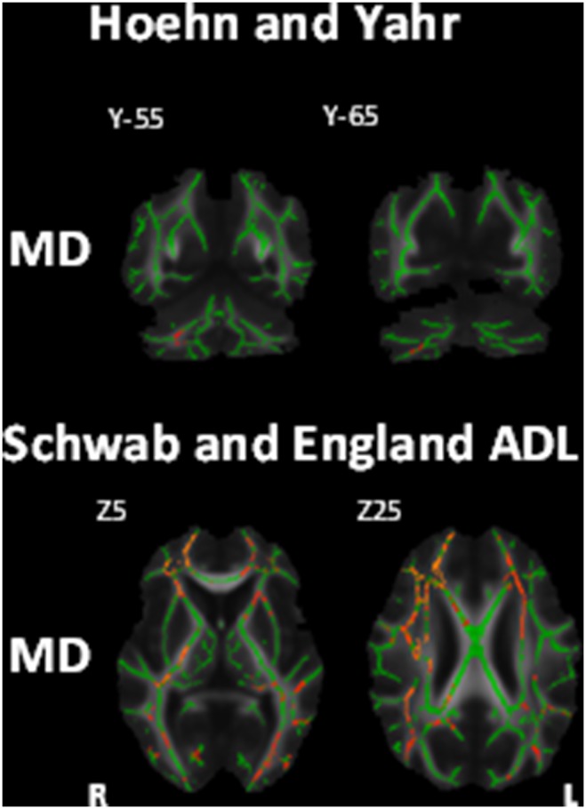
Although often clinically indistinguishable in the early stages, Parkinson's disease (PD), Multiple System Atrophy (MSA) and Progressive Supranuclear Palsy (PSP) have distinct neuropathological changes. The aim of the current study was to identify white matter tract neurodegeneration characteristic of each of the three syndromes. Tract-based spatial statistics (TBSS) was used to perform a whole-brain automated analysis of diffusion tensor imaging (DTI) data to compare differences in fractional anisotropy (FA) and mean diffusivity (MD) between the three clinical groups and healthy control subjects. Further analyses were conducted to assess the relationship between these putative indices of white matter microstructure and clinical measures of disease severity and symptoms. In PSP, relative to controls, changes in DTI indices consistent with white matter tract degeneration were identified in the corpus callosum, corona radiata, corticospinal tract, superior longitudinal fasciculus, anterior thalamic radiation, superior cerebellar peduncle, medial lemniscus, retrolenticular and anterior limb of the internal capsule, cerebral peduncle and external capsule bilaterally, as well as the left posterior limb of the internal capsule and the right posterior thalamic radiation. MSA patients also displayed differences in the body of the corpus callosum corticospinal tract, cerebellar peduncle, medial lemniscus, anterior and superior corona radiata, posterior limb of the internal capsule external capsule and cerebral peduncle bilaterally, as well as the left anterior limb of the internal capsule and the left anterior thalamic radiation. No significant white matter abnormalities were observed in the PD group. Across groups, MD correlated positively with disease severity in all major white matter tracts. These results show widespread changes in white matter tracts in both PSP and MSA patients, even at a mid-point in the disease process, which are not found in patients with PD.
Conflict of interest statement
Figures
Similar articles
-
Disease-specific structural changes in thalamus and dentatorubrothalamic tract in progressive supranuclear palsy.Neuroradiology. 2015 Nov;57(11):1079-91. doi: 10.1007/s00234-015-1563-z. Epub 2015 Aug 8. Neuroradiology. 2015. PMID: 26253801 Free PMC article.
-
Alterations of mean diffusivity in brain white matter and deep gray matter in Parkinson's disease.Neurosci Lett. 2013 Aug 29;550:64-8. doi: 10.1016/j.neulet.2013.06.050. Epub 2013 Jul 3. Neurosci Lett. 2013. PMID: 23831353
-
Diffusion tensor imaging differentiates vascular parkinsonism from parkinsonian syndromes of degenerative origin in elderly subjects.Eur J Radiol. 2014 Nov;83(11):2074-9. doi: 10.1016/j.ejrad.2014.07.012. Epub 2014 Jul 24. Eur J Radiol. 2014. PMID: 25154005
-
The role of diffusion tensor imaging and fractional anisotropy in the evaluation of patients with idiopathic normal pressure hydrocephalus: a literature review.Neurosurg Focus. 2016 Sep;41(3):E12. doi: 10.3171/2016.6.FOCUS16192. Neurosurg Focus. 2016. PMID: 27581308 Review.
-
The value of novel MRI techniques in Parkinson-plus syndromes: diffusion tensor imaging and anatomical connectivity studies.Rev Neurol (Paris). 2014 Apr;170(4):266-76. doi: 10.1016/j.neurol.2013.10.013. Epub 2014 Mar 20. Rev Neurol (Paris). 2014. PMID: 24656811 Review.
Cited by
-
Cortical gray and subcortical white matter associations in Parkinson's disease.Neurobiol Aging. 2017 Jan;49:100-108. doi: 10.1016/j.neurobiolaging.2016.09.015. Epub 2016 Sep 28. Neurobiol Aging. 2017. PMID: 27776262 Free PMC article.
-
Microstructural white matter alterations in patients with drug induced parkinsonism.Hum Brain Mapp. 2017 Dec;38(12):6043-6052. doi: 10.1002/hbm.23809. Epub 2017 Sep 12. Hum Brain Mapp. 2017. PMID: 28901627 Free PMC article.
-
Magnetic resonance imaging and tensor-based morphometry in the MPTP non-human primate model of Parkinson's disease.PLoS One. 2017 Jul 24;12(7):e0180733. doi: 10.1371/journal.pone.0180733. eCollection 2017. PLoS One. 2017. PMID: 28738061 Free PMC article.
-
Disease-specific structural changes in thalamus and dentatorubrothalamic tract in progressive supranuclear palsy.Neuroradiology. 2015 Nov;57(11):1079-91. doi: 10.1007/s00234-015-1563-z. Epub 2015 Aug 8. Neuroradiology. 2015. PMID: 26253801 Free PMC article.
-
Evaluation of white matter microstructure in patients with Parkinson's disease using microscopic fractional anisotropy.Neuroradiology. 2020 Feb;62(2):197-203. doi: 10.1007/s00234-019-02301-1. Epub 2019 Nov 4. Neuroradiology. 2020. PMID: 31680195
References
-
- Litvan I, Bhatia KP, Burn DJ, Goetz CG, Lang AE, et al. (2003) SIC Task Force appraisal of clinical diagnostic criteria for parkinsonian disorders. Movement Disorders 18: 467–486. - PubMed
-
- Braak H, Braak E (2000) Pathoanatomy of Parkinson’s disease. Journal of Neurology 247: II3–II10. - PubMed
-
- Braak H, Tredici KD, Rüb U, de Vos RA, Jansen Steur EN, et al. (2003) Staging of brain pathology related to sporadic Parkinson’s disease. Neurobiology of aging 24: 197–211. - PubMed
-
- Hauw J-J, Daniel S, Dickson D, Horoupian D, Jellinger K, et al. (1994) Preliminary NINDS neuropathologic criteria for Steele-Richardson-Olszewski syndrome (progressive supranuclear palsy). Neurology 44: 2015–2015. - PubMed
Publication types
MeSH terms
Grants and funding
LinkOut - more resources
Full Text Sources
Other Literature Sources
Medical
Miscellaneous
