

IL-17A and serum amyloid A are elevated in a cigarette smoke cessation model associated with the persistence of pigmented macrophages, neutrophils and activated NK cells
- PMID: 25405776
- PMCID: PMC4236152
- DOI: 10.1371/journal.pone.0113180
IL-17A and serum amyloid A are elevated in a cigarette smoke cessation model associated with the persistence of pigmented macrophages, neutrophils and activated NK cells
Abstract
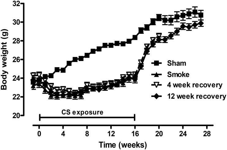
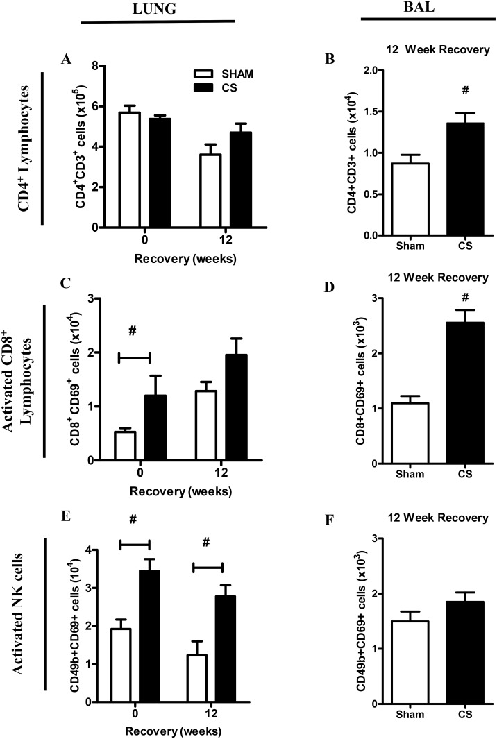
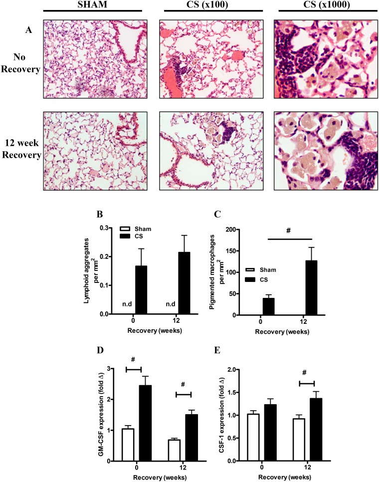
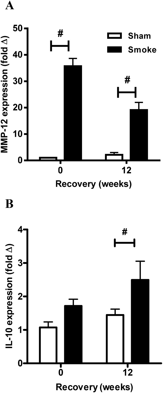
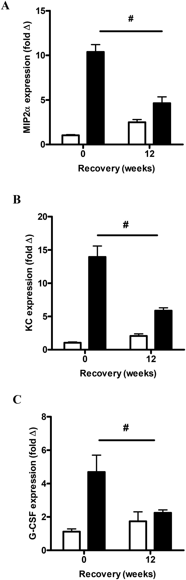
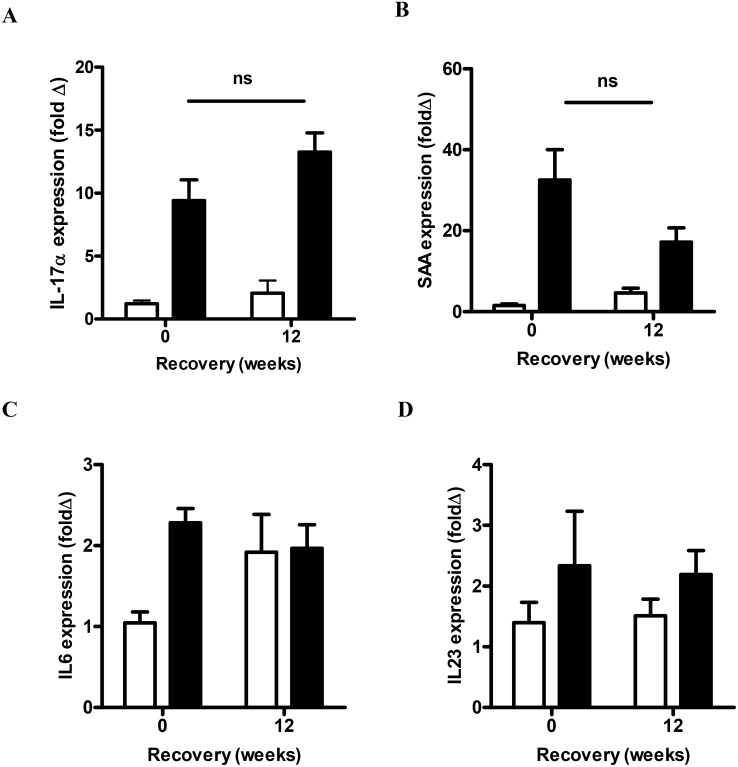
While global success in cessation advocacy has seen smoking rates fall in many developed countries, persistent lung inflammation in ex-smokers is an increasingly important clinical problem whose mechanistic basis remains poorly understood. In this study, candidate effector mechanisms were assessed in mice exposed to cigarette smoke (CS) for 4 months following cessation from long term CS exposure. BALF neutrophils, CD4+ and CD8+ T cells and lung innate NK cells remained significantly elevated following smoking cessation. Analysis of neutrophil mobilization markers showed a transition from acute mediators (MIP-2α, KC and G-CSF) to sustained drivers of neutrophil and macrophage recruitment and activation (IL-17A and Serum Amyoid A (SAA)). Follicle-like lymphoid aggregates formed with CS exposure and persisted with cessation, where they were in close anatomical proximity to pigmented macrophages, whose number actually increased 3-fold following CS cessation. This was associated with the elastolytic protease, MMP-12 (macrophage metallo-elastase) which remained significantly elevated post-cessation. Both GM-CSF and CSF-1 were significantly increased in the CS cessation group relative to the control group. In conclusion, we show that smoking cessation mediates a transition to accumulation of pigmented macrophages, which may contribute to the expanded macrophage population observed in COPD. These macrophages together with IL-17A, SAA and innate NK cells are identified here as candidate persistence determinants and, we suggest, may represent specific targets for therapies directed towards the amelioration of chronic airway inflammation.
Conflict of interest statement
Figures

Similar articles
-
Innate cellular sources of interleukin-17A regulate macrophage accumulation in cigarette- smoke-induced lung inflammation in mice.Clin Sci (Lond). 2015 Nov;129(9):785-96. doi: 10.1042/CS20140703. Epub 2015 Jul 1. Clin Sci (Lond). 2015. PMID: 26201093 Free PMC article.
-
Cigarette smoke induces molecular responses in respiratory tissues of ApoE(-/-) mice that are progressively deactivated upon cessation.Toxicology. 2013 Dec 6;314(1):112-24. doi: 10.1016/j.tox.2013.09.013. Epub 2013 Oct 1. Toxicology. 2013. PMID: 24096154
-
Serum amyloid A promotes lung neutrophilia by increasing IL-17A levels in the mucosa and γδ T cells.Am J Respir Crit Care Med. 2013 Jul 15;188(2):179-86. doi: 10.1164/rccm.201211-2139OC. Am J Respir Crit Care Med. 2013. PMID: 23627303 Free PMC article.
-
Increased surfactant protein-D and foamy macrophages in smoking-induced mouse emphysema.Respirology. 2007 Mar;12(2):191-201. doi: 10.1111/j.1440-1843.2006.01009.x. Respirology. 2007. PMID: 17298450 Review.
-
Signal transduction pathways linking the activation of alveolar macrophages with the recruitment of neutrophils to lungs in chronic obstructive pulmonary disease.Exp Lung Res. 2009 Aug;35(6):439-85. doi: 10.1080/01902140902759290. Exp Lung Res. 2009. PMID: 19842832 Review.
Cited by
-
Changes in the expression level of IL-17A and p53-fibrinolytic system in smokers with or without COPD.Mol Biol Rep. 2018 Dec;45(6):2835-2841. doi: 10.1007/s11033-018-4398-y. Epub 2018 Sep 24. Mol Biol Rep. 2018. PMID: 30250995
-
Granulocyte-CSF links destructive inflammation and comorbidities in obstructive lung disease.J Clin Invest. 2018 Jun 1;128(6):2406-2418. doi: 10.1172/JCI98224. Epub 2018 Apr 30. J Clin Invest. 2018. PMID: 29708507 Free PMC article.
-
Enhanced lung inflammatory response in whole-body compared to nose-only cigarette smoke-exposed mice.Respir Res. 2021 Mar 17;22(1):86. doi: 10.1186/s12931-021-01680-5. Respir Res. 2021. PMID: 33731130 Free PMC article.
-
Quantitative Computed Tomography Assessment of Pectoralis and Erector Spinae Muscle Area and Disease Severity in Chronic Obstructive Pulmonary Disease Referred for Lung Volume Reduction.COPD. 2021 Apr;18(2):191-200. doi: 10.1080/15412555.2021.1897560. Epub 2021 Mar 19. COPD. 2021. PMID: 33736550 Free PMC article.
-
Emerging Biological Functions of IL-17A: A New Target in Chronic Obstructive Pulmonary Disease?Front Pharmacol. 2021 Jul 2;12:695957. doi: 10.3389/fphar.2021.695957. eCollection 2021. Front Pharmacol. 2021. PMID: 34305606 Free PMC article. Review.
References
-
- Barnes PJ (2008) Immunology of asthma and chronic obstructive pulmonary disease. Nat Rev Immunol 8: 183–192. - PubMed
-
- Hogg JC, Chu F, Utokaparch S, Woods R, Elliott WM, et al. (2004) The nature of small-airway obstruction in chronic obstructive pulmonary disease. N Engl J Med 350: 2645–2653. - PubMed
-
- Saetta M, Di Stefano A, Turato G, Facchini FM, Corbino L, et al. (1998) CD8+ T-lymphocytes in peripheral airways of smokers with chronic obstructive pulmonary disease. Am J Respir Crit Care Med 157: 822–826. - PubMed
-
- Rutgers SR, Postma DS, ten Hacken NH, Kauffman HF, van Der Mark TW, et al. (2000) Ongoing airway inflammation in patients with COPD who Do not currently smoke. Chest 117: 262S. - PubMed
-
- Willemse BW, ten Hacken NH, Rutgers B, Lesman-Leegte IG, Postma DS, et al. (2005) Effect of 1-year smoking cessation on airway inflammation in COPD and asymptomatic smokers. Eur Respir J 26: 835–845. - PubMed
Publication types
MeSH terms
Substances
Grants and funding
LinkOut - more resources
Full Text Sources
Other Literature Sources
Medical
Research Materials
Miscellaneous
