. 2015 Mar;45(3):758-72.
doi: 10.1002/eji.201344412. Epub 2014 Dec 23.

NLRC5 interacts with RIG-I to induce a robust antiviral response against influenza virus infection

Affiliations

NLRC5 interacts with RIG-I to induce a robust antiviral response against influenza virus infection

Priya Ranjan et al. Eur J Immunol. 2015 Mar.

Abstract

The NLR protein, NLRC5 is an important regulator of MHC class I gene expression, however, the role of NLRC5 in other innate immune responses is less well defined. In the present study, we report that NLRC5 binds RIG-I and that this interaction is critical for robust antiviral responses against influenza virus. Overexpression of NLRC5 in the human lung epithelial cell line, A549, and normal human bronchial epithelial cells resulted in impaired replication of influenza virus A/Puerto Rico/8/34 virus (PR8) and enhanced IFN-β expression. Influenza virus leads to induction of IFN-β that drives RIG-I and NLRC5 expression in host cells. Our results suggest that NLRC5 extends and stabilizes influenza virus induced RIG-I expression and delays expression of the viral inhibitor protein NS1. We show that NS1 binds to NLRC5 to suppress its function. Interaction domain mapping revealed that NLRC5 interacts with RIG-I via its N-terminal death domain and that NLRC5 enhanced antiviral activity in an leucine-rich repeat domain independent manner. Taken together, our findings identify a novel role for NLRC5 in RIG-I-mediated antiviral host responses against influenza virus infection, distinguished from the role of NLRC5 in MHC class I gene regulation.

Keywords: Influenza; Interferon; NLRC5; NS1; RIG-I Antiviral.


Conflict of interest statement

Conflict of interest: The authors declare no financial or commercial conflict of interest.

Figures

Figure 1.
Figure 1.
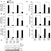
NLRC5 overexpression inhibits influenza virus PR8 replication and induces RIG-I and IFN-β expression in A549 cells. A549 cells were transfected with 2 μg of vector alone, myc-NLRC5 or flag-CIITA expression vector. Twenty-four hours post-transfection, the cells were infected with PR8 virus (MOI 0, 0.01, 0.1, and 1.0) for 24 h. (A) Supernatants were tested for viral titers by plaque assay using MDCK cells. (B–F) The expression of (B) NP vRNA, (C) NP mRNA, (D) IFN-β mRNA, (E) RIG-I mRNA, and (F) IFN-α was analyzed by real-time RT-PCR, relative to β-actin. Data shown are mean + SD of three samples per group, pooled from three independent experiments carried out in duplicate. (F) Expression of myc-NLRC5 and NS1 was analyzed by immunoblotting and the immunoblot shown is from one single experiment representative of three independent experiments. β-Actin was used as a loading control. ANOVA was performed to compare vector control versus myc-NLRC5 or flag-CIITA-transfected A549 cells and p values <0.05 are indicated with an asterisk.
Figure 2.
Figure 2.
Viral NS1 counteracts endogenous NLRC5, RIG-I, and IFN-β expression. A549 cells were infected with PR8 or PR8ΔNS1 (MOI 1.0) for 0, 3, 6, 12, and 24 h and the expression of (A) NP vRNA, (B) NP mRNA, (C) NLRC5 mRNA, (D) RIG-I mRNA, (E) IFN-β mRNA, and (F) IFN-α was analyzed by real-time RT-PCR, relative to β-actin. Data shown are mean + SD of three samples per group, pooled from three independent experiments carried out in duplicate. ANOVA was performed to compare PR8-infected versus PR8ΔNS1-infected A549 cells and p values <0.05 are indicated with an asterisk.
Figure 3.
Figure 3.
NS1 complementation inhibits PR8ΔNS1-induced NLRC5. A549 cells were cotransfected with 2 μg of vector or myc-NS1 expression vector and IFN-β promoter LUC reporter using lipofectamine 2000. Twenty-four hours post-transfection, these cells were infected with PR8ΔNS1 for another 24 h. (A, B) Cells were harvested and mRNA expression of (A) NLRC5 and (B) RIG-I was determined by real-time RT-PCR, relative to β-actin. (C) IFN-β induction was assayed by LUC reporter assay. (D) NLRC5, RIG-I, and myc-NS1 expression was analyzed by immunoblotting. The immunoblot shown is from one single experiment representative of three independent experiments. β-Actin was used as a loading control. Data shown are mean + SD of three samples per group, pooled from three independent experiments carried out in duplicate. ANOVA was performed to compare vector control versus myc-NS1-transfected A549 cells and p values <0.05 are indicated with an asterisk.
Figure 4.
Figure 4.
NS1ΔPR8 virus induces NLRC5, IFN-β, RANTES expression, and NFκB activation in a RIG-I-dependent manner. The expression of endogenous NLRC5 or RIG-I was silenced using gene-specific NLRC5 or RIG-I siRNA in A549 cells followed by infection with PR8 or PR8ΔNS1 (MOI 1.0). Cells were also cotransfected with NFκB promoter LUC reporter using lipofectamine 2000. (A–C) Cells were harvested 24 h postinfection to assess the expression of (A) NLRC5 mRNA, (B) IFN-β mRNA, and (C) RIG-I mRNA, relative to β-actin by real-time RT-PCR. (D) Cells were analyzed for endogenous RIG-I, NLRC5, and β-actin (loading control) protein expression by immunoblotting and the immunoblot shown is from one single experiment representative of three independent experiments. Cell supernatants were assayed for (E) IFN-β and (F) RANTES by ELISA. (G) NFκB activation was measured by LUC reporter assay. Data shown are mean + SD of three samples per group, pooled from three independent experiments carried out in duplicate. ANOVA was performed to compare PR8-infected versus PR8ΔNS1-infected A549 cells and p values <0.05 are indicated with an asterisk.
Figure 5.
Figure 5.
LPS-induced IFN-β induction and NFκB activation remain unchanged in the presence or absence of NLRC5. (A–C) The expression of endogenous NLRC5 in A549 cells was silenced using gene-specific NLRC5 siRNA. (D–F) Alternatively, A549 cells were transfected with 2 μg of vector alone or myc-NLRC5 expression vector. (A–F) Cells were also cotransfected with IFN-β promoter or NFκB promoter LUC reporter using lipofectamine 2000 and treated with indicated dose of LPS. (A–F) Cells were harvested 24 h post-LPS treatment to assess (A and D) IFN-β induction and (B and E) NFκB activation by LUC reporter assay. (C and F) Cells were analyzed for myc-NLRC5, RIG-I, and β-actin (loading control) protein expression by immunoblotting and the immunoblot shown is from one single experiment representative of two independent experiments. Data are shown as mean + SD of three samples per group, pooled from three independent experiments carried out in duplicate. ANOVA was performed to compare control siRNA versus NLRC5 siRNA treated A549 cells or vector versus NLRC5-transfected A549 cells and p values <0.05 are indicated with an asterisk; ns: not significant.
Figure 6.
Figure 6.
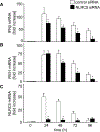
NLRC5 is required for robust IFN-β and RIG-I expression. A549 cells transfected with control siRNA or NLRC5 siRNA were infected with NS1-del PR8 (MOI 1.0). (A–C) Cells were harvested 0, 24, 48, 72, and 96 h postinfection and analyzed for (A) IFN-β, (B) RIG-I, and (C) NLRC5 mRNA expression, relative to β-actin by real-time RT-PCR. Data are shown as mean + SD of three samples per group, pooled from three independent experiments carried out in duplicate. ANOVA was performed to compare control siRNA versus NLRC5 siRNA treated A549 cells and p values <0.05 are indicated with an asterisk.
Figure 7.
Figure 7.
The NLRC5 death domain and nucleotide-binding domain is critical for NLRC5-mediated antiviral function. A549 cells were transfected with vector alone or with myc-tagged wtNLRC5, NLRC5-K234A, NLRC5-ISO3, NLRC5-ΔDD, NLRC5-DD, or LRR domain of NLRC5 and subsequently infected with PR8 (MOI 1.0) for 24 h. (A) The upper panel shows the schematic representation of the NLRC5 constructs used and the lower panel shows expression of NLRC5 constructs in the cells by immunoblotting. β-Actin was used as a loading control. (B, C) Supernatants were collected and cells were harvested to determine (B) viral titers by plaque assay and (C) IFN-β mRNA expression, relative to β-actin by real-time RT-PCR. (D) Secretion of CCL5 (RANTES) in cell supernatants was measured by ELISA. Data are shown as mean + SD of three samples per group, pooled from three independent experiments carried out in duplicate. ANOVA was performed to compare control vector versus myc-NLRC5 expression vectors transfected A549 cells and p values <0.05 are indicated with an asterisk.
Figure 8.
Figure 8.
NLRC5 stabilizes RIG-I. (A) A549 cells transfected with vector alone or myc-wtNLRC5 were infected with PR8 (MOI 1.0) for 0, 3, 6, 9, 12, 18, 24, 48, and 72 h and harvested for NLRC5, RIG-I, and NS1 expression and coimmunoprecipitation assay. β-Actin was used as a loading control. Cell lysates from 6, 12, and 24 h were immunoprecipitated with anti-myc, anti-NS1, or anti-RIG-I antibodies and immunoprecipitates were analyzed for the presence of RIG-I, NS1, and NLRC5 by immunoblotting. (B) To map the domain responsible for NLRC5 interaction with RIG-I and NS1, A549 cells were transfected for 24 h with myc-vector alone or with myc-tagged wtNLRC5, NLRC5-K234A, NLRC5-ISO3, NLRC5-ΔDD, NLRC5-DD, or NLRC5-LRR mutants and then infected with PR8 (MOI 1.0) for 3 or 9 h. Cell lysates were immunoprecipitated with anti-myc, anti-NS1, or anti-RIG-I antibodies and immunoprecipitates were analyzed for the presence of RIG-I, NS1, and NLRC5 by immunoblotting. β-Actin was used as a loading control. The input for the immunoblot was about 5% of the total cell lysate. (C) A549 cells transfected with wtNLRC5 were infected with PR8 (MOI 1.0) in the presence or absence of actinomycin D (5 μg/mL)/cyclohexamide (20 μg/mL) combination. Cell lysates were analyzed for RIG-I and NLRC5 expression at 0, 3, 6, and 9 h postinfection by immunoblotting. Data shown are from one single experiment representative of two independent experiments.
Figure 9.
Figure 9.
Schematic representation of the role of NLRC5 in influenza virus infection. NLRC5 induces a RIG-I-dependent robust antiviral response against influenza virus and induced type I IFN. Type I IFN upregulated NLRC5, RIG-I expression, and subsequently its own production (left). Absence of NLRC5 results in a weaker type-I IFN response. NS1 can suppress the NLRC5-mediated antiviral response by interacting with the RIG-I/NLRC5 complex or by sequestering NLRC5 and preventing its interaction with RIG-I (top right). While 5′PPP-RNA is a natural ligand for RIG-I, it is not known if NLRC5 binds to viral RNA or undergoes conformational changes (bottom left and right). (Protein–protein interactions shown in the figure do not represent the specific domains responsible for binding.)

Similar articles

Cited by

References

    1. Akira S, Uematsu S. and Takeuchi O, Pathogen recognition and innate immunity. Cell 2006. 124: 783–801. - PubMed
    1. Kawai T. and Akira S, TLR signaling. Cell Death Differ. 2006. 13: 816–825. - PubMed
    1. Ranjan P, Bowzard JB, Schwerzmann JW, Jeisy-Scott V, Fujita T. and Sambhara S, Cytoplasmic nucleic acid sensors in antiviral immunity. Trends Mol. Med 2009. 15: 359–368. - PubMed
    1. Takahasi K, Yoneyama M, Nishihori T, Hirai R, Kumeta H, Narita R, Gale M. et al., Nonself RNA-sensing mechanism of RIG-I helicase and activation of antiviral immune responses. Mol. Cell 2008. 29: 428–440. - PubMed
    1. Lamkanfi M. and Kanneganti TD, Regulation of immune pathways by the NOD-like receptor NLRC5. Immunobiology 2012. 217: 13–16. - PMC - PubMed

Publication types

MeSH terms

Substances

LinkOut - more resources