. 2015 Feb;35(3):498-513.
doi: 10.1128/MCB.01079-14. Epub 2014 Nov 17.

MUNC, a long noncoding RNA that facilitates the function of MyoD in skeletal myogenesis

Affiliations

MUNC, a long noncoding RNA that facilitates the function of MyoD in skeletal myogenesis

Adam C Mueller et al. Mol Cell Biol. 2015 Feb.

Abstract

An in silico screen for myogenic long noncoding RNAs (lncRNAs) revealed nine lncRNAs that are upregulated more than 10-fold in myotubes versus levels in myoblasts. One of these lncRNAs, MyoD upstream noncoding (MUNC, also known as DRR(eRNA)), is encoded 5 kb upstream of the transcription start site of MyoD, a myogenic transcription factor gene. MUNC is specifically expressed in skeletal muscle and exists as in unspliced and spliced isoforms, and its 5' end overlaps with the cis-acting distal regulatory region (DRR) of MyoD. Small interfering RNA (siRNA) of MUNC reduced myoblast differentiation and specifically reduced the association of MyoD to the DRR enhancer and myogenin promoter but not to another MyoD-dependent enhancer. Stable overexpression of MUNC from a heterologous promoter increased endogenous MyoD, Myogenin, and Myh3 (myosin heavy chain, [MHC] gene) mRNAs but not the cognate proteins, suggesting that MUNC can act in trans to promote gene expression but that this activity does not require an induction of MyoD protein. MUNC also stimulates the transcription of other genes that are not recognized as MyoD-inducible genes. Knockdown of MUNC in vivo impaired murine muscle regeneration, implicating MUNC in primary satellite cell differentiation in the animal. We also discovered a human MUNC that is induced during differentiation of myoblasts and whose knockdown decreases differentiation, suggesting an evolutionarily conserved role of MUNC lncRNA in myogenesis. Although MUNC overlaps with the DRR enhancer, our results suggest that MUNC is not a classic cis-acting enhancer RNA (e-RNA) acting exclusively by stimulating the neighboring MyoD gene but more like a promyogenic lncRNA that acts directly or indirectly on multiple promoters to increase myogenic gene expression.


Figures

FIG 1
FIG 1
(A) Workflow (left to right) of the computational screen that identified potential long noncoding RNAs induced during muscle differentiation. (B) UCSC Genome Browser screenshot showing the locations of Pol II ChIP-Seq, H3K4me3 ChIP-Seq, and RNA-Seq signals. Chr7, chromosome 7.
FIG 2
FIG 2
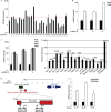
Predicted muscle-specific noncoding RNAs are upregulated in myotubes. (A) qPCR confirmation of predicted noncoding RNAs induced in myotubes versus myoblasts. Nine predicted RNA-Seq fragments at seven independent genomic loci were >10-fold induced in myotubes (MT) versus levels in myoblasts (MB). (B) qPCR analysis of MyoD transfected transdifferentiating 10T1/2 fibroblasts for several predicted myogenically regulated lncRNAs when cells were transferred to low-serum differentiation medium (DM) versus growth medium (GM). (C) RT-qPCR of lncRNAs from screen in differentiating C2C12 myoblasts on the indicated days after the switch to low-serum differentiation medium. (D) RT-qPCR of lncRNAs 2 and 3 (or MUNC) from mouse embryos (embryonic days 7, 11, 15, and 17) and murine tissues showing high expression in skeletal muscle. (E) Schematic of MUNC genomic region upstream of the MyoD1 locus. MUNC overlaps the previously characterized distal regulatory region (DRR) enhancer and putative noncoding transcripts 2 and 3 (pNC2 and pNC3). (F) RT-qPCR analysis of MUNC expression in primary myoblasts and myotubes. MUNC 1F-1R and 2F-2R primers measure the primary unspliced MUNC while the 1F-2R primers measure spliced MUNC (Fig. 3E). Data represent means ± standard errors of the means (n = 3).
FIG 3
FIG 3
Characterization of MyoD upstream noncoding transcript. (A) Schematic of primers used for 5′ RACE PCR and PCR primer walking to determine ends of MUNC transcript. (B) 5′ end mapping of MUNC: PCR product generated by 5′ RACE PCR on cap-captured DM4 C2C12 RNA with the R-B′ (nested) primer. (C) 3′ end mapping of MUNC: PCR products with 1F and indicated R primers on genomic DNA (positive control) and DM4 C2C12 cDNA. Only the R-B primer gave a product on the cDNA, putting it at the 3′ end of MUNC. (D) PCR on genomic DNA (positive control) and cDNA from DM4 C2C12 cells confirms the 5′ end of MUNC and the presence of unspliced and spliced isoforms. Primers F-A plus R-B produced two products on cDNA: genome-length unspliced (∼1,000 bases) and spliced (∼500 bases) cDNAs. (E) PCR products amplified by 1F-2R primers. Lane 1, genomic DNA from C2C12 with extension time of 60 s; lane 2, cDNA from DM3 C2C12 with an extension time of 20 s; lane 3, negative control (no DNA) with an extension time of 20 s. (F) Sequences of unspliced and spliced MUNC forms amplified by F-A and R-B primers. Bold represents the 5′ splice site, and bold italic represents the 3′ splice site. Coordinates are according to the UCSC Genome Browser (assembly of July 2007).
FIG 4
FIG 4
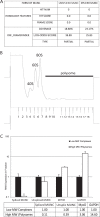
Unspliced and spliced MUNC forms are predicted to have low coding potential and are not associated with polysomes during differentiation. (A) Analysis obtained from the Coding Potential Calculator based on evolutionary conservation and open reading frame (ORF) attributes (http://cpc.cbi.pku.edu.cn/). Both forms of MUNC are likely to be noncoding transcripts. (B) Polysome fractionation profile of differentiating C2C12 cells. Monosomes, fractions 3 to 8; polysomes, fractions 9 to 18. (C) qRT-PCR of polysome fractions and monosome fractions. Spliced and unspliced (Unsplic) MUNCs are depleted from the polysome fraction while the mRNAs for MyoD and GAPDH are enriched in the polysome fraction. Data represent means ± standard errors of the means (n = 3). MW, molecular weight.
FIG 5
FIG 5
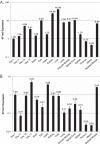
Tissue expression of two other myogenically upregulated long noncoding RNA transcripts, 9 and 13 (lnc9 and lnc13, respectively). Panels A and B show qRT-PCR results of the indicated transcripts across a panel of embryonic and adult mouse tissue samples. Values are normalized to expression of RPS13, a housekeeping gene with low tissue variability, and plotted relative to expression in day 7 embryonic tissue. Data represents means ± standard errors of the means (n = 3). MT, myotube.
FIG 6
FIG 6
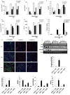
MUNC knockdown represses the induction of myogenic differentiation markers and impairs myotube formation in culture. (A to E) qRT-PCR measuring induction of unspliced and spliced MUNC (I and J) and myogenic markers MyoD, myogenin, and Myh3 (MHC) during differentiation of C2C12 cells incubated with either control siRNA or siRNA targeting the 5′ or 3′ end of MUNC. MUNC levels were normalized to those of GAPDH. Data represent means ± standard errors of the means (n = 3). Note log scale of the y axis in panels A to D. (F) Fusion index of differentiating C2C12 cells shows that MUNC knockdown impairs myotube formation. The fusion index was calculated by dividing the number of nuclei contained within multinucleated cells by the number of total nuclei in a field. (G) Immunofluorescence of MHC (green) and MyoD (blue) in differentiating C2C12 cells. C2C12 cells incubated with control siRNA show much greater formation of MHC-positive, multinucleated cells on differentiation days 3 and 5 than cells incubated with siRNA targeting MUNC. Data represent means ± standard errors of the means (n = 3). (H) Western blot analysis of MHC, myogenin, and MyoD in differentiating C2C12 cells. Independent siRNA targeting MUNC reduces expression of these myogenic proteins. (I to M) qRT-PCR measuring induction of unspliced and spliced forms of MUNC (I and J) and myogenic markers MyoD, myogenin, and Myh3 (K to M) during differentiation of primary murine myoblasts derived from DRR+/− and DRR−/− mice. Expression levels are normalized to those of GAPDH. Data represent means ± standard errors of the means (n = 3).
FIG 7
FIG 7
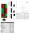
Global gene expression changes that occur during skeletal myogenesis are inhibited by MUNC depletion. (A) Hierarchical clustering of the 400 genes that varied the most upon differentiation of C2C12 cells. The cells were treated with either MUNC or control silencer RNAs in both GM and DM. Signals are scaled to Z scores of the rows. I and II, clusters I and II, respectively. (B) Clusters I and II represent genes that are enriched in GO terms associated with cell cycle and growth (I) and with muscle-specific processes (II). (C) Deviation from a perfect correlation coefficient of 1 of gene expression profiles of GM versus DM3 cells with either siControl or siMUNC as measured by the Affymetrix exon array analysis. (D and E) Mean expression fold change of the 50 most upregulated or downregulated genes in control differentiating cells. This fold change is suppressed in siMUNC cells. Values are means ± standard errors of the means (n = 5). (F) Values on the y axis represent fold change of genes known to be induced (top) or repressed (bottom) by MyoD (25). Values on the x axis represent the fold change of the same genes after knockdown of MUNC (left, repressed by MUNC; right, induced by MUNC). Boxes represent the following: I, genes induced by MyoD but downregulated by MUNC; II, genes induced by MyoD but not regulated by MUNC; III, genes downregulated by MyoD but not affected by MUNC. (G) Description of the genes belonging to the three subclasses (boxes I to III) identified in panel F. FDR, false discovery rate.
FIG 8
FIG 8
MUNC is required for MyoD binding at certain target sites and not others. MUNC expression is dependent on MyoD. Graphs represent results of MyoD and myogenin (MyoG) ChIP at the MyoD distal regulatory region (A) Myogenin promoter region (B), and MyoD core enhancer region (C). Cells were treated with either control siRNA or siMUNC and incubated in differentiation medium for 72 h. Data represent means ± standard errors of the means (n = 3). (D to F) MUNC expression level in proliferating C2C12 cells (GM) and in differentiating cells (differentiation day 4, DM4) under control conditions (siRNA targeting luciferase GL2 [siGL2]) and after MyoD knockdown (siMyoD). Cells were transfected with siRNA and harvested 48 h later (GM) or retransfected (at DM0 and DM2) and incubated in differentiation medium for 4 days (DM4). Data represent means ± standard errors of the means (n = 3).
FIG 9
FIG 9
Stable overexpression of MUNC enhances RNA of myogenic markers but not that of the corresponding proteins. qRT-PCR expression of MUNC isoforms and myogenic markers following C2C12 transfection with linearized vectors encoding the WT unspliced form of MUNC, an unspliceable form of MUNC with a point mutation preventing RNA splicing, and a spliced form of MUNC. Measurements were performed on proliferating cells (GM) and differentiating cells after 3 days in differentiation medium (DM3). Expression data of unspliced MUNC (A and F), spliced MUNC (B and G), MyoD (C and H), myogenin (D and I), and Myh3 (E and J) RNAs are shown. Data are normalized to GAPDH expression and then normalized again in each panel to the level in vector-transfected cells in GM or DM. Data represent means ± standard errors of the means (n = 3). (K) Western blot analysis showing level of MyoD and MyoG proteins in C2C12 cells overexpressing MUNC in GM. Actin was used as a loading control. (L) The same experiment as shown in panel K except performed in cells after 3 days in DM.
FIG 10
FIG 10
MUNC knockdown reduces myogenic marker expression during skeletal muscle regeneration in adult mice and impairs regeneration. MUNC is conserved between humans and mice. (A and B) qRT-PCR expression of MUNC isoforms (A) and myogenic markers (B) following knockdown of MUNC in three adult mouse TA muscles. Mice were injected twice with siMUNC (Invitrogen in vivo Silencer/Invivofectamine complexes), and RNA was harvested 5 days following the first injection. Control siRNA was injected into the contralateral leg. 1F-1R and 2F-2R measure the 5′ and 3′ regions of the unspliced MUNC, while 1F-2R measure spliced MUNC. Data represent means ± standard errors of the means (n = 3). (C and D) qRT-PCR expression of MUNC isoforms (C) and myogenic markers (D) after knockdown in adult mouse TA muscle 14 days after injury with cardiotoxin. Mice were injected with cardiotoxin and with then siMUNC (Invitrogen in vivo Silencer/Invivofectamine complexes) twice, on days 2 and 5 following injury. RNA was harvested 14 days following the injury. Control siRNA was injected into the contralateral leg. Data represent means ± standard errors of the means (n = 4). (E) Representative hematoxylin and eosin (H&E)-, desmin-, and laminin-stained sections of regenerating mouse TA muscle 14 days after cardiotoxin injection and control or MUNC in vivo siRNA knockdown. DAPI, 4′,6′-diamidino-2-phenylindole. (F) Quantitation of myofiber cross-sectional area. Data represents means ± standard errors of the means (n = 4). For statistical analysis, Student's t test was used. *, P < 0.05 (significant); **, P > 0.05 (not significant). (G) Induction of expression of human MUNC (hMUNC) RNA during differentiation of LHCN cells. (H and I) qRT-PCR analysis of human MUNC RNA (H) and myogenic markers (I) in LHCN human myoblasts after transfection with siRNA targeting human MUNC (si-hMUNC) and 7 days of in low-serum differentiation medium. Data are normalized to beta-actin expression. Data represent means ± standard errors of the means (n = 3).
FIG 11
FIG 11
Alignment between the human DRR sequence plus downstream 1,000 bp (chromosome 11, positions 17714232 to 17716026) and the mouse MUNC locus from 400 bp upstream of the TSS to end of MUNC (chromosome 7, positions 46371003 to 46372492). Blue in the human sequence indicates DRR; red in the mouse sequence indicates MUNC exon 1 and MUNC exon 2.

Similar articles

Cited by

References

    1. Wang KC, Chang HY. 2011. Molecular mechanisms of long noncoding RNAs. Mol Cell 43:904–914. doi:10.1016/j.molcel.2011.08.018. - DOI - PMC - PubMed
    1. Wapinski O, Chang HY. 2011. Long noncoding RNAs and human disease. Trends Cell Biol 21:354–361. doi:10.1016/j.tcb.2011.04.001. - DOI - PubMed
    1. Clark MB, Amaral PP, Schlesinger FJ, Dinger ME, Taft RJ, Rinn JL, Ponting CP, Stadler PF, Morris KV, Morillon A, Rozowsky JS, Gerstein MB, Wahlestedt C, Hayashizaki Y, Carninci P, Gingeras TR, Mattick JS. 2011. The reality of pervasive transcription. PLoS Biol 9:e1000625. doi:10.1371/journal.pbio.1000625. - DOI - PMC - PubMed
    1. Derrien T, Johnson R, Bussotti G, Tanzer A, Djebali S, Tilgner H, Guernec G, Martin D, Merkel A, Knowles DG, Lagarde J, Veeravalli L, Ruan X, Ruan Y, Lassmann T, Carninci P, Brown JB, Lipovich L, Gonzalez JM, Thomas M, Davis CA, Shiekhattar R, Gingeras TR, Hubbard TJ, Notredame C, Harrow J, Guigó R. 2012. The GENCODE v7 catalog of human long noncoding RNAs: analysis of their gene structure, evolution, and expression. Genome Res 22:1775–1789. doi:10.1101/gr.132159.111. - DOI - PMC - PubMed
    1. Guttman M, Garber M, Levin JZ, Donaghey J, Robinson J, Adiconis X, Fan L, Koziol MJ, Gnirke A, Nusbaum C, Rinn JL, Lander ES, Regev A. 2010. Ab initio reconstruction of cell type-specific transcriptomes in mouse reveals the conserved multi-exonic structure of lincRNAs. Nat Biotechnol 28:503–510. doi:10.1038/nbt.1633. - DOI - PMC - PubMed

Publication types