. 2014 Nov 13;10(10):e1004514.
doi: 10.1371/journal.ppat.1004514. eCollection 2014 Oct.

PUL21a-Cyclin A2 interaction is required to protect human cytomegalovirus-infected cells from the deleterious consequences of mitotic entry

Affiliations

PUL21a-Cyclin A2 interaction is required to protect human cytomegalovirus-infected cells from the deleterious consequences of mitotic entry

Martin Eifler et al. PLoS Pathog. .

Abstract

Entry into mitosis is accompanied by dramatic changes in cellular architecture, metabolism and gene expression. Many viruses have evolved cell cycle arrest strategies to prevent mitotic entry, presumably to ensure sustained, uninterrupted viral replication. Here we show for human cytomegalovirus (HCMV) what happens if the viral cell cycle arrest mechanism is disabled and cells engaged in viral replication enter into unscheduled mitosis. We made use of an HCMV mutant that, due to a defective Cyclin A2 binding motif in its UL21a gene product (pUL21a), has lost its ability to down-regulate Cyclin A2 and, therefore, to arrest cells at the G1/S transition. Cyclin A2 up-regulation in infected cells not only triggered the onset of cellular DNA synthesis, but also promoted the accumulation and nuclear translocation of Cyclin B1-CDK1, premature chromatin condensation and mitotic entry. The infected cells were able to enter metaphase as shown by nuclear lamina disassembly and, often irregular, metaphase spindle formation. However, anaphase onset was blocked by the still intact anaphase promoting complex/cyclosome (APC/C) inhibitory function of pUL21a. Remarkably, the essential viral IE2, but not the related chromosome-associated IE1 protein, disappeared upon mitotic entry, suggesting an inherent instability of IE2 under mitotic conditions. Viral DNA synthesis was impaired in mitosis, as demonstrated by the abnormal morphology and strongly reduced BrdU incorporation rates of viral replication compartments. The prolonged metaphase arrest in infected cells coincided with precocious sister chromatid separation and progressive fragmentation of the chromosomal material. We conclude that the Cyclin A2-binding function of pUL21a contributes to the maintenance of a cell cycle state conducive for the completion of the HCMV replication cycle. Unscheduled mitotic entry during the course of the HCMV replication has fatal consequences, leading to abortive infection and cell death.


Conflict of interest statement

The authors have declared that no competing interests exist.

Figures

Figure 1
Figure 1. HCMV-pUL21a interacts with Cyclin A2.
(A) Alignment of two putative Cyclin A2-binding sites in pUL21a with validated RXL/Cy motifs of human Cyclin A2-CDK substrates and inhibitors. Identical residues are highlighted in black, conserved residues in grey, phenylalanine/leucin residues at positions +1 or +2 relative to the RXL motif in red or yellow. (B) Schematic of pUL21a showing the relative location of RXL sequence motifs, the APC/C-binding site and a minimal consensus CDK phosphorylation site (SP). (C, D) Recombinant Strep-His-tagged versions of pp150, pUL21a wild-type (wt), pUL21a-RRLAFARAAF (RXL1mut) and pUL21a-RRLFQARAFQ (RXL2mut) mutants were purified and immobilized to Ni-NTA agarose beads. The beads were incubated with HEK293 cell lysates containing ectopically expressed HA-Cyclin A2 wildtype (CycA2-wt), HA-Cyclin A2 hydrophobic patch mutant (CycA2-hpm) or HA-Cyclin B1 (CycB1). The input lysates and the pulled down material were analyzed by immunoblotting (IB) for the presence of pUL21a and HA-tagged cyclins. (E) Cyclin A2-associated kinase activity was immunoprecipitated from HEK293 lysates and incubated with the substrate protein pp150 and radioactively labeled γ-P32-ATP. Where indicated, equal amounts of pUL21a-wt or pUL21-RXL2mut were added to the reaction, as controlled by Coomassie staining (lower panel, IgG-LC: Immunoglobulin G light chain). The phosphorylation products were analyzed by autoradiography (upper panel).
Figure 2
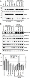
Figure 2. Interaction with pUL21a leads to Cyclin A2 protein degradation.
HEK293 cells were transfected with the indicated pUL21a-wt and mutant expression plasmids (pUL21a-PRAA: APCmut) or an empty vector control. Where indicated, MG132 was added to the cells at 24 h post transfection. Cells were harvested at 48 h post transfection and processed for the following assays. (A) Cyclin A2-CDK complexes were immunoprecipitated (IP) from extracts of transfected, MG132-treated cells and analyzed by immunoblotting for the presence of Cyclin A2, CDK2 and HA-pUL21a. (B) Whole cell lysates of transfected cells were analyzed by immunoblotting for expression of the indicated proteins. (C) Total RNA preparations of transfected cells were analyzed by quantitative RT-PCR for Cyclin A2 and APC5 mRNA expression. Error bars indicate the standard deviations of triplicate samples.
Figure 3
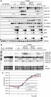
Figure 3. The pUL21a-RXL/Cy motif is required for Cyclin A2 repression by HCMV.
Density arrested human embryonic lung (HEL) fibroblasts were infected with HCMV reconstituted from TB40-BAC4-wt or derivatives carrying the indicated UL21a mutations. Cells were harvested at regular intervals and analyzed for viral and cellular gene expression by immunoblotting (A) and ribonuclease protection assay (B). Cell culture supernatants were analyzed for the number of IE protein-forming units (IU) by virus titration (C). Results are represented as mean values plus standard deviations of triplicate samples.
Figure 4
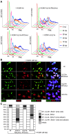
Figure 4. Cyclin A2 expression relieves the block of cellular DNA synthesis in HCMV-infected cells.
(A) Cells were infected as described in the legend to figure 3. Cell cycle progression was monitored by flow cytometric analysis of cellular DNA content (n =  haploid number of chromosomes). (B) To distinguish cellular from viral DNA replication, infected cells were labeled with BrdU at 48 h post infection (hpi). The sites of BrdU incorporation were stained by immunofluorescence and compared with the localization of pUL44-containing viral replication compartments, using confocal microscopy. In the right column, merged images of the middle columns are shown. (C) Quantitative analysis of cells undergoing cellular and/or viral DNA replication at 48 hpi. Mitotic cells were identified by DAPI co-staining. The results represent at least 200 randomly selected IE1/2-positive cells.
Figure 5
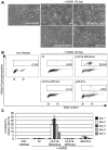
Figure 5. Cyclin A2 up-regulation results in mitotic entry of infected cells.
Cells were infected as described in the legend to figure 3. (A) At 72 hpi, their morphology was examined by phase-contrast microscopy to detect cell rounding. (B, C) Cells were further stained with propidium iodide, IE1/IE2-and phospho-histone H3(Ser10)-specific antibodies and analyzed by flow cytometry to determine the proportion of infected cells with (pre-)mitotic chromosome condensation. The results of one representative experiment (A, B) and of five independent experiments (C) are shown.
Figure 6
Figure 6. Disruption of pUL21a-Cyclin A2 interaction facilitates nuclear translocation of Cyclin B1-CDK1 and nuclear envelope breakdown in HCMV-infected cells.
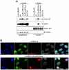
Cells were infected as described in the legend to figure 3. (A) At 48 hpi, nuclear and cytoplasmic fractions were prepared and analyzed for the presence of Cyclin B1 and CDK1 protein. The purity of both fractions was controlled by immunoblot detection of Lamin A/C and β-Tubulin. (B) In parallel, cells were analyzed by immunofluorescence microscopy for markers of chromosome condensation (histone H3 phosphorylation at serine 10) and nuclear envelope integrity (Lamin A/C). Shown are representative images. All cells were confirmed to be HCMV-positive by IE1/IE2 antibody co-staining.
Figure 7
Figure 7. Lack of Cyclin A2 degradation promotes metaphase arrest, aberrant spindle formation and chromosomal instability in HCMV-infected cells.
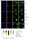
Cells were infected with HCMV-wt, HCMV-UL21a-RXL2mut or HCMV-ΔUL21a. Infected cells were examined by immunofluorescence microscopy for localization and structural organization of chromosomal material (DAPI staining), centromeres (CENP-A staining) and mitotic spindles (α-Tubulin). Representative images are shown in (A). Chromosomes lacking centromeres and accordingly have lost spindle attachment are marked by arrowheads. Quantitative data based on at least 100 mitotic cells per sample are presented in (B). Non-mitotic cells were not included in this quantitative analysis.
Figure 8
Figure 8. Generalized chromosome shattering in HCMV-UL21a-RXL2mut-infected cells.
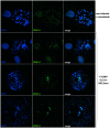
Metaphase spreads from nocodazole-treated, non-infected cells were subjected to CENP-A immunofluorescence and DAPI staining and compared to equally prepared material of cells 3 days post HCMV-UL21a-RXL2mut-infection. Representative images are shown.
Figure 9
Figure 9. Precocious separation of sister chromatids and progressive chromosome fragmentation predominate the chromosomal appearance of HCMV-UL21a-RXL2mut-infected cells.
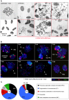
(A) Metaphase spreads from nocodazole-treated, non-infected cells were subjected to Giemsa staining and compared to equally prepared chromosomal material of HCMV-wt and HCMV-UL21a-RXL2mut-infected cells at 2 to 4 days post infection (dpi). Where indicated, magnified views of the encircled areas #1 and #2 in the adjacent image of RXL2mut-infected cells are shown. Scale bars: 5 µm. (B) Metaphase spreads were analyzed by fluorescence in situ hybridization (FISH) using whole chromosome painting probes for chromosomes 1 and 3. DNA was counterstained with DAPI. Typical examples of cells with intact metaphase chromosomes (IMC), fragmented chromosomes (FC), incomplete chromosome condensation (ICC) or precocious separation of sister chromatids (PSC) are shown and labeled accordingly. (C) Quantitative evaluation of FISH analysis based on at least 100 mitotic HCMV-UL21a-RXL2mut-infected cells per sample. Non-mitotic cells were not included in the analysis.
Figure 10
Figure 10. Selective down-regulation of IE2 protein in mitotic cells.
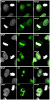
Cells were infected with HCMV-UL21a-RXL2mut and analyzed at 72 hpi by confocal immunofluorescence microscopy for the expression and subcellular localization of IE1 and IE2 proteins. DAPI staining enabled discrimination of mitotic from non-mitotic cells. Representative images are shown. The right column displays the merged fluorescence channels.
Figure 11
Figure 11. Impaired viral DNA replication in mitotic cells.
Cells were infected with HCMV-UL21a-RXL2mut and prepared at 48 hpi for immunofluorescence microscopy. Chromatin condensation (DAPI), localization of viral DNA replication compartments (pUL44) and DNA synthesis (BrdU) were analyzed and representative images are shown. The right column displays the merged fluorescence channels. Arrows point to the small pUL44-positive areas in metaphase cells. Scale bars: 5 µm.
Figure 12
Figure 12. An RXL-based molecular interface between HCMV and Cyclin A2.
The interface consists of two classical Cyclin A2 interaction motifs that have been acquired by viral gene products pp150 and pUL21a. The tegument protein pp150 is a substrate of Cyclin A2-dependent phosphorylation and blocks viral gene expression when Cyclin A2-CDK activity is high. Once viral gene expression has started, the early-late protein pUL21a blocks Cyclin A2-CDK activity by Cyclin A2 degradation.
Figure 13
Figure 13. Crosstalk and antagonism between Cyclin A2 and APC/C-inhibitory functions of HCMV-pUL21a.
The UL21a gene product conveys to independent cell cycle activities, inhibition of Cyclin A2-dependent protein phosphorylation and inhibition of APC/C-dependent protein ubiquitination. Cyclin A2 inhibition, by impinging on Cyclin B1 accumulation and CDK1 nuclear translocation, prevents HCMV-infected cells from entering mitosis. APC/C inhibition leads to a block of metaphase-anaphase transition (left panel). If the Cyclin A2-inhibitory function is selectively impaired by UL21a-RXL mutation, the remaining APC/C-inhibitory function favors the accumulation of Cyclin A2, Cyclin B1, nuclear CDK1 and hence mitotic entry, while anaphase entry is still blocked (middle panel). If both functions are ablated by UL21a knockout, mitotic entry is still possible but much less efficient. Once in mitosis, the HCMV-ΔUL21a-infected cells can proceed to ana- and telophase. Green or red arrows represent the net effects of the indicated HCMV-UL21a variants on G2/M and metaphase/anaphase transitions.

Similar articles

Cited by

References

    1. Britt W (2008) Manifestations of human cytomegalovirus infection: proposed mechanisms of acute and chronic disease. Curr Top Microbiol Immunol 325: 417–470. - PubMed
    1. Johnsen JI, Baryawno N, Soderberg-Naucler C (2011) Is human cytomegalovirus a target in cancer therapy? Oncotarget 2: 1329–1338. - PMC - PubMed
    1. Schreiber A, Harter G, Schubert A, Bunjes D, Mertens T, et al. (2009) Antiviral treatment of cytomegalovirus infection and resistant strains. Expert Opin Pharmacother 10: 191–209. - PubMed
    1. Yu Y, Clippinger AJ, Alwine JC (2011) Viral effects on metabolism: changes in glucose and glutamine utilization during human cytomegalovirus infection. Trends Microbiol 19: 360–367. - PMC - PubMed
    1. Fliss PM, Brune W (2012) Prevention of cellular suicide by cytomegaloviruses. Viruses 4: 1928–1949. - PMC - PubMed

Publication types

MeSH terms

Grants and funding

This work was supported by grant WI2043/3-1 from the Deutsche Forschungsgemeinschaft (http://www.dfg.de) to LW and CH. The funders had no role in study design, data collection and analysis, decision to publish, or preparation of the manuscript.

LinkOut - more resources