. 2015 Apr;72(8):1613-29.
doi: 10.1007/s00018-014-1777-y. Epub 2014 Nov 13.

MEK1 transduces the prion protein N2 fragment antioxidant effects

Affiliations

MEK1 transduces the prion protein N2 fragment antioxidant effects

C L Haigh et al. Cell Mol Life Sci. 2015 Apr.

Abstract

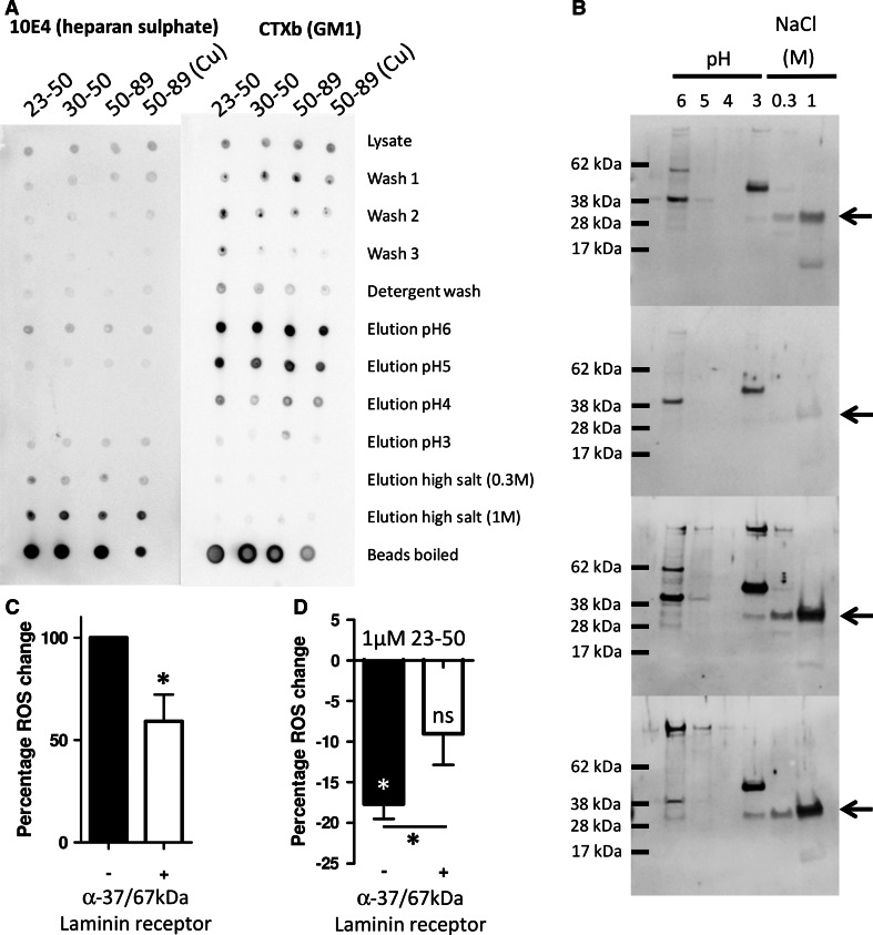
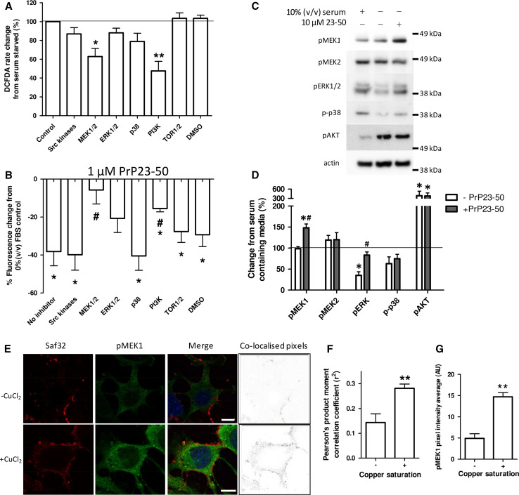
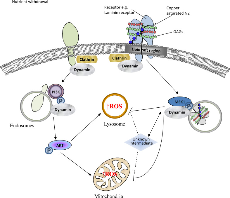
The prion protein (PrP(C)) when mis-folded is causally linked with a group of fatal neurodegenerative diseases called transmissible spongiform encephalopathies or prion diseases. PrP(C) normal function is still incompletely defined with such investigations complicated by PrP(C) post-translational modifications, such as internal cleavage, which feasibly could change, activate, or deactivate the function of this protein. Oxidative stress induces β-cleavage and the N-terminal product of this cleavage event, N2, demonstrates a cellular protective response against oxidative stress. The mechanisms by which N2 mediates cellular antioxidant protection were investigated within an in vitro cell model. N2 protection was regulated by copper binding to the octarepeat domain, directing the route of internalisation, which stimulated MEK1 signalling. Precise membrane interactions of N2, determined by copper saturation, and involving both the copper-co-ordinating octarepeat region and the structure conferred upon the N-terminal polybasic region by the proline motif, were essential for the correct engagement of this pathway. The phenomenon of PrP(C) post-translational modification, such as cleavage and copper co-ordination, as a molecular "switch" for activation or deactivation of certain functions provides new insight into the apparent multi-functionality of PrP(C).


Figures

Fig. 1
Fig. 1
Copper saturation of N2 influences its anti-oxidant function, cell surface interactions and trafficking. a Schematic of the PrP N2 cleavage fragment (residues 23–89) with antibody binding sites and the P26/28A mutation shown. The octarepeat (copper-binding) region is indicated in blue. b Log10 dose–response of the intracellular ROS reduction activity displayed by copper-saturated (filled bars) and zinc-saturated (hollow bars) PrP23-89 following serum deprivation. c Measurement of MTS metabolism 24 h after serum deprivation and peptide addition. d Antibody detection of the PrP23-89 fragment at the cell surface (non-permeabilised cells) and inside the cell 15 min after peptide addition with or without copper saturation. Antibody signal is shown in green and blue staining indicates DAPI within the nucleus. Graphs show quantification of the detected PrP23-89 signal with white bars indicating apo-PrP23-89 and blue bars indicating copper-saturated PrP23-89 staining. e Staining of CF10 cells in the absence of peptide addition to control for non-specific antibody binding. f Immunofluorescent detection of PrP23-89 inside of cells 90 min post-peptide addition with or without copper. g Immunofluorescent detection of PrP23-89P26/28A at the cell surface, with and without copper saturation, 15 min after peptide addition. Scale bars = 25 µm. *p < 0.05, **p < 0.01, ***p < 0.001
Fig. 2
Fig. 2
Inhibition of internalisation reduces stress-induced intracellular ROS and inhibits the protective action of PrP23-50. a DCFDA assay for the detection of intracellular ROS produced in response to serum deprivation. b Cells were stressed by serum deprivation with or without the internalisation inhibitors T-A23 and Dynasore. Changes are shown normalised to 0 % (v/v) serum control values as 100 % ROS production. c Measurement of the percentage ROS reduction exerted by PrP23-50 with and without internalisation inhibition. Results are shown as percentage reduction from the serum-deprived ∓ inhibitor control. Asterisks denote significantly different from no peptide control (*p < 0.05, **p < 0.01, ***p < 0.001) and hash shows significantly different from the no inhibitor action of PrP23-50 (# < 0.05). d Incorporation of the lipophilic dye laurdan into cell membranes before treatment with PrP23-50 (or a RS peptide control) in serum-free media
Fig. 3
Fig. 3
Copper-saturated PrP23-89 reduces the lysosomal and mitochondrial ROS caused by serum deprivation. a Representative plates of RedoxSensor Red changes due to serum deprivation and as influenced by PrP23-89 with and without copper saturation. RedoxSensor Red fluorescence was co-localised with MitoTracker green to indicate compartmental partitioning. Scale bars = 25 µM. Quantification of the cytosolic [co-localised with MitoTracker; (b)] and lysosomal (c) ROS fractions shown relative to 10 % (v/v) serum control. d Representative plates of mitoSOX fluorescence production in response to serum deprivation. Scale bars = 25 µM. e Quantification of mitoSOX fluorescence intensity. *p < 0.05, **p < 0.01
Fig. 4
Fig. 4
Downstream targeting of lysosomes does not play a prominent role in 23–50 ROS reduction and PrP23-89 is not trafficked to this destination. a Effect of the inhibition of NADPH oxidase ROS signalling and lysosomal acidity on serum-deprived ROS production as assayed by DCFDA fluorescence. Data are plotted as a percentage of the serum-deprived control. b 23–50 ROS reduction response after inhibition of NADPH oxidases or lysosomal acidification. Results are shown as percentage reduction from the serum-deprived ∓ inhibitor control. *p < 0.05. Co-localisation of 1 µM PrP23-89 (∓ copper saturation) with lamp-1 lysosomal marker (c) and SOD2 mitochondrial marker (d). Blue = DAPI nuclei staining. Scale bar = 20 µm
Fig. 5
Fig. 5
The PrP N-terminus pulls down a complex containing heparan sulphate, lipid rafts and the 37/67 kDa laminin receptor. a Dot blots for HS and GM1 of pull downs from cell lysate using PrP N-terminal fragments. b The fractions were western blotted for the 37/67kDA laminin receptor (expected size of the 37/67 kDa laminin receptor band is indicated by arrows). c DCFDA assay of the effect of blocking the 37/67 kDa laminin receptor with antibodies on the induction of ROS during serum deprivation. d Assessment of the activity of PrP23-50 in reducing intracellular ROS produced in response to serum deprivation following targeting of the 37/67 kDa laminin receptor with antibodies. *p < 0.05
Fig. 6
Fig. 6
PrP23-50 influences MEK-1, ERK1/2, and PI3K signalling pathways and copper-saturated N2 shows increased co-localisation with pMEK1. a Inhibitors of cellular signalling pathways were assayed for their ability to interfere with the ROS produced in response to serum deprivation. The inhibitors used were as follows; Src kinases—500 nM PP2; MEK—10 µM U0126; ERK—10 µM 3-(2-Aminoethyl)-5-((4-ethoxyphenyl)methylene)-2,4-thiazolidinedione hydrochloride; p38—10 µM SB203580; PI3K—1 µM wortmannin; TOR1/2—1 µM rapamycin; DMSO control—0.1 % (v/v); the target protein is indicated in the graph; DCFDA assay results are shown relative to the serum-deprived 100 % control value. b PrP23-50 ROS reduction was assessed by DCFDA fluorescence and shown as percentage reduction from the serum-deprived ± inhibitor control. *p < 0.05 from the no peptide control, # p < 0.05 from the no inhibitor action of PrP23-50. c Western blots and d corresponding densitometry of changes in phosphorylation (activation) of central signal transduction intermediates in response to serum deprivation with and without PrP23-50. Graphical data are presented relative to serum control. e Co-localisation of PrP23-89 with pMEK1. PrP23-89, as detected by saf32, is shown in red and pMEK1 staining is shown in green, with DAPI staining of the nuclei in blue. The heat map (right plates) shows co-localising pixels. Scale bars = 10 µM. f Pearson’s product moment correlation co-efficient analysis of the apo- and copper-saturated PrP23-89 co-localisation. **p < 0.01. g Quantification of pixel intensity for the pMEK1 staining. **p < 0.01
Fig. 7
Fig. 7
Schematic representation of intracellular signalling pathways influenced by N2 during cellular protection against oxidative stress. Upon serum withdrawal, an intracellular ROS increase is stimulated within mitochondria and lysosomes. The ROS reaction is signalled through internalisation/trafficking of membrane complexes and involves central intermediates of the AKT and MAPK signal transduction pathways. Correct engagement of a lipid-raft membrane complex containing glycosaminoglycans and the 37/67 kDa laminin receptor by copper-loaded the PrP N2 fragment modulates intracellular trafficking and activates MEK1 signalling to oppose ROS production

Similar articles

Cited by

References

    1. Haigh CL, Brown DR. Prion protein reduces both oxidative and non-oxidative copper toxicity. J Neurochem. 2006;98(3):677–689. doi: 10.1111/j.1471-4159.2006.03906.x. - DOI - PubMed
    1. Brown DR, Schulz-Schaeffer WJ, Schmidt B, Kretzschmar HA. Prion protein-deficient cells show altered response to oxidative stress due to decreased SOD-1 activity. Exp Neurol. 1997;146(1):104–112. doi: 10.1006/exnr.1997.6505. - DOI - PubMed
    1. Klamt F, Dal-Pizzol F, Conte da Frota ML, Jr, Walz R, Andrades ME, da Silva EG, Brentani RR, Izquierdo I, Fonseca Moreira JC. Imbalance of antioxidant defense in mice lacking cellular prion protein. Free Radic Biol Med. 2001;30(10):1137–1144. doi: 10.1016/S0891-5849(01)00512-3. - DOI - PubMed
    1. Rachidi W, Vilette D, Guiraud P, Arlotto M, Riondel J, Laude H, Lehmann S, Favier A. Expression of prion protein increases cellular copper binding and antioxidant enzyme activities but not copper delivery. J Biol Chem. 2003;278(11):9064–9072. doi: 10.1074/jbc.M211830200. - DOI - PubMed
    1. Senator A, Rachidi W, Lehmann S, Favier A, Benboubetra M. Prion protein protects against DNA damage induced by paraquat in cultured cells. Free Radic Biol Med. 2004;37(8):1224–1230. doi: 10.1016/j.freeradbiomed.2004.07.006. - DOI - PubMed

Publication types

MeSH terms

LinkOut - more resources