Histone deacetylase 7 (Hdac7) suppresses chondrocyte proliferation and β-catenin activity during endochondral ossification
- PMID: 25389289
- PMCID: PMC4281714
- DOI: 10.1074/jbc.M114.596247
Histone deacetylase 7 (Hdac7) suppresses chondrocyte proliferation and β-catenin activity during endochondral ossification
Abstract
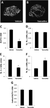
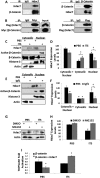
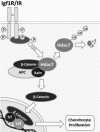
Histone deacetylases (Hdacs) regulate endochondral ossification by suppressing gene transcription and modulating cellular responses to growth factors and cytokines. We previously showed that Hdac7 suppresses Runx2 activity and osteoblast differentiation. In this study, we examined the role of Hdac7 in postnatal chondrocytes. Hdac7 was highly expressed in proliferating cells within the growth plate. Postnatal tissue-specific ablation of Hdac7 with a tamoxifen-inducible collagen type 2a1-driven Cre recombinase increased proliferation and β-catenin levels in growth plate chondrocytes and expanded the proliferative zone. Similar results were obtained in primary chondrocyte cultures where Hdac7 was deleted with adenoviral-Cre. Hdac7 bound β-catenin in proliferating chondrocytes, but stimulation of chondrocyte maturation promoted the translocation of Hdac7 to the cytoplasm where it was degraded by the proteasome. As a result, β-catenin levels and transcription activity increased in the nucleus. These data demonstrate that Hdac7 suppresses proliferation and β-catenin activity in chondrocytes. Reducing Hdac7 levels in early chondrocytes may promote the expansion and regeneration of cartilage tissues.
Keywords: ATDC5 Cells; Beta-catenin (β-catenin); Cartilage; Growth Plate; Histone Deacetylase (HDAC); Histone Deacetylase 7 (Hdac7); Insulin.
© 2015 by The American Society for Biochemistry and Molecular Biology, Inc.
Figures


Similar articles
-
Histone deacetylase 7 controls endothelial cell growth through modulation of beta-catenin.Circ Res. 2010 Apr 16;106(7):1202-11. doi: 10.1161/CIRCRESAHA.109.213165. Epub 2010 Mar 11. Circ Res. 2010. PMID: 20224040
-
Vital Roles of β-catenin in Trans-differentiation of Chondrocytes to Bone Cells.Int J Biol Sci. 2018 Jan 1;14(1):1-9. doi: 10.7150/ijbs.23165. eCollection 2018. Int J Biol Sci. 2018. PMID: 29483820 Free PMC article.
-
Developmental regulation of Wnt/beta-catenin signals is required for growth plate assembly, cartilage integrity, and endochondral ossification.J Biol Chem. 2005 May 13;280(19):19185-95. doi: 10.1074/jbc.M414275200. Epub 2005 Mar 10. J Biol Chem. 2005. PMID: 15760903
-
Regulatory mechanisms for the development of growth plate cartilage.Cell Mol Life Sci. 2013 Nov;70(22):4213-21. doi: 10.1007/s00018-013-1346-9. Epub 2013 May 4. Cell Mol Life Sci. 2013. PMID: 23640571 Free PMC article. Review.
-
The cartilage extracellular matrix as a transient developmental scaffold for growth plate maturation.Matrix Biol. 2016 May-Jul;52-54:363-383. doi: 10.1016/j.matbio.2016.01.008. Epub 2016 Jan 23. Matrix Biol. 2016. PMID: 26807757 Review.
Cited by
-
Epigenetic Regulation of Skeletal Tissue Integrity and Osteoporosis Development.Int J Mol Sci. 2020 Jul 12;21(14):4923. doi: 10.3390/ijms21144923. Int J Mol Sci. 2020. PMID: 32664681 Free PMC article. Review.
-
Concise Review: The Regulatory Mechanism of Lysine Acetylation in Mesenchymal Stem Cell Differentiation.Stem Cells Int. 2020 Jan 28;2020:7618506. doi: 10.1155/2020/7618506. eCollection 2020. Stem Cells Int. 2020. PMID: 32399051 Free PMC article. Review.
-
Interplay between genetics and epigenetics in osteoarthritis.Nat Rev Rheumatol. 2020 May;16(5):268-281. doi: 10.1038/s41584-020-0407-3. Epub 2020 Apr 9. Nat Rev Rheumatol. 2020. PMID: 32273577 Review.
-
TET1 Directs Chondrogenic Differentiation by Regulating SOX9 Dependent Activation of Col2a1 and Acan In Vitro.JBMR Plus. 2020 Jun 26;4(8):e10383. doi: 10.1002/jbm4.10383. eCollection 2020 Aug. JBMR Plus. 2020. PMID: 33134768 Free PMC article.
-
Hdac3 Deficiency Increases Marrow Adiposity and Induces Lipid Storage and Glucocorticoid Metabolism in Osteochondroprogenitor Cells.J Bone Miner Res. 2016 Jan;31(1):116-28. doi: 10.1002/jbmr.2602. Epub 2015 Aug 20. J Bone Miner Res. 2016. PMID: 26211746 Free PMC article.
References
-
- Kronenberg H. M. (2003) Developmental regulation of the growth plate. Nature 423, 332–336 - PubMed
-
- Razidlo D. F., Whitney T. J., Casper M. E., McGee-Lawrence M. E., Stensgard B. A., Li X., Secreto F. J., Knutson S. K., Hiebert S. W., Westendorf J. J. (2010) Histone deacetylase 3 depletion in osteo/chondroprogenitor cells decreases bone density and increases marrow fat. PLoS One 5, e11492. - PMC - PubMed
Publication types
MeSH terms
Substances
Grants and funding
LinkOut - more resources
Full Text Sources
Molecular Biology Databases
Research Materials

