. 2015 May 1;523(7):1038-1053.
doi: 10.1002/cne.23706. Epub 2015 Feb 17.

Connectivity of pacemaker neurons in the neonatal rat superficial dorsal horn

Affiliations

Connectivity of pacemaker neurons in the neonatal rat superficial dorsal horn

Jie Li et al. J Comp Neurol. .

Abstract

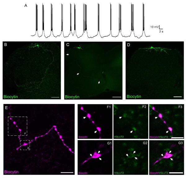
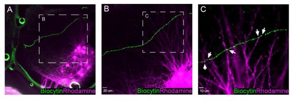
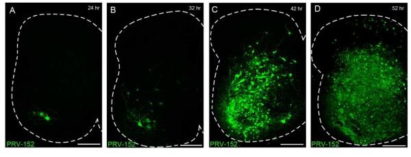
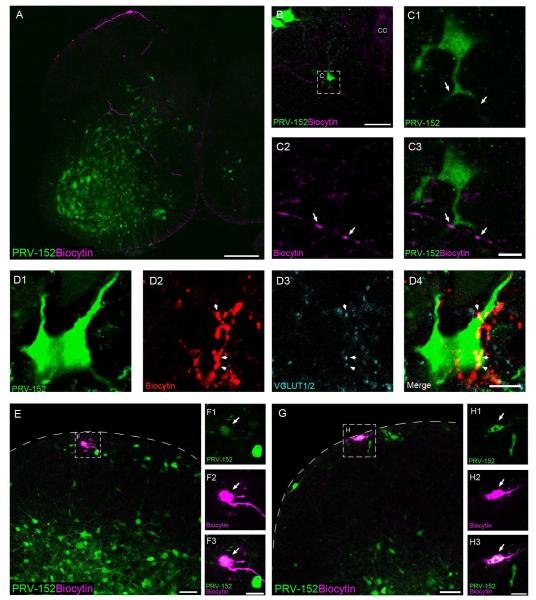
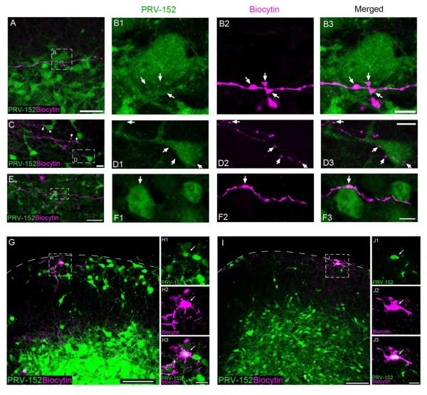
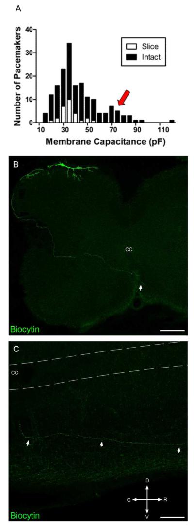
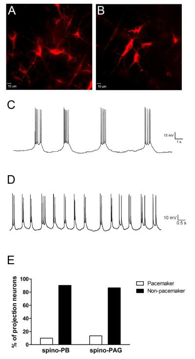
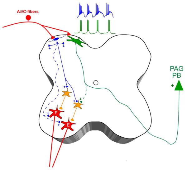
Pacemaker neurons with an intrinsic ability to generate rhythmic burst-firing have been characterized in lamina I of the neonatal spinal cord, where they are innervated by high-threshold sensory afferents. However, little is known about the output of these pacemakers, as the neuronal populations that are targeted by pacemaker axons have yet to be identified. The present study combines patch-clamp recordings in the intact neonatal rat spinal cord with tract-tracing to demonstrate that lamina I pacemaker neurons contact multiple spinal motor pathways during early life. Retrograde labeling of premotor interneurons with the trans-synaptic pseudorabies virus PRV-152 revealed the presence of burst-firing in PRV-infected lamina I neurons, thereby confirming that pacemakers are synaptically coupled to motor networks in the spinal ventral horn. Notably, two classes of pacemakers could be distinguished in lamina I based on cell size and the pattern of their axonal projections. Whereas small pacemaker neurons possessed ramified axons that contacted ipsilateral motor circuits, large pacemaker neurons had unbranched axons that crossed the midline and ascended rostrally in the contralateral white matter. Recordings from identified spino-parabrachial and spino-periaqueductal gray neurons indicated the presence of pacemaker activity within neonatal lamina I projection neurons. Overall, these results show that lamina I pacemakers are positioned to regulate both the level of activity in developing motor circuits and the ascending flow of nociceptive information to the brain, thus highlighting a potential role for pacemaker activity in the maturation of pain and sensorimotor networks in the central nervous system.

Keywords: AB_1587626; AB_2301751; AB_2336883; burst-firing; lamina I; premotor interneuron; spinal cord; synapse.


Figures

Figure 1
Figure 1
Lamina I pacemakers send glutamatergic projections to multiple regions of the intact neonatal rat spinal cord. A, Example of the rhythmic burst-firing that characterizes pacemaker activity in the immature superficial dorsal horn (SDH). B, C, D, Composite images constructed from 11 (B), 5 (C) or 8 (D) overlaid serial sections (each 20 μm thick). While some pacemaker axons display extensive branching in the deep dorsal horn before reaching the lateral edge of the gray matter (B; scale bar = 100 μm), other pacemakers send relatively unbranched axons around the lateral edge of the dorsal horn and across the anterior commissure (C; scale bar = 200 μm). A third group of pacemakers was characterized by axons which ramified exclusively within the superficial laminae of the dorsal horn (D; scale bar = 100 μm). E, Image (consisting of a z-stack of 11 optical sections at a thickness of 0.5 μm) depicting a pacemaker axon located in the deep dorsal horn with several prominent boutons (see boxed regions). Scale bar = 10 μm. F, G, Single optical sections (0.5 μm) showing a higher magnification of the boxed regions indicated in panel E, demonstrating that the axonal boutons of small pacemaker neurons (magenta) co-localize (arrows) with VGLUT2 immunoreactivity (green). Scale bars = 5 μm.
Figure 2
Figure 2
Putative axodendritic contacts between pacemaker neurons and spinal motor neurons. A, Low magnification image showing a biocytin-filled axon (green) of an identified lamina I pacemaker neuron that travels through the deep dorsal horn of the spinal cord in a ventromedial direction. A pool of lumbar motor neurons (magenta) has been retrogradely labeled via rhodamine dextran applied to the L4 ventral root. Scale bar = 50 μm. B, Higher magnification of the boxed region in panel A illustrating the dense network of motor neuron dendrites which extend dorsally in proximity to the pacemaker axon. Scale bar = 20 μm. C, Single optical section (0.5 μm) showing a higher magnification of boxed region in panel B. Pacemaker axonal varicosities (green) are in close apposition to rhodamine-labeled motor neuron dendrites (magenta) in the deep dorsal horn of the neonatal spinal cord (arrows). Scale bar = 10 μm.
Figure 3
Figure 3
Visualization of spinal motor pathways via the retrograde transport of PRV-152. A-D, Time course of PRV-152 labeling following injection into the left hindlimb quadriceps. Motor neurons are labeled beginning at 24 - 28 hours post-infection (p.i.) (A), with GFP expression appearing in premotor interneurons between 32 - 44 hours p.i. (B, C). D, At approximately 48 - 52 hours p.i., PRV-infected cells are clearly observed in the superficial dorsal horn. Scale bars = 200 μm.
Figure 4
Figure 4
Lamina I pacemakers synapse onto flexor motor pathways in the spinal cord. A, Example of a pacemaker axon (magenta) coursing through the deep dorsal horn and ventral horn of the neonatal spinal cord, where it lies in close proximity to numerous GFP-expressing cells following the injection of PRV-152 into the biceps femoris. Scale bar = 200 μm. B, Image depicting a pacemaker axon projecting towards the anterior commissure that makes putative contacts with the dendrites of a GFP-expressing cell in the ipsilateral ventral horn. CC = central canal; scale bar = 50 μm. C, Higher magnification of the boxed region in panel B. Arrows indicate sites where the biocytin-filled axonal boutons (magenta) co-localized with GFP (green) in a single 0.5 μm optical section. Scale bar = 10 μm. D, Single optical sections (0.5 μm) illustrating an example of pacemaker axonal boutons (D2) positive for VGLUT1/2 immunoreactivity (D3) making putative contacts with a PRV-152 infected cell (D1) in the deep dorsal horn (D4). Scale bar = 10 μm. E-H, Examples of electrophysiologically identified lamina I pacemaker neurons that were infected with PRV-152 at 52 hours following virus injection into the biceps femoris, as evidenced by the co-localization (F3, H3) of biocytin labeling (F2, H2; magenta) and GFP expression (F1, H1; green). Scale bars = 20 μm. F, H, Single optical sections (0.5 μm thickness) showing higher magnification of the boxed regions in panels E and G, respectively. Scale bars = 10 μm.
Figure 5
Figure 5
Pacemakers engage spinal networks innervating hindlimb extensor muscles in the neonatal rat. A, Pacemaker axonal boutons (magenta) are in close apposition to a GFP-expressing soma (green) in the deep dorsal horn following PRV-152 injection into the quadriceps muscles. Scale bar = 50 μm. B, Single 0.5 μm optical sections showing higher magnification of the boxed region in panel A, illustrating putative sites of contact between the pacemaker axon and the PRV-infected cell (arrows). Scale bar = 10 μm. C, A pacemaker axon branches extensively in the vicinity of numerous PRV-infected cells located in the deep dorsal horn. Scale bar = 20 μm. D, Higher magnification of the boxed region shown in C (single 0.5 μm optical sections) reveals multiple putative contacts between the axon of the pacemaker neuron and a GFP-expressing cell (arrows). Scale bar = 10 μm. E, Example of a pacemaker axon in close association with a PRV-infected soma in the spinal ventral horn. Scale bar = 50 μm. F, Single 0.5 μm optical sections showing higher magnification of the boxed region in E. Scale bar = 10 μm. G-J, Examples of electrophysiologically identified lamina I pacemaker neurons that were infected with PRV-152 at 52 hours following virus injection into the left quadriceps, as evidenced by the co-localization (H3, J3) of biocytin labeling (H2, J2; magenta) and GFP expression (H1, J1; green). Scale bars = 100 μm. H, J, Single optical sections (0.5 μm thickness) showing higher magnification of boxed regions in panels G and I, respectively. Scale bars = 20 μm.
Figure 6
Figure 6
The intact spinal cord contains a population of large pacemaker neurons with distinct axonal projection patterns. A, Histogram showing the distribution of pacemaker neurons as a function of membrane capacitance in the spinal cord slice (n = 28) vs. intact spinal cord preparations (n = 172). Arrow indicates a population of larger pacemakers identified in the intact spinal cord (black) which was not observed when recording from the spinal cord slice preparation (white; see also Li and Baccei 2011). B, Representative example of a large pacemaker neuron (recorded using the intact spinal cord preparation) whose axon projects to the contralateral side (arrow) via the anterior commissure. Composite images were constructed from 10 overlaid serial transverse sections (each 50 μm thick). Scale bar = 200 μm. C, Parasagittal section (50 μm) of the spinal cord on the side contralateral to the pacemaker soma, illustrating the rostral trajectory taken by the pacemaker axon after crossing the anterior commissure. CC = central canal; D = Dorsal; V = Ventral; R = Rostral; C = Caudal. Scale bar = 100 μm.
Figure 7
Figure 7
Ascending lamina I projection neurons can exhibit pacemaker activity within the intact spinal cord network during early life. A, B, Representative confocal images of retrogradely-labeled lamina I projection neurons within the intact spinal cord preparation following DiI injection into the parabrachial nucleus (PB). Scale bars = 10 μm. C, Example of the rhythmic burst-firing which was observed in an identified spino-PB projection neuron during the neonatal period. D, A subset of pacemaker projection neurons exhibited shorter bursts of activity which were often characterized by spike doublets. E, There were no significant differences in the prevalence of pacemaker activity between the spino-PB (n = 41) and spino-PAG (n = 37) populations of projection neurons during the first postnatal week (p = 0.729; Fisher’s exact test).
Figure 8
Figure 8
Working model of pacemaker connectivity in the developing spinal cord. Lamina I of the neonatal dorsal horn is proposed to contain two distinct populations of pacemaker neurons, which are both innervated by high-threshold primary afferents (i.e. Aδ/C-fibers). Small pacemakers (blue) correspond to glutamatergic interneurons which project to the superficial laminae and/or to the deep dorsal and ventral horns, where they synapse onto premotor interneurons (orange) within flexor (F) and extensor (E) motor pathways. Meanwhile, the majority of large pacemakers (green) project their axons into the contralateral white matter and correspond to ascending projection neurons targeting supraspinal sites of nociceptive processing such as the parabrachial nucleus (PB) and periaqueductal gray (PAG). Dotted lines represent potential synaptic connections which require further investigation.

Similar articles

Cited by

References

    1. Al-Khater KM, Todd AJ. Collateral projections of neurons in laminae I, III, and IV of rat spinal cord to thalamus, periaqueductal gray matter, and lateral parabrachial area. J Comp Neurol. 2009;515:629–46. - PMC - PubMed
    1. Beggs S, Torsney C, Drew LJ, Fitzgerald M. The postnatal reorganization of primary afferent input and dorsal horn cell receptive fields in the rat spinal cord is an activity-dependent process. Eur J Neurosci. 2002;16:1249–58. - PubMed
    1. Blumberg MS, Lucas DE. Dual mechanisms of twitching during sleep in neonatal rats. Behav Neurosci. 1994;108:1196–202. - PubMed
    1. Busetto G, Buffelli M, Cangiano L, Cangiano A. Effects of evoked and spontaneous motoneuronal firing on synapse competition and elimination in skeletal muscle. J Neurocytol. 2003;32:795–802. - PubMed
    1. Card JP, Rinaman L, Lynn RB, Lee BH, Meade RP, Miselis RR, Enquist LW. Pseudorabies virus infection of the rat central nervous system: ultrastructural characterization of viral replication, transport, and pathogenesis. J Neurosci. 1993;13:2515–39. - PMC - PubMed

Publication types

MeSH terms

LinkOut - more resources