. 2015 Jan;156(1):346-59.
doi: 10.1210/en.2014-1524.

Role of retinoic acid and platelet-derived growth factor receptor cross talk in the regulation of neonatal gonocyte and embryonal carcinoma cell differentiation

Affiliations

Role of retinoic acid and platelet-derived growth factor receptor cross talk in the regulation of neonatal gonocyte and embryonal carcinoma cell differentiation

Gurpreet Manku et al. Endocrinology. 2015 Jan.

Abstract

Neonatal gonocytes are direct precursors of spermatogonial stem cells, the cell pool that supports spermatogenesis. Although unipotent in vivo, gonocytes express pluripotency genes common with embryonic stem cells. Previously, we found that all-trans retinoic acid (RA) induced the expression of differentiation markers and a truncated form of platelet-derived growth factor receptor (PDGFR)β in rat gonocytes, as well as in F9 mouse embryonal carcinoma cells, an embryonic stem cell-surrogate that expresses somatic lineage markers in response to RA. The present study is focused on identifying the signaling pathways involved in RA-induced gonocyte and F9 cell differentiation. Mitogen-activated protein kinase kinase (MEK) 1/2 activation was required during F9 cell differentiation towards somatic lineage, whereas its inhibition potentiated RA-induced Stra8 expression, suggesting that MEK1/2 acts as a lineage specification switch in F9 cells. In both cell types, RA increased the expression of the spermatogonial/premeiotic marker Stra8, which is in line with F9 cells being at a stage before somatic-germline lineage specification. Inhibiting PDGFR kinase activity reduced RA-induced Stra8 expression. Interestingly, RA increased the expression of PDGFRα variant forms in both cell types. Together, these results suggest a potential cross talk between RA and PDGFR signaling pathways in cell differentiation. RA receptor-α inhibition partially reduced RA effects on Stra8 in gonocytes, indicating that RA acts in part via RA receptor-α. RA-induced gonocyte differentiation was significantly reduced by inhibiting SRC (v-src avian sarcoma [Schmidt-Ruppin A-2] viral oncogene) and JAK2/STAT5 (Janus kinase 2/signal transducer and activator of transcription 5) activities, implying that these signaling molecules play a role in gonocyte differentiation. These results suggest that gonocyte and F9 cell differentiation is regulated via cross talk between RA and PDGFRs using different downstream pathways.


Figures
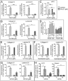
Figure 1.
Figure 1.. Effects of PDGFR signaling pathways inhibitors on F9 cell differentiation.
F9 cells were treated for 72 hours with 0.1μM RA and the indicated inhibitors, alone or in combination with RA. A and B, mRNA levels of 2 differentiation markers of somatic lineage differentiation (Collagen IV and Laminin B1) were quantified using qPCR analysis for F9 cells treated with or without RA and the PDGFR kinase inhibitors AG370 and AG1295. C, mRNA levels of cMYC were quantified for F9 cells treated with or without RA and AG370. D, Levels of F9 cell proliferation in response to varying concentrations of PDGF-AA and PDGF-BB were determined using a BrdU ELISA proliferation assay. E–G, Effects of PLCγ inhibition (U73122), PI3K inhibition (Wortmannin), and MEK1/2 inhibition (U0126) on RA-induced differentiation of F9 cells. H, The mRNA levels of the primitive endoderm markers S1P1 and Moesin were quantified by qPCR analysis. Results shown represent the mean ± SEM of at least 3 independent experiments for each condition (*, P < .05; **, P < .01; ***, P < .001).
Figure 2.
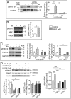
Figure 2.. RA-induced somatic differentiation in F9 cells requires ERK1/2 pathway activation.
F9 cells were treated for 72 hours with 0.1μM RA, 50μM AG370, 10μM U0126, and 250μM cAMP alone or in the different combinations indicated. The proteins were examined by immunoblot analysis, signal intensity analysis of immunoreactive bands was quantified by densitometry, and the results were normalized against GAPDH or tubulin levels (loading control). A, Protein levels of Laminin B1 in F9 cells treated with or without RA and U0126. B, Immunoblot and quantitative analysis of MEK1/2 phosphorylation levels relative to total MEK1 expression in F9 cells treated with or without RA. C, Immunoblot and quantitative analysis of ERK1/2 phosphorylation levels relative to total ERK1/2 expression in F9 cells treated with or without RA and cAMP. D, Immunoblot and quantitative analysis of ERK1/2 phosphorylation levels relative to total ERK1/2 expression in F9 cells treated with or without RA, AG370, and U0126. The results shown represent the mean ± SEM of at least 3 independent experiments for each condition (*, P < .05; **, P < .01; ***, P < .001).
Figure 3.
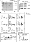
Figure 3.. RA-dependent formation of a 45-kDa variant PDGFRα in F9 cells requires MEK1/2 activation.
RA induces the formation of truncated forms of PDGFRα in differentiating F9 cells, which are dependent on PDGFR and MEK1/2 activation. F9 cells were treated for 72 hours with or without RA and AG370. A, Representative immunoblot shown of results obtained for PDGFRα expression in RA-treated F9 cells with or without AG370 treatment. B, Signal density analysis of the immunoblot in A was performed. Results were normalized to GAPDH (loading control). C, Immunoblot analysis of F9 cells treated for 72 hours with or without RA and Cathepsin L inhibitor I. Representative blot shown. D and E, Representative immunoblot shown alongside signal density analysis of the 45-kDa variant PDGFRα expression in F9 cells treated with or without RA and the MEK1/2 inhibitor U0126. Results were normalized to GAPDH (loading control). Results shown represent the mean ± SEM of at least 3 independent experiments for each condition (*, P < .05; **, P < .01; ***, P < .001).
Figure 4.
Figure 4.. Confirmation of F9 cell PDGFRα proteins identity by LC-MS analysis.
LC-MS analysis of variant PDGFRα expression in RA-induced differentiation of F9 cells. F9 cells were treated for 72 hours with or without 0.1μM RA. A, Immunoblot analysis of PDGFRα in F9 cells treated with or without RA, nonimmunoprecipitated. B, Immunoblot analysis of PDGFRα in F9 cells treated with or without RA, immunoprecipitated. C, LC-MS analysis of immunoprecipitated bands of interest and the position/coverage of the identified peptides within the sequence of intact PDGFRα protein.
Figure 5.
Figure 5.. The RA-induced expression of Stra8 in F9 cells requires PDGFR activation and is potentiated by MEK1/2 inhibition.
RA cross talk with several PDGFR-related signaling pathways in differentiating F9 cells. F9 cells were treated for 72 hours with or without 0.1μM RA and a variety of different inhibitors. A, mRNA levels of STRA8 gene expression in F9 cells treated with or without RA and AG370. B, mRNA levels of STRA8 gene expression in F9 cells treated with or without RA and MEK1/2 inhibitor U0126. C, mRNA levels of STRA8 gene expression in F9 cells treated with or without RA and RAS inhibitor FTS. Results shown represent the mean ± SEM of at least 3 independent experiments for each condition (*, P < .05; **, P < .01).
Figure 6.
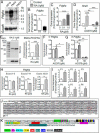
Figure 6.. RA induces the expression of a PDGFRα variant form in PND3 gonocytes.
A, Northern blot analysis of PDGFRα expression in gestational day 18, PND3, and PND21 whole rat testes. GAPDH used as loading control. B, mRNA levels of PDGFRα in PND3 gonocytes (G3) compared with levels in PND8 spermatogonia (G8) using primers specific to the 3′-end of the PDGFRα sequence. C, mRNA levels of PDGFRα in gonocytes treated with RA in a concentration-dependent manner. D, mRNA levels of STRA8 gene expression in gonocytes treated for 24 hours with or without RA and AG370. E, Immunoblot and signal density analysis of PDGFRα expression of gonocytes treated with RA for 72 hours. F and G, mRNA levels of PDGFRα in gonocytes treated with or without RA, specifically aimed at the 5′ (F) (exon 3) and 3′ (G) (exon 21) ends of the PDGFRα sequence. H, mRNA levels of PDGFRα in gonocytes treated with or without RA examined for all exons. Expression grouped by exon-specific areas of the PDGFRα sequence. qPCR analysis was performed for each exon separately, and the data were further pooled to cover 3 main areas, the 5′ end, central exons, and 3′-end of the sequence. I, a similar approach was used to determine the expression levels of 6 sequences located in intron 12, exons 12 and 13, in control and RA-treated gonocytes. J, Representation of intron 12 to exon 13 rat sequences showing in gray shading the regions that were examined and found to be increased by RA treatment. Red box, potential ATG start codon. K, Representation of predicted TF binding sites in rat intron 12 of PDGFRa. Results shown in B–H represent the mean ± SEM of at least 3 independent experiments for each condition. Data in I are from 2 independent experiments (*, P < .05; **, P < .01; ***, P < .001).
Figure 7.
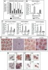
Figure 7.. Identification of RAR and signaling pathways involved in RA-induced gonocytes differentiation.
A, qPCR quantification b of RARs and RXRs transcripts expressed in PND3 gonocytes and PND8 spermatogonia, B, Effect of the RARα inhibitor BMS195614 on the RA-induced increase in Stra8 mRNA expression in gonocytes treated for 24 hours. Results shown represent the mean ± SEM of at least 3 independent experiments for each condition (*, P < .05; ***, P < .001). C, Effects of SRC, JAK2, and STAT5 inhibitors on Stra8 expression. Gonocytes were treated for 24 hours with or without 1.0μM RA and/or the inhibitors SU6656 (SRC family of kinases), AG490 (JAK2), or Pimozide (STAT5). Stra8 mRNA levels were measured by qPCR. Results shown represent the mean ± SEM of at least 3 independent experiments for each condition (*, P < .05; **, P < .01; ***, P < .001). D–F, Immunohistochemical (D) and immunocytochemical (E and F) analyses of protein expression levels (native and phosphorylated forms) of the SRC kinase family members SRC, FYN, and LCK in PND3 testes (D) and isolated PND3 gonocytes treated with or without RA (E and F). Representative images shown.
Figure 8.
Figure 8.. Summary diagram of the signaling pathways cross talking with RA.
The results of this study indicate that RA cross talk with PDGFR-related signaling pathways in differentiating F9 embryonal carcinoma cells and PND3 gonocytes.

Similar articles

Cited by

References

    1. Culty M. Gonocytes, the forgotten cells of the germ cell lineage. Birth Defects Res C Embryo Today. 2009;876:1–26. - PubMed
    1. Culty M. Gonocytes, from the fifties to the present: is there a reason to change the name? Biol Reprod. 2013;89(2):46. - PubMed
    1. Orth JM, Boehm R. Functional coupling of neonatal rat Sertoli cells and gonocytes in coculture. Endocrinology. 1990;127:2812–2820. - PubMed
    1. McGuinness MP, Orth JM. Reinitiation of gonocyte mitosis and movement of gonocytes to the basement membrane in testes of newborn rats in vivo and in vitro. Anat Rec. 1992;233:527–537. - PubMed
    1. McGuinness MP, Orth JM. Gonocytes of male rats resume migratory activity postnatally. Eur J Cell Biol. 1992;59:196–210. - PubMed

Publication types

MeSH terms

Substances

Grants and funding

This work was supported in part by a Natural Sciences and Engineering Research Council of Canada Discovery Grant 386038-2013 and an award from the Royal Victoria Hospital Foundation, Montreal (M.C.); and by funds from the Centre for the Study of Reproduction, McGill University, the Division of Endocrinology and Metabolism (McGill University Health Centre), and the Réseau Québecois en Reproduction (G.M.). The Research Institute of McGill University Health Centre is supported in part by a Center grant from Le Fonds de la Recherche en Santé du Quebec.