. 2015 Jan 15;89(2):1182-94.
doi: 10.1128/JVI.02745-14. Epub 2014 Nov 5.

Three-dimensional structure of a protozoal double-stranded RNA virus that infects the enteric pathogen Giardia lamblia

Affiliations

Three-dimensional structure of a protozoal double-stranded RNA virus that infects the enteric pathogen Giardia lamblia

Mandy E W Janssen et al. J Virol. .

Abstract

Giardia lamblia virus (GLV) is a small, nonenveloped, nonsegmented double-stranded RNA (dsRNA) virus infecting Giardia lamblia, the most common protozoan pathogen of the human intestine and a major agent of waterborne diarrheal disease worldwide. GLV (genus Giardiavirus) is a member of family Totiviridae, along with several other groups of protozoal or fungal viruses, including Leishmania RNA viruses and Trichomonas vaginalis viruses. Interestingly, GLV is more closely related than other Totiviridae members to a group of recently discovered metazoan viruses that includes penaeid shrimp infectious myonecrosis virus (IMNV). Moreover, GLV is the only known protozoal dsRNA virus that can transmit efficiently by extracellular means, also like IMNV. In this study, we used transmission electron cryomicroscopy and icosahedral image reconstruction to examine the GLV virion at an estimated resolution of 6.0 Å. Its outermost diameter is 485 Å, making it the largest totivirus capsid analyzed to date. Structural comparisons of GLV and other totiviruses highlighted a related "T=2" capsid organization and a conserved helix-rich fold in the capsid subunits. In agreement with its unique capacity as a protozoal dsRNA virus to survive and transmit through extracellular environments, GLV was found to be more thermoresistant than Trichomonas vaginalis virus 1, but no specific protein machinery to mediate cell entry, such as the fiber complexes in IMNV, could be localized. These and other structural and biochemical findings provide a basis for future work to dissect the cell entry mechanism of GLV into a "primitive" (early-branching) eukaryotic host and an important enteric pathogen of humans.

Importance: Numerous pathogenic bacteria, including Corynebacterium diphtheriae, Salmonella enterica, and Vibrio cholerae, are infected with lysogenic bacteriophages that contribute significantly to bacterial virulence. In line with this phenomenon, several pathogenic protozoa, including Giardia lamblia, Leishmania species, and Trichomonas vaginalis are persistently infected with dsRNA viruses, and growing evidence indicates that at least some of these protozoal viruses can likewise enhance the pathogenicity of their hosts. Understanding of these protozoal viruses, however, lags far behind that of many bacteriophages. Here, we investigated the dsRNA virus that infects the widespread enteric parasite Giardia lamblia. Using electron cryomicroscopy and icosahedral image reconstruction, we determined the virion structure of Giardia lamblia virus, obtaining new information relating to its assembly, stability, functions in cell entry and transcription, and similarities and differences with other dsRNA viruses. The results of our study set the stage for further mechanistic work on the roles of these viruses in protozoal virulence.


Figures

FIG 1
FIG 1
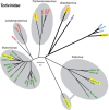
Phylogenetic relationships among taxonomically approved and tentatively assigned members of family Totiviridae. A maximum-likelihood phylogenetic tree was reconstructed from sequences encompassing the predicted CP+RdRp products of each analyzed virus, as described in Materials and Methods. Branches with support values <70% were collapsed to the preceding node, and some terminal branches within the genus Victorivirus are not labeled for simplicity but were all supported at ≥97%. Viruses are labeled with abbreviated names (see Materials Methods for key and GenBank accession numbers), and those for which 3D structures have been determined are highlighted in yellow. Protozoal viruses are labeled in red, fungal viruses are labeled in green, arthropod viruses are labeled in blue, and the one vertebrate virus is labeled in purple. Approved Totiviridae members are grouped within gray ovals according to current genus assignments as labeled; fungal virus UmV-H1 is currently grouped in genus Totivirus as shown but is divergent and appears to warrant reclassification. The black circle and thicker branch lines indicate an apparent subclade of viruses that can undergo efficient extracellular transmission. The scale bar indicates the number of substitutions per aligned position.
FIG 2
FIG 2
Gels and electron micrographs of purified GLV virions. (A) Gel analyses. SDS-PAGE and Coomassie blue staining (left) shows the GLV capsid protein (a) migrating near 100 kDa and the GLV CP/RdRp (b) near 190 kDa. Minor protein bands near 85 and ∼28 kDa are thought to be contaminants or degradation products. Agarose gel electrophoresis and ethidium bromide staining (right) shows a single band for GLV dsRNA (c). (B) Negative-stain TEM. Virions were stained with uranyl acetate to assess particle quality. The scale bar also applies to panel C. (C) Cryo-TEM. Unstained, vitrified virions are shown in one field used for image reconstruction. Examples of full and empty GLV particles are highlighted with black and white arrows, respectively. White dotted arrows point to putative strands of free dsRNA in the solvent background, also seen in the enlarged inset at higher contrast.
FIG 3
FIG 3
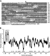
Mass spectrometry to assess protein contents of GLV virions. (A) Sequence of GLV CP precursor is shown. Arrow indicates position of the N-proximal processing cleavage (overlining, N-terminal sequencing results [10]). Sequences represented in the peptides recovered from LC-MS/MS after trypsin or chymotrypsin cleavage of GLV virions are indicated by gray shading. Predominantly uncharged regions as defined in the Discussion are underlined. The highly charged region near the CP N terminus is indicated by dotted underlines. (B) A polarity plot was generated as described in Materials and Methods. Highlighted features (arrow, underlines) are translated from panel A.
FIG 4
FIG 4
Icosahedral 3D image reconstruction of GLV virions. (A) Space-filling stereo view of the GLV particle surface as viewed down an icosahedral 2-fold axis (magenta). One icosahedral 3-fold axis (black) and one icosahedral 5-fold axis (blue) are marked. The map is color-coded by radius (dark green, outermost; red, innermost). (B) Central (equatorial), 1-pixel (1.09-Å)-thick section through the virion density map shown in grayscale (black and white corresponding to highest and lowest densities, respectively). Symmetry axes (icosahedral two-, three-, and 5-fold) are marked, as well as close contacts between capsid and RNA densities (arrowheads). (C) Radial sections through the capsid, centered at indicated radii and each 1 pixel (1.09 Å) thick. The map is coded in grayscale as in panel B. The outlines of one CP-A (orange) and one CP-B (cyan) subunit are shown in each radial section.
FIG 5
FIG 5
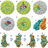
Segmentation of the GLV virion capsid and possible assembly intermediates. (A) Densities corresponding to CP-A and CP-B subunits according to segmentation analysis are colored yellow and light green, respectively. (B) Subunits are labeled to identify contacts between them. Colors are the same as in panel A, plus subunit A1 in orange, subunit B1 in cyan, subunit A2 in brown, subunit B2 in magenta, subunit A3 in red, subunit B3 in blue, and subunit B4 in green. (C and D) The most plausible assembly intermediates are shown: compact decamer (C) and compact tetramer (D). Colors are the same as in panel B. (E) One of the AB dimers in the GLV capsid in which the two subunits are more parallel in orientation. (F) Rotated 90° clockwise and enlarged view of the AB dimer in panel E. The black arrow indicates the region of CP-A that approaches the icosahedral 5-fold channel, and the dotted ellipse indicates the comparable region of CP-B, which has a different orientation. (G) Superimposition of segmented A and B subunits shown in four different orientations, beginning with a view as seen from outside the capsid (as in panel E) and subsequently rotated three more times, each by 90° clockwise. Predominant differences occur at the tip of each of the two subunits closest to the icosahedral 5-fold axis (red arrow) and at the AB interface near the icosahedral 2-fold axis (black/gray arrows).
FIG 6
FIG 6
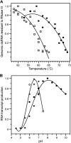
Different responses of GLV and TVV1 virions to temperature and pH. Experiments were performed as detailed in Materials and Methods: GLV (closed symbols), TVV1 (open symbols). (A) Normalized amount of dsRNA resistant to RNase III as a function of temperature. The data points from three (GLV) or four (TVV) experiments with each virus were superimposed in the graph and used to generate the quadratic fit. (B) RNA transcriptase activity as a function of pH. The data points from two experiments with each virus, using two different buffers (Tris-acetate [diamonds] and Tris-HCl [circles]) were superimposed in the graph and used to generate the polynomial fit.
FIG 7
FIG 7
Comparison of GLV to four other taxonomically approved or tentatively assigned totiviruses. All particles are viewed along an icosahedral 2-fold axis, and the surfaces are color coded by radius according to the spectrum shown at right, which lists radii in angstroms. IMNV is tentatively assigned to family Totiviridae.
FIG 8
FIG 8
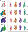
Comparison of the CP subunits of GLV to those of ScV-L-A, HvV190S, and TVV1. (A) Structure of the A1B1 dimer as defined in Fig. 5. Top to bottom, as seen from outside the particle: ScV-L-A (A, red; B, gray), GLV (A, orange; B, cyan), TVV1 (A, purple; B, blue), and HvV190S (A, green; B, steel blue). The structures shown are from segmentation of cryo-TEM maps except for the X-ray crystal structures shown for ScV-L-A. (B) Structure of the A subunits of ScV-L-A, GLV, TVV1, and HvV190S, viewed from four different orientations each. The rotations indicated are around the y axis. Color scheme for the subunits is translated from panel A. Helices in the A subunit of ScV-L-A are shown as coiled ribbons, whereas putative helices in the A subunits of GLV, HvV190S, and TVV1 are depicted by tubes of similar color as their respective density maps. Four putative, long α-helices, located and oriented similarly in the A subunits of these four viruses, are shown in blue.

Similar articles

Cited by

References

    1. Walker G, Dorrell RG, Schlacht A, Dacks JB. 2011. Eukaryotic systematics: a user's guide for cell biologists and parasitologists. Parasitology 138:1638–1663. doi:10.1017/S0031182010001708. - DOI - PubMed
    1. Upcroft P, Upcroft JA. 2001. Drug targets and mechanisms of resistance in the anaerobic protozoa. Clin Microbiol Rev 14:150–164. doi:10.1128/CMR.14.1.150-164.2001. - DOI - PMC - PubMed
    1. Cotton JA, Beatty JK, Buret AG. 2011. Host parasite interactions and pathophysiology in Giardia infections. Int J Parasitol 41:925–933. doi:10.1016/j.ijpara.2011.05.002. - DOI - PubMed
    1. Lane S, Lloyd D. 2002. Current trends in research into the waterborne parasite Giardia. Crit Rev Microbiol 28:123–147. doi:10.1080/1040-840291046713. - DOI - PubMed
    1. Stark D, Barratt JL, van Hal S, Marriott D, Harkness J, Ellis JT. 2009. Clinical significance of enteric protozoa in the immunosuppressed human population. Clin Microbiol Rev 22:634–650. doi:10.1128/CMR.00017-09. - DOI - PMC - PubMed

Publication types

MeSH terms

LinkOut - more resources