. 2014 Dec 19;289(51):35296-313.
doi: 10.1074/jbc.M114.562959. Epub 2014 Oct 29.

Aggregation properties of the small nuclear ribonucleoprotein U1-70K in Alzheimer disease

Affiliations

Aggregation properties of the small nuclear ribonucleoprotein U1-70K in Alzheimer disease

Ian Diner et al. J Biol Chem. .

Abstract

Recent evidence indicates that U1-70K and other U1 small nuclear ribonucleoproteins are Sarkosyl-insoluble and associate with Tau neurofibrillary tangles selectively in Alzheimer disease (AD). Currently, the mechanisms underlying the conversion of soluble nuclear U1 small nuclear ribonucleoproteins into insoluble cytoplasmic aggregates remain elusive. Based on the biochemical and subcellular distribution properties of U1-70K in AD, we hypothesized that aggregated U1-70K itself or other biopolymers (e.g. proteins or nucleic acids) interact with and sequester natively folded soluble U1-70K into insoluble aggregates. Here, we demonstrate that total homogenates from AD brain induce soluble U1-70K from control brain or recombinant U1-70K to become Sarkosyl-insoluble. This effect was not dependent on RNA and did not correlate with detergent-insoluble Tau levels as AD homogenates with reduced levels of these components were still capable of inducing U1-70K aggregation. In contrast, proteinase K-treated AD homogenates and Sarkosyl-soluble AD fractions were unable to induce U1-70K aggregation, indicating that aggregated proteins in AD brain are responsible for inducing soluble U1-70K aggregation. It was determined that the C terminus of U1-70K, which harbors two disordered low complexity (LC) domains, is necessary for U1-70K aggregation. Moreover, both LC1 and LC2 domains were sufficient for aggregation. Finally, protein cross-linking and mass spectrometry studies demonstrated that a U1-70K fragment harboring the LC1 domain directly interacts with aggregated U1-70K in AD brain. Our results support a hypothesis that aberrant forms of U1-70K in AD can directly sequester soluble forms of U1-70K into insoluble aggregates.

Keywords: Neurodegenerative Disease; Protein Aggregation; RNA; Spliceosome; Tau Protein (Tau).


Figures

FIGURE 1.
FIGURE 1.
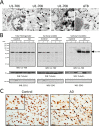
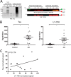
Subcellular and biochemical distribution properties for U1-70K in AD. A, immunogold transmission electron microscopy of U1-70K or Tau neurofibrillary tangles (AT8) in AD frontal cortex. Immunogold positive staining for U1-70K (left panel, scale bar, 0.5 μm) is observed in both the nucleus (Nu), denoted by arrowheads, and cytoplasm (Cy), denoted by arrows. The asterisk denotes twisted-ribbon tangle-like structures positive for U1-70K in the cytoplasm (middle panels), resembling AT8-positive NFTs shown at higher magnification (right panel). The nuclear envelope (NE) is also denoted. B, U1-70K displays a near complete solubility shift in AD brain by Western blot (WB). Total brain homogenates prepared from control (n = 3) and AD (n = 3) frontal cortex (left panel) were fractionated into Sarkosyl-soluble (middle panel) and Sarkosyl-insoluble fractions (right panel) and blotted for U1-70K. Upon fractionation U1-70K from AD brain almost exclusively partitions into the Sarkosyl-insoluble fraction. Tubulin served as a relative loading control and early endosomal antigen 1 (EEA1) served as a soluble protein marker. C, immunohistochemisty using antibodies against U1-70K in both control (left panel) and AD brain (right panel) tissue (scale bar, 50 μm; higher magnification shown in the insets). Arrows highlight cytoplasmic U1-70K tangles, and arrowheads highlight U1-70K-positive nuclei.
FIGURE 2.
FIGURE 2.
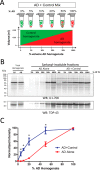
AD homogenates induce the aggregation of normal soluble U1-70K from control human brain. A, frontal cortex homogenates of AD and control brains were mixed in six ratios with increasing proportions of AD homogenate. The mixtures were incubated 4 h at 4 °C and fractionated. B, representative WB for U1-70K (top panel) of Sarkosyl-insoluble fractions from control and AD (AD + Control) mixtures or from matching quantities of AD homogenates alone (AD alone). TDP-43 insolubility was consistent across control and AD Sarkosyl-insoluble fractions and was therefore used as a loading control (bottom panel). C, quantification of U1-70K in the insoluble fractions from AD+Control mixtures revealed a nonlinear increase in the signal of Sarkosyl-insoluble U1-70K, consistent with the sequestration of soluble U1-70K from control brain into the Sarkosyl-insoluble fraction. In contrast, the AD alone series revealed a roughly proportional relationship between the amount of AD brain homogenate and the Sarkosyl-insoluble U1-70K signal. Analysis of three biological replicates indicates significant enrichment of U1-70K in AD+Control series compared with the AD alone series. Statistical significance (*) was calculated using Student's t test (p < 0.05).
FIGURE 3.
FIGURE 3.
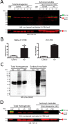
AD homogenates, but not CBD homogenates, induce the aggregation of soluble recombinant U1-70K. A, HEK lysate with overexpressed Myc-tagged rU1-70K (full-length) was incubated with control (n = 3) and AD (n = 3) brain homogenates for 4 h at 4 °C and fractionated. Sarkosyl-insoluble fractions were analyzed by quantitative WB for native U1-70K (red) and Myc-tagged rU1-70K (green). HEK lysate alone did not harbor insoluble rU1-70K, and only AD homogenate was able to induce rU1-70K aggregation (top panel). TDP-43 (red) served as a loading control (bottom panel). B, densitometry analysis revealed a significant increase in both native and rU1-70K in the Sarkosyl-insoluble fractions of the AD + HEK mixtures compared with the control + HEK mixtures. Statistical significance (*) was calculated using Student's t test (p < 0.05). C, both CBD and to a greater extent AD homogenate had increased levels of Sarkosyl-insoluble phosphorylated Tau (Ser(P)-404) levels compared with control brain homogenate. D, compared with AD homogenate, a CBD homogenate was unable to induce rU1-70K into an aggregated state. TDP-43 served as a loading control (bottom panel).
FIGURE 4.
FIGURE 4.
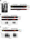
U1-70K aggregation is not dependent on RNA and requires Sarkosyl-insoluble proteins in AD homogenate. A, AD brain homogenate and HEK lysate overexpressing rU1-70K were pretreated with RNase A prior to performing co-aggregation experiments. Total RNA extracts were analyzed by urea-PAGE to confirm RNA degradation (left panel). HEK lysate with overexpressed rU1-70K (± RNase) was incubated alone or with control and AD homogenates (± RNase) for 4 h at 4 °C and fractionated. WB analysis of Sarkosyl-insoluble fractions revealed equivalent levels of native U1-70K (red) and rU1-70K (green) in both RNase-treated and -untreated samples (right panel). TDP-43 served as a loading control (bottom panel). B, AD homogenate was pretreated with PK for 1 h at 37 °C and then inhibited with the addition of excess PMSF. The PK-treated AD homogenate was incubated with rU1-70K lysate for 4 h at 4 °C and fractionated. Ponceau S staining of the transfer membrane indicates near complete digestion of protein in the PK-treated samples (bottom panel). PK digestion abolished the ability of AD homogenate to seed rU1-70K. C, rU1-70K HEK lysate was incubated with control and AD brain homogenates as well as the Sarkosyl-soluble fraction of AD brain (AD sol.) as before. The resulting Sarkosyl-insoluble fractions were subjected to Western blot analysis for native U1-70K (red) and rU1-70K (green), suggesting that the Sarkosyl-insoluble protein fraction is responsible for inducing rU1-70K aggregation (top panel). Ponceau S staining of the transfer membrane shows equal protein loading across total control homogenate, AD homogenate, and Sarkosyl-soluble AD fractions (bottom panel).
FIGURE 5.
FIGURE 5.
U1-70K and Tau do not always co-aggregate in AD. A, AD case with low levels of Sarkosyl-insoluble Tau (AD-LT) was subjected to WB analysis for Tau, along with a typical “high Tau” AD case (AD) and a control case (left panel). The control (Ctl), AD, and AD-LT cases were incubated with rU1-70K lysate for a co-aggregation assay. The resulting Sarkosyl-insoluble fractions were subjected to WB analysis for native U1-70K (red) and rU1-70K (green) (top right panel). Both the AD and AD-LT homogenates were equally capable of inducing rU1-70K aggregation, in contrast to the control (Ctl) homogenate. The rU1-70K lysate alone (HEK) did not harbor any Sarkosyl-insoluble rU1-70K. TDP-43 served as the loading control (bottom right panel). B, U1-70K and Tau levels in the Sarkosyl-insoluble proteome were quantified by peptide ion intensities across 18 individual cases representing five controls (each run in technical replicate) and 13 AD cases. Both U1-70K and Tau levels were significantly enriched in the Sarkosyl-insoluble fractions of AD cases compared with control cases (*, p value < 0.01, Student's t test). C, coefficient of determination across AD (n = 13) reveals a weak correlation between Tau and U1-70K levels in the insoluble fractions. Protein signal intensities were normalized to the individual AD cases with the maximum signal of U1-70K and Tau, respectively.
FIGURE 6.
FIGURE 6.
U1-70K is highly disordered and has C-terminal low complexity domains. A, DISOPRED algorithm predicts intrinsically disordered regions within the N-terminal (amino acids 50–100) and C-terminal (amino acids 220–437) portion of U1-70K, which harbors two distinct low complexity domains, LC1 (amino acids 231–308) and LC2 (amino acids 317–407). B, primary amino acid sequence of U1-70K with the N-terminal (green), RNA recognition motif (RRM) domain (red), and two LC domains (orange) are highlighted. Regions predicted to be intrinsically disordered are underlined.
FIGURE 7.
FIGURE 7.
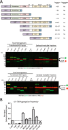
Intrinsically disordered C terminus is necessary and sufficient for U1-70K aggregation. A, to determine which region(s) or amino acid sequences of U1-70K are required for aggregation, we assessed the ability of AD brain homogenate to induce the aggregation of full-length rU1-70K and a number of GST-tagged N- and C-terminal truncations (top panel). Two representative Western blots are shown. Each rU1-70K fragment was expressed in HEK cells, and individual lysates harbored equivalent amounts of recombinant protein (inputs). Each rU1-70K lysate was incubated with AD brain homogenate (bottom panels, 7th lanes) for 12 h at 4 °C prior to fractionation. The Sarkosyl-insoluble fractions were subjected to WB analysis for native U1-70K (red) and rU1-70K (green). rU1-70K residues 182–437 also displays a C-terminal fragment (∼37 kDa) caused by in vivo proteolysis. B, aggregation propensity of the full-length rU1-70K and various N- and C-terminal truncations was determined by quantifying the recombinant (green) signal ratio of Sarkosyl-insoluble/input fractions. The rU1-70K fragment 182–310 demonstrated the highest degree of aggregation. The dashed line represents the baseline level for full-length rU1-70K (residues 1–437) aggregation. Co-aggregation assays were performed in duplicate for each rU1-70K fragment (± S.E.).
FIGURE 8.
FIGURE 8.
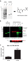
SDA-labeled rU1-70K fragments harboring the LC1 domain directly interact with aggregated U1-70K in AD brain. A, GST-tagged N- or C-terminal rU1-70K fragments (residues 1–99 and 182–310) were expressed in HEK cells, affinity-purified with glutathione-agarose, and assessed for purity by SDS-PAGE and Coomassie Blue staining (left panel). Purified fragments were derivatized with the amine-reactive photo-activatable cross-linker NHS-diazirine (SDA) (right panel). B, co-aggregation assays using Sarkosyl-insoluble fractions from AD brain were performed with unlabeled or SDA-labeled purified rU1-70K fragments (residues 1–99 or 182–310). Following UV treatment and fractionation, the cross-linked products were analyzed by Western blotting for both rU1-70K (green) and native U1-70K (red) (top panel). Higher molecular weight cross-linked species were only observed in co-aggregation experiments with SDA-labeled 182–310-residue rU1-70K fragment. Notably, lower molecular weight C-terminal fragments in the 182–310-residue rU1-70K co-aggregation assays were also observed to form cross-linked products. TDP-43 was used as a loading control (bottom panel). C, approximately 60% of native monomeric U1-70K from AD brain was cross-linked in co-aggregation assays using SDA-labeled 182–310-residue rU1-70K fragments compared with the control unlabeled fragments (no SDA). In contrast, ∼14% of native TDP-43 was cross-linked in these same experiments, which were performed in technical quadruplicate (n = 4). Student's t test was performed for significance (p < 0.05).
FIGURE 9.
FIGURE 9.
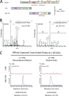
Mass spectrometry analysis of affinity-captured cross-linked products confirm a direct interaction between rU1-70K and aggregated U1-70K in AD brain. A, co-aggregation assays using Sarkosyl-insoluble fractions from AD brain were performed with dual-labeled (SDA+biotin) purified rU1-70K fragments (amino acids (aa) 1–99 or 182–310). Following UV cross-linking and fractionation, the cross-linked products were affinity-captured with streptavidin magnetic beads. The eluted products were resolved by SDS-PAGE, and the gel regions of >60 kDa were excised, subjected to in-gel trypsin digestion, and analyzed by LC-MS/MS. Both rU1-70K and native U1-70K peptides from AD brain were identified by MS/MS and quantified by XIC. B, representative MS/MS spectra for triply charged U1-70K peptides mapping to amino acids 219–231 (YDERPGPSPLPHR) or 145–155 (GYAFIEYEHER). C, XIC for U1-70K peptide sequence 219–231 (m/z = 507.59) and 145–155 (m/z = 471.89) from high molecular weight species cross-linked with rU1-70K fragments 1–99 (negative control) and 182–310. Notably, the peptide derived from native U1-70K (residues 145–155) was only detected in the 182–310-residue rU1-70K cross-linked sample, consistent with a direct interaction with aggregated U1-70K in AD brain. Peptide signal intensities were normalized to levels in the 182–310-residue samples (100% maximum intensity).

Similar articles

Cited by

References

    1. Taylor J. P., Hardy J., Fischbeck K. H. (2002) Toxic proteins in neurodegenerative disease. Science 296, 1991–1995 - PubMed
    1. Ross C. A., Poirier M. A. (2004) Protein aggregation and neurodegenerative disease. Nat. Med. 10, S10–S17 - PubMed
    1. Querfurth H. W., LaFerla F. M. (2010) Alzheimer's disease. New Engl. J. Med. 362, 329–344 - PubMed
    1. Sperling R. A., Aisen P. S., Beckett L. A., Bennett D. A., Craft S., Fagan A. M., Iwatsubo T., Jack C. R., Jr., Kaye J., Montine T. J., Park D. C., Reiman E. M., Rowe C. C., Siemers E., Stern Y., Yaffe K., Carrillo M. C., Thies B., Morrison-Bogorad M., Wagster M. V., Phelps C. H. (2011) Toward defining the preclinical stages of Alzheimer's disease: recommendations from the National Institute on Aging-Alzheimer's Association workgroups on diagnostic guidelines for Alzheimer's disease. Alzheimer's Dementia 7, 280–292 - PMC - PubMed
    1. O'Brien R. J., Resnick S. M., Zonderman A. B., Ferrucci L., Crain B. J., Pletnikova O., Rudow G., Iacono D., Riudavets M. A., Driscoll I., Price D. L., Martin L. J., Troncoso J. C. (2009) Neuropathologic Studies of the Baltimore Longitudinal Study of Aging (BLSA). J. Alzheimer's Dis. 18, 665–675 - PMC - PubMed

Publication types

MeSH terms