. 2015 Jun 15;211(12):1977-86.
doi: 10.1093/infdis/jiu592. Epub 2014 Oct 28.

Severity of retinopathy parallels the degree of parasite sequestration in the eyes and brains of malawian children with fatal cerebral malaria

Affiliations

Severity of retinopathy parallels the degree of parasite sequestration in the eyes and brains of malawian children with fatal cerebral malaria

Valentina Barrera et al. J Infect Dis. .

Abstract

Background: Malarial retinopathy (MR) has diagnostic and prognostic value in children with Plasmodium falciparum cerebral malaria (CM). A clinicopathological correlation between observed retinal changes during life and the degree of sequestration of parasitized red blood cells was investigated in ocular and cerebral vessels at autopsy.

Methods: In 18 Malawian children who died from clinically defined CM, we studied the intensity of sequestration and the maturity of sequestered parasites in the retina, in nonretinal ocular tissues, and in the brain.

Results: Five children with clinically defined CM during life had other causes of death identified at autopsy, no MR, and scanty intracerebral sequestration. Thirteen children had MR and died from CM. MR severity correlated with percentage of microvessels parasitized in the retina, brain, and nonretinal tissues with some neuroectodermal components (all P < .01). In moderate/severe MR cases (n = 8), vascular congestion was more intense (ρ = 0.841; P < .001), sequestered parasites were more mature, and the quantity of extraerythrocytic hemozoin was higher, compared with mild MR cases (n = 5).

Conclusions: These data provide a histopathological basis for the known correlation between degrees of retinopathy and cerebral dysfunction in CM. In addition to being a valuable tool for clinical diagnosis, retinal observations give important information about neurovascular pathophysiology in pediatric CM.

Keywords: Plasmodium falciparum malaria; cerebral malaria; clinicopathological correlation; histopathology; malarial retinopathy; microvascular congestion; neurovasculature; parasite sequestration; pediatric coma; vascular pathology.


Figures

Figure 1.
Figure 1.
Clinical and gross pathology photographs for patient 3, who had moderate/severe MR. A, Color fundus montage image from the left eye. Retinal whitening, appearing as patches of opacification, can be seen in all retinal quadrants. Vessel discoloration, characterized by orange color changes, was seen (black arrows), frequently in association with vessel tortuosity (asterisks). Two hemorrhages are present at the retinal periphery (white arrows). B, Gross photograph, taken after death, of the enucleated eye of patient 3, showing retinal vessel discoloration and hemorrhages (black and white arrows, respectively).
Figure 2.
Figure 2.
Quantitative assessment of parasitized red blood cell (pRBC) sequestration in retinal and cerebral microvasculature. A, Correlation between the overall score for clinical fundoscopy (Table 1) and the percentage of parasitized capillaries (n = 18; P < .001). No malarial retinopathy (MR); (No; n = 5) open circles; mild MR (n = 5), light gray circles; moderate/severe (m/s) MR (n = 8), dark gray circles. B, Bar chart showing percentages of arterioles, capillaries, and venules affected by pRBC sequestration across the 3 MR classification groups. Results are shown as means ± SDs for each individual MR group. P ≤ .001 for all tests across groups per type of vessel, except P = .8 for the comparison of means of arterioles in patients with no MR vs those with mild MR. Data were normally distributed (Shapiro-Wilk test). Means were compared using analysis of variance (with the Bonferroni post-hoc test). C, Correlation between the percentage of retinal and cerebral capillaries parasitized in the same patients as in panels A and B (n = 18; P < .001).
Figure 3.
Figure 3.
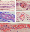
Photomicrographs of parasitized retinal capillaries and venules. A, Representative image of retina cross-sections from a moderate/severe MR specimen (patient 3) containing pigmented (mature-form) parasitized red blood cells (pRBCs). Cross-section showing capillaries and venules affected by parasite sequestration are shown (arrows, intense sequestration; arrowheads, low or no sequestration). B, High-power image of 1 representative venule with numerous pRBCs. A prominent endothelial cell nucleus is shown (arrow). C, A Giemsa-stained venule illustrating pigmented (mature-form) pRBCs (patient 7). D, Parasitized arteriole in a cross-section stained by hematoxylin-eosin, with a lower number of pRBCs (arrow; patient 8). The vessel wall is characterized by a thick, pink-stained muscle layer. E, Highly parasitized venules with adjacent superficial retinal hemorrhage (patient 4). Section was stained by hematoxylin-eosin. Nonparasitized RBCs stain red, while pigmented pRBCs contain hemozoin, which appears as dark brown spots. Scale bars: 100 μm (A); 20 μm (BD), and 50 μm (E).
Figure 4.
Figure 4.
Differential parasitized red blood cell (pRBC) sequestration in ocular microvasculature. AG, Box plots showing the percentage of parasitized capillaries (white boxes) and venules (gray boxes) in the different ocular tissues. A and C–G, There were 5 specimens in the group with no malaria retinopathy (MR), 5 in the mild MR group, and 8 in the moderate/severe (m/s) MR group. B, There were 4 specimens in the group with no MR, 4 in the group with mild MR, and 8 in the group with m/s MR n = 8. Data were compared using analysis of variance (with the Bonferroni post-hoc test). *P < .05 and **P < .01, when comparing no MR or mild MR vs m/s MR per vessel type. P < .01 for all tests comparing no MR vs mild MR per vessel type, except for choroidal capillaries and episcleral and extraocular muscle vessels. Boxes denote medians and 25th and 75th percentiles. Outliers are reported as white or grey dots outside the boxes. H, Representative image of a cross-section of a parasitized capillary from a ciliary body (patient 4), stained by hematoxylin-eosin. Two pigmented mature pRBCs, probably schizonts, are shown. The parasitophorous vacuoles of the parasites inside the RBCs are clearly visible (arrows), with dark brown dots of hemozoin and blue nuclei of new growing parasites (arrowhead). Ciliary muscle fiber is indicated by an asterisk. Scale bar: 20 μm.
Figure 5.
Figure 5.
Congestion in retinal venules affected by parasite sequestration. A, Scatterplots and ranking correlation between the percentage of parasitized venules and number of parasitized red blood cells (pRBCs) sequestered (n = 18). B, Correlation between the number of pRBCs plus nonparasitized RBCs and the percentage of parasitized venules (n = 17; P = .001). Linear regression was performed after natural logarithm loge-(loge) transformation of dependent variables. The coefficient was 1.911. The fit line, R2, and 95% confidence interval for the mean are reported on the plot. Patient 18 was removed from the analysis because values for the dependent and independent variables were 0. To evaluate the quality of the model, residual diagnostics were accomplished, and confounders were excluded. C, Highly congested venule from a moderate/severe malarial retinopathy (MR) case (patient 3). The section was stained by hematoxylin-eosin. Vessel wall (thin and attenuated) and endothelium nucleus are indicated by arrows and an arrowhead, respectively. Scale bar: 50 μm.
Figure 6.
Figure 6.
Maturation stage of parasitized red blood cells (pRBCs) in retinal microvasculature. A, Representative image of retinal venules containing unpigmented (immature form, with parasite nucleus stained blue inside the infected RBC) stage pRBCs detected in patient 5. B and C, Box plots showing the relative number of unpigmented (white boxes) and pigmented (gray boxes) pRBCs sequestered in 100 retinal capillaries (B) and venules (C) across classification groups. There were 5 specimens in the group with no malaria retinopathy (MR), 5 in the mild MR group, and 8 in the moderate/severe (m/s) MR group. D, Representative image of retinal microvessel with extraerythrocytic hemozoin (HZ) from patient 2, who had m/s MR (E). Bar graphs showing the percentage of retinal capillaries and venules containing extraerythrocytic HZ across classification groups. Results are reported as means ± SD for each individual MR group. F, Box plots showing the percentage of parasitized retinal capillaries in the same patients as in panel E, but clustered by CM classification. CM1, sequestration of pRBCs in cerebral capillaries; CM2, sequestration of pRBCs in cerebral capillaries plus intravascular and perivascular pathology (Supplementary Table 1); CM3, sequestration of <21% of cerebral capillaries. Boxes denote medians and 25th and 75th percentiles. Data were analyzed by the Kruskal–Wallis test within classification groups. *P ≤ .05 and **P ≤ .01. Scale bars: 50 μm (A); 20 μm (D).

Similar articles

Cited by

  • Parasite histones are toxic to brain endothelium and link blood barrier breakdown and thrombosis in cerebral malaria.
    Moxon CA, Alhamdi Y, Storm J, Toh JMH, McGuinness D, Ko JY, Murphy G, Lane S, Taylor TE, Seydel KB, Kampondeni S, Potchen M, O'Donnell JS, O'Regan N, Wang G, García-Cardeña G, Molyneux M, Craig AG, Abrams ST, Toh CH. Moxon CA, et al. Blood Adv. 2020 Jul 14;4(13):2851-2864. doi: 10.1182/bloodadvances.2019001258. Blood Adv. 2020. PMID: 32579667 Free PMC article.
  • Grading fluorescein angiograms in malarial retinopathy.
    MacCormick IJ, Maude RJ, Beare NA, Borooah S, Glover S, Parry D, Leach S, Molyneux ME, Dhillon B, Lewallen S, Harding SP. MacCormick IJ, et al. Malar J. 2015 Sep 24;14:367. doi: 10.1186/s12936-015-0897-7. Malar J. 2015. PMID: 26403288 Free PMC article.
  • Pathophysiology and neurologic sequelae of cerebral malaria.
    Schiess N, Villabona-Rueda A, Cottier KE, Huether K, Chipeta J, Stins MF. Schiess N, et al. Malar J. 2020 Jul 23;19(1):266. doi: 10.1186/s12936-020-03336-z. Malar J. 2020. PMID: 32703204 Free PMC article. Review.
  • Ocular Pathology of Cerebral Malaria.
    White VA, Barrera V, MacCormick IJC. White VA, et al. Methods Mol Biol. 2022;2470:749-763. doi: 10.1007/978-1-0716-2189-9_55. Methods Mol Biol. 2022. PMID: 35881387
  • A retinal model of cerebral malaria.
    Paquet-Durand F, Beck SC, Das S, Huber G, Le Chang, Schubert T, Tanimoto N, Garcia-Garrido M, Mühlfriedel R, Bolz S, Hoffmann W, Schraermeyer U, Mordmüller B, Seeliger MW. Paquet-Durand F, et al. Sci Rep. 2019 Mar 5;9(1):3470. doi: 10.1038/s41598-019-39143-z. Sci Rep. 2019. PMID: 30837488 Free PMC article.

References

    1. Newton CR, Taylor TE, Whitten RO. Pathophysiology of fatal falciparum malaria in African children. Am J Trop Med Hyg 1998; 58:673–83. - PubMed
    1. Taylor TE, Fu WJ, Carr RA, et al. Differentiating the pathologies of cerebral malaria by postmortem parasite counts. Nat Med 2004; 10:143–5. - PubMed
    1. Hero M, Harding SP, Riva CE, Winstanley PA, Peshu N, Marsh K. Photographic and angiographic characterization of the retina of Kenyan children with severe malaria. Arch Ophthalmol 1997; 115:997–1003. - PubMed
    1. Maccormick IJ, Beare NA, Taylor TE, et al. Cerebral malaria in children: using the retina to study the brain. Brain 2014; 137(Pt 8):2119–42. - PMC - PubMed
    1. Essuman VA, Ntim-Amponsah CT, Astrup BS, et al. Retinopathy in severe malaria in Ghanaian children--overlap between fundus changes in cerebral and non-cerebral malaria. Malar J 2010; 9:232. - PMC - PubMed

Publication types